. 2024 Jul 25;13(12):103130.
doi: 10.1016/j.eats.2024.103130. eCollection 2024 Dec.

Long Head of Biceps Tenodesis for Maintaining Inherent Length and Uniform Tension at the Bicipital Groove: Suprapectoral Double-Row Technique With All-Suture Anchors

Affiliations

Long Head of Biceps Tenodesis for Maintaining Inherent Length and Uniform Tension at the Bicipital Groove: Suprapectoral Double-Row Technique With All-Suture Anchors

Ayyappan V Nair et al. Arthrosc Tech. .

Abstract

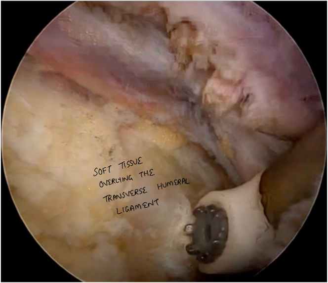
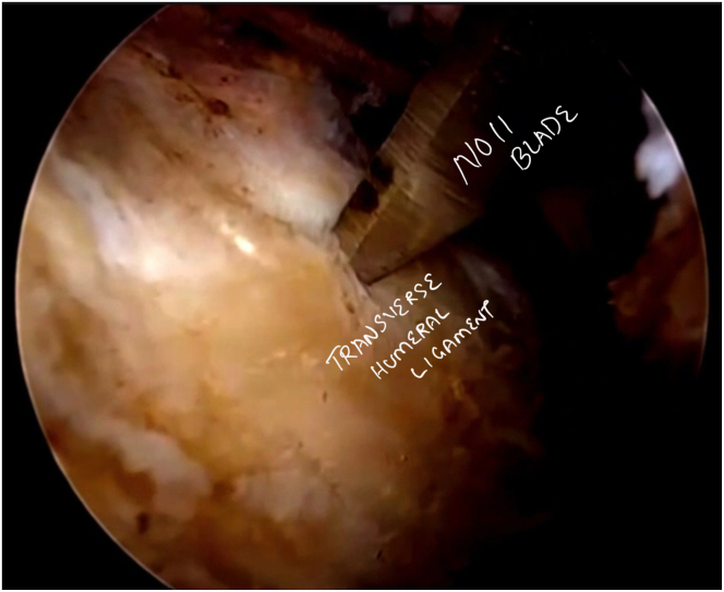
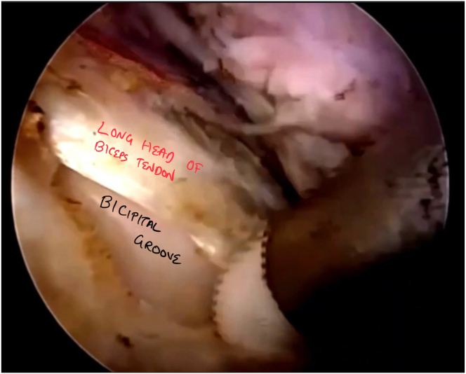
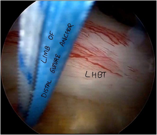
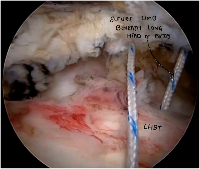
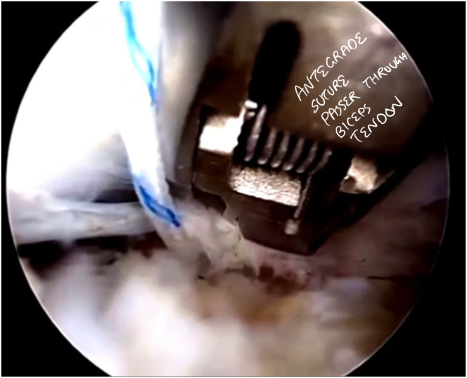
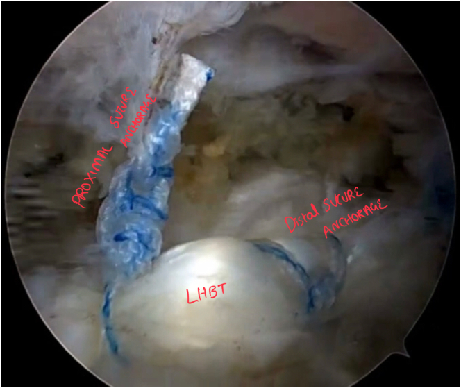
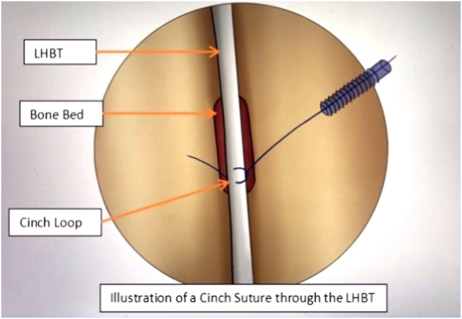
The clinicopathologic conditions of the long head of the biceps tendon vary, encompassing tendinitis, peritendinous inflammation, hypertrophy, and partial or complete tears. These symptoms are typically linked with SLAP tears and instability of the long head of the biceps tendon, often resulting in partial displacement or complete dislocation. The choice between tenotomy and tenodesis depends on varied factors. The choice of suprapectoral or subpectoral tenodesis is a difficult decision and should be weighed on a tailored basis. Our approach to suprapectoral tenodesis presents a blend of factors, maintaining uniform tension in the bicipital groove, limiting the number of portals for arthroscopy, re-tensioning the tendon after initial anchor placement, and preserving the inherent length of the functional biceps tendon without compromising its quality.


Figures

Fig 1
Fig 1
The patient is placed in the lateral decubitus position (right side), with the arthroscopic electrocautery device inserted through the midaxillary portal and viewing performed through the midlateral portal. This setup is used for clearing the tissues overlying the bicipital groove, making the transverse humeral ligament more evident during arthroscopy.
Fig 2
Fig 2
The patient is placed in the lateral decubitus position (right side). A No. 11 arthroscopic blade is introduced through the midaxillary portal, with viewing performed through the midlateral portal. The transverse humeral ligament is incised using a No. 11 blade over the biceps groove to visualize the underlying long head of the biceps tendon.
Fig 3
Fig 3
The patient is placed in the lateral decubitus position (right side). The arthroscopic electrocautery device is inserted through the midaxillary portal, with viewing performed through the midlateral portal. The long head of the biceps tendon is released with clearance made beside the lateral ridge of the bicipital groove. This approach helps avoid damage to the subscapularis muscle.
Fig 4
Fig 4
The patient is placed in the lateral decubitus position (right side). The arthroscopic electrocautery device is introduced via the midaxillary portal, with viewing performed through the midlateral portal. The first suture anchor is placed behind the long head of the biceps tendon after displacement of the tendon medially after bone bed preparation. Microfracturing the bone bed helps achieve good bone-to-tendon healing.
Fig 5
Fig 5
The patient is placed in the lateral decubitus position (right side). The bird-beak device is introduced via the midaxillary portal, with viewing performed through the midlateral portal. The bird-beak device is used to pass the FiberWire beneath the long head of the biceps tendon (LHBT).
Fig 6
Fig 6
The patient is placed in the lateral decubitus position (right side). An arthroscopic antegrade suture passer is introduced through the midaxillary portal, with viewing performed through the midlateral portal. The antegrade suture passer makes way for the cinch sutures to be made by use of an overlay technique for tendon apposition over the bicipital groove.
Fig 7
Fig 7
The patient is placed in the lateral decubitus position (right side), with viewing performed through the midlateral portal. The final construct is shown, with double-row anchor suture apposition, ensuring excellent abutment of the tendon. (LHBT, long head of biceps tendon.)
Fig 8
Fig 8
Cinched loop suture construct over biceps tendon. In this surgical technique, the suture material is looped around the biceps tendon within the groove and then cinched down to create tension, effectively securing the tendon in place. (LHBT, long head of biceps tendon.)

Similar articles

References

    1. Friedman J.L., FitzPatrick J.L., Rylander L.S., Bennett C., Vidal A.F., McCarty E.C. Biceps tenotomy versus tenodesis in active patients younger than 55 years: Is there a difference in strength and outcomes? Orthop J Sports Med. 2015;3 - PMC - PubMed
    1. Hassan S., Patel V. Biceps tenodesis versus biceps tenotomy for biceps tendinitis without rotator cuff tears. J Clin Orthop Trauma. 2019;10:248–256. - PMC - PubMed
    1. Jenkins S.M., Hwang I.M., Del Sol S.R., et al. Subpectoral biceps tenodesis using an all-suture anchor. Arthrosc Tech. 2022;11:e555–e562. - PMC - PubMed
    1. Ergün S., Cırdı Y.U., Baykan S.E., Akgün U., Karahan M. Clinical outcome comparison of suprapectoral and subpectoral tenodesis of the long head of the biceps with concomitant rotator cuff repair: A systematic review. Shoulder Elb. 2022;14:6–15. - PMC - PubMed
    1. Maier D., Izadpanah K., Jaeger M., Ogon P., Südkamp N.P. Tenoscopic suprapectoral biceps tenodesis. Arthrosc Tech. 2016;5:e55–e62. - PMC - PubMed

LinkOut - more resources