Review
. 2024 Dec 23:4:1396588.
doi: 10.3389/fneph.2024.1396588. eCollection 2024.

Vascular injury in glomerulopathies: the role of the endothelium

Affiliations
Review

Vascular injury in glomerulopathies: the role of the endothelium

Géssica Sabrine Braga Barbosa et al. Front Nephrol. .

Abstract

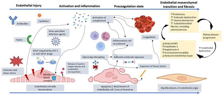
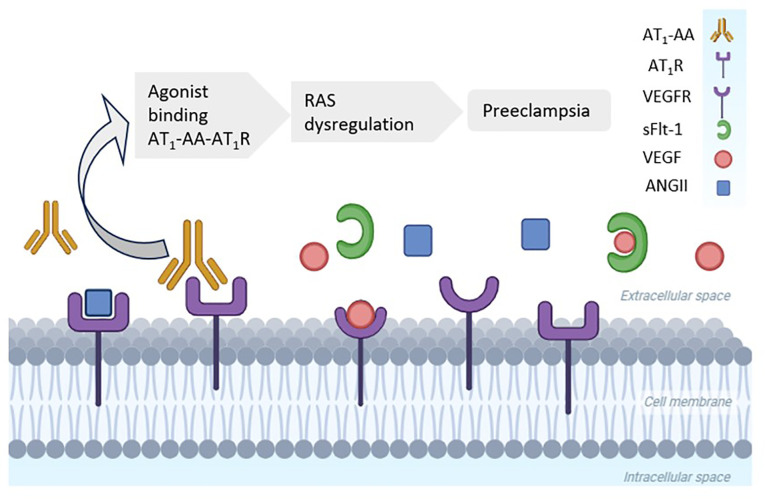
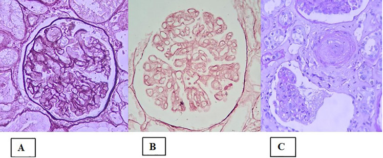
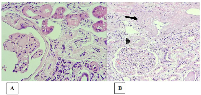
In glomerulopathies, endothelial dysfunction and the presence of histological vascular lesions such as thrombotic microangiopathy, arteriolar hyalinosis, and arteriosclerosis are related to a severe clinical course and worse renal prognosis. The endothelial cell, which naturally has anti-inflammatory and anti-thrombotic regulatory mechanisms, is particularly susceptible to damage caused by various etiologies and can become dysfunctional due to direct/indirect injury or a deficiency of protective factors. In addition, endothelial regulation and protection involve participation of the complement system, factors related to angiogenesis, the renin-angiotensin system (RAS), endothelin, the glycocalyx, the coagulation cascade, interaction between these pathways, interactions between glomerular structures (the endothelium, mesangium, podocyte, and basement membrane) and interstitial structures (tubules, arterioles and small vessels). Dysregulation of those components is also associated with the progression of renal fibrosis, since endothelial cell damage promotes endothelial-to-mesenchymal transition. Although the potential mechanisms of vascular injury have been widely described in diabetic kidney disease, hypertensive nephrosclerosis, and hemolytic uremic syndrome, they require further elucidation in other glomerulopathies. A better understanding of the pathogenesis of vascular injury in patients with glomerular diseases could contribute to the development of specific treatments for such injury.

Keywords: arteriolar hyalinosis; arteriosclerosis; glomerular endothelial cell; glomerulopathy; thrombotic microangiopathy.


Conflict of interest statement

The authors declare that the research was conducted in the absence of any commercial or financial relationships that could be construed as a potential conflict of interest. The author(s) declared that they were an editorial board member of Frontiers, at the time of submission. This had no impact on the peer review process and the final decision.

Figures

Figure 1
Figure 1
Representation of the process of endothelial dysfunction resulting from injury to a glomerular endothelial cell by toxins, cytokines, antibodies, infectious agents, VEGF depletion, ischemia, or another factor. Cellular injury promotes the release of reactive oxygen species and endothelial microparticles. There is activation of the complement system, which can culminate in the production of the membrane attack complex and endothelial lysis. In addition, anaphylatoxins from the complement system, such as C3a and C5a, promote recruitment of inflammatory cells, amplifying the inflammatory state. The disruption of the glycocalyx and the endothelial injury itself promote exposure of adhesion molecules, which increases the connection with other inflammatory mediators. There is also exposure of tissue factor with activation of prothrombotic mechanisms, which favors microthrombi affecting the microcirculation. The coagulation system is also activated through dysregulation of the complement system, which favors platelet aggregation via cytokines. Given that the insult persists, and endothelial repair is impaired, there can be loss of fenestrations and endothelial cell detachment, with reduced vascular permeability. The persistent dysfunction evolves with a change from the endothelial to the mesenchymal phenotype, contributing to renal fibrosis. Furthermore, there is increased expression of the angiotensin II and endothelin-1 receptors, which act by promoting vasoconstriction by decreasing nitric oxide (NO) production, thus increasing inflammation and fibrosis. The interaction between the endothelium, podocyte, and tubulointerstitium is dysregulated, increasing proteinuria and contributing to glomerulosclerosis and tubulointerstitial fibrosis, including impairment of arterioles and small vessels. Finally, there is accelerated progression to chronic kidney disease because uremic toxins perpetuate the state of endothelial dysfunction. VEGF, vascular endothelial growth factor; sFlt-1, soluble fms-like tyrosine kinase-1. (Created with BioRender.com).
Figure 2
Figure 2
High levels of agonist angiotensin II (AngII) type 1 receptor autoantibody (AT1-AA) in women with preeclampsia. AT1-AA binds to the angiotensin type 1 receptor (AT1R), whereas soluble fms-like tyrosine kinase-1 (sFlt-1) blocks the vascular endothelial growth factor receptor (VEGFR). Both processes promote a hypertensive status in preeclampsia. RAS, renin–angiotensin system. (Created with BioRender.com).
Figure 3
Figure 3
Histological lesions in the presentation of thrombotic microangiopathy. (A) Global mesangiolysis; (B) Extensive double contours of the glomerular basement membrane (two-layer appearance); (C) Concentric myointimal proliferation in an “onion-skin” pattern, with mucoid edema and lumen obliteration. There is also a glomerular tuft with a retracted appearance and a wrinkled basement membrane (lower portion of the image).
Figure 4
Figure 4
Arteriolosclerosis and renal arteriosclerosis. (A) Bulky, circumferential mural hyaline deposits in the arteriole walls (upper portion of the image), in a patient with diabetes and nodular glomerulosclerosis (lower left corner of the image); (B) Moderate intimal fibrosis in the interlobular arterial branch (arrow) and intense arteriolar hyalinosis (arrowhead).

Similar articles

References

    1. Jourde-Chiche N, Fakhouri F, Dou L, Bellien J, Burtey S, Frimat M, et al. . Endothelium structure and function in kidney health and disease. Nat Rev Nephrol. (2019) 15(2):87–108. doi: 10.1038/s41581-018-0098-z - DOI - PubMed
    1. Neves PDMM, Souza RA, Torres FM, Reis FA, Pinheiro RB, Dias CB, et al. . Evidences of histologic thrombotic microangiopathy and the impact in renal outcomes of patients with IgA nephropathy. PloS One. (2020) 15(11):e0233199. doi: 10.1371/journal.pone.0233199 - DOI - PMC - PubMed
    1. Manenti L, Gnappi E, Vaglio A, Allegri L, Noris M, Bresin E, et al. . Atypical haemolytic uraemic syndrome with underlying glomerulopathies. A case series and a review of the literature. Nephrol Dialysis Transplant. (2013) 28(9):2246–59. doi: 10.1093/ndt/gft220 - DOI - PubMed
    1. Chen F-F, Yu X-J, Wang H, Zhang X, Tan Y, Qu Z, et al. . Clinical value of the renal pathologic scoring system in complement-mediated thrombotic microangiopathy. Ren Fail. (2023) 45(1):2161396. doi: 10.1080/0886022X.2022.2161396 - DOI - PMC - PubMed
    1. Veríssimo R, Mateus C, Laranjinha I, Manso RT, Dickson J, Gonçalves M, et al. . Thrombotic microangiopathy triggered by podocytopathy. Clin Nephrol Case Stud. (2021) 9:110–6. doi: 10.5414/CNCS110534 - DOI - PMC - PubMed

LinkOut - more resources