

Three-dimensional spiral-shaping method of microcatheter for paraclinoid aneurysms: assessment using silicone models
- PMID: 39780921
- PMCID: PMC11704776
- DOI: 10.18999/nagjms.86.4.655
Three-dimensional spiral-shaping method of microcatheter for paraclinoid aneurysms: assessment using silicone models
Abstract
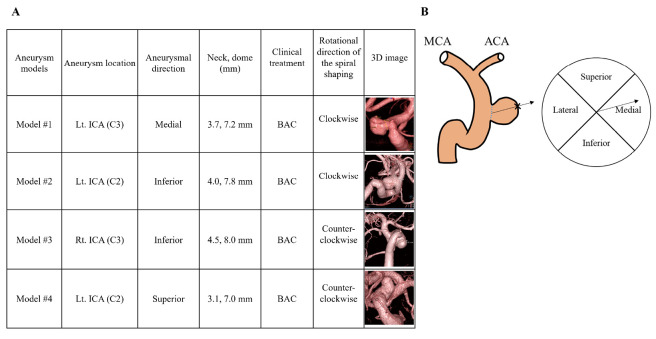
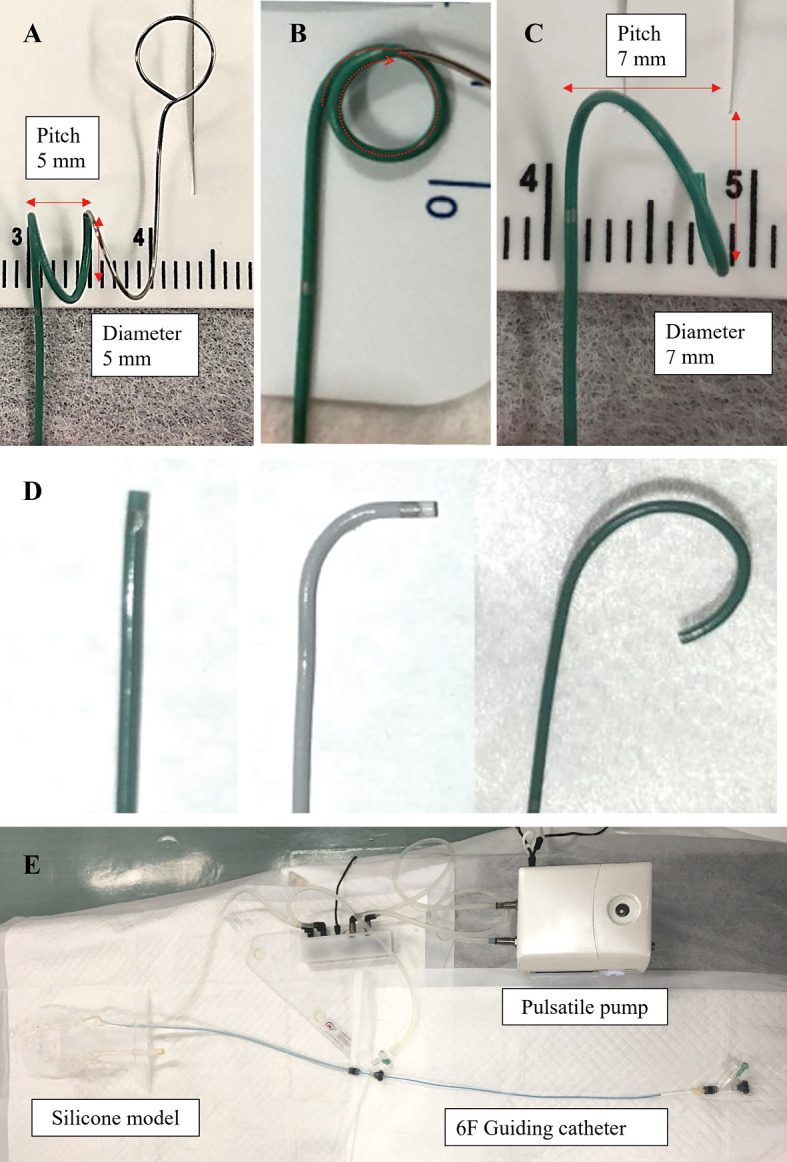
Selecting an appropriate microcatheter tip shape for paraclinoid aneurysms is difficult. Therefore, we devised an original simple and uniform three-dimensional (3D) spiral-shaping method of microcatheter and validated the characteristics and usefulness of this method for coil embolization of paraclinoid aneurysms using patient-specific silicone models. These silicone models were produced based on clinical data from four patients with four paraclinoid aneurysms that underwent endovascular treatment using the 3D spiral-shaping method. These models were classified into four types: superior, medial, inferior, and lateral corresponding to the aneurysm protrusion and locations (C3 or C2 segments by Fisher's classification). Employing a pulsatile pump setup, two operators assessed the following items: navigation methods (pull and wire guiding), catheterization times, microcatheter tip position in the aneurysm, and the feasibility of inserting a framing coil by simple technique compared with three other shapes (straight, 90, pigtail). Three-dimensional spiral-shaped microcatheter could be placed in the medial and inferior type models of C3 segments and superior type model of C2 segment by the pullback method. Catheterization times using a 3D spiral-shaped catheter were significantly shorter than other shaped ones in the superior type models. No significant difference was found in another silicone model. Three-dimensional spiral- and pigtail-shaped catheters tended to position the tip at the center of the aneurysm. In conclusion, 3D spiral-shaped microcatheter was especially effective for the superior projected aneurysm at the C2 segment. The 3D spiral-shaping method can provide easy and secure navigation of the microcatheter into the paraclinoid aneurysms, ensuring optimal positioning for coil insertion.
Keywords: coil; embolization; microcatheter; paraclinoid aneurysm; silicone model.
Conflict of interest statement
The authors have no conflicts of interest to disclose.
Figures

Similar articles
-
Geometric Classification of Paraclinoid Aneurysms for Microcatheter Superselection in Coil Embolization.Turk Neurosurg. 2020;30(5):651-657. doi: 10.5137/1019-5149.JTN.27371-19.2. Turk Neurosurg. 2020. PMID: 32996576
-
Technique for shaping microcatheter tips in coil embolization of paraclinoid aneurysms using full-scale volume rendering images of 3D rotational angiography.Minim Invasive Neurosurg. 2009 Aug;52(4):201-3. doi: 10.1055/s-0029-1239588. Epub 2009 Oct 16. Minim Invasive Neurosurg. 2009. PMID: 19838977
-
Simple and Reproducible Microcatheter Shaping Method for Coil Embolization of Medially-directed Paraclinoid Internal Carotid Artery Aneurysms.J Neuroendovasc Ther. 2020;14(4):119-125. doi: 10.5797/jnet.oa.2019-0046. Epub 2020 Feb 27. J Neuroendovasc Ther. 2020. PMID: 37520175 Free PMC article.
-
Use of the 0.027-inch VIA microcatheter for delivery of Pipeline Flex: a technical note.J Neurointerv Surg. 2017 Jul;9(7):689-693. doi: 10.1136/neurintsurg-2016-012971. Epub 2017 Feb 1. J Neurointerv Surg. 2017. PMID: 28151413 Review.
-
[Basic Setup and Coil Embolization Technique for Ruptured Cerebral Aneurysms].No Shinkei Geka. 2024 Sep;52(5):995-1002. doi: 10.11477/mf.1436205009. No Shinkei Geka. 2024. PMID: 39285549 Review. Japanese.
References
-
- Fischer E. Die Lageabweichungen der vorderen hirnarterie im gefassbild. Zentralbl Neurochir. 1938;3:300–313.
MeSH terms
Substances
LinkOut - more resources
Full Text Sources
Miscellaneous