. 2025 Jan 7:29:0127.
doi: 10.34133/bmr.0127. eCollection 2025.

Functionalized Periosteum-Derived Microsphere-Hydrogel with Sequential Release of E7 Short Peptide/miR217 for Large Bone Defect Repairing

Affiliations

Functionalized Periosteum-Derived Microsphere-Hydrogel with Sequential Release of E7 Short Peptide/miR217 for Large Bone Defect Repairing

Jun Yao et al. Biomater Res. .

Abstract

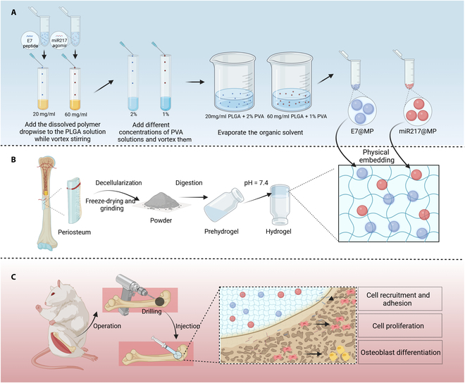
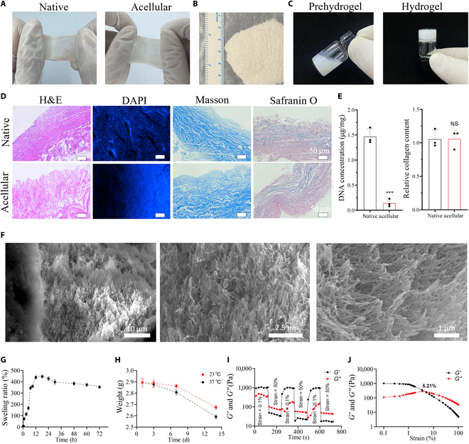
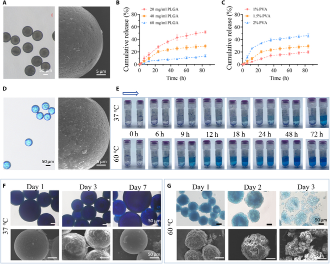
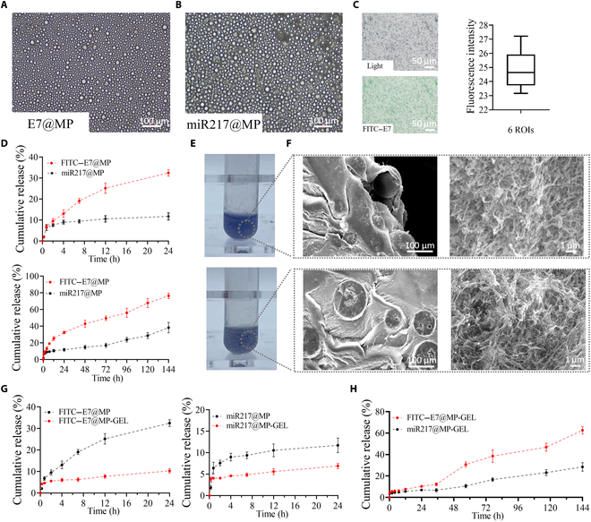
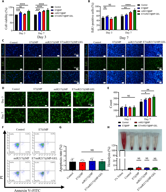
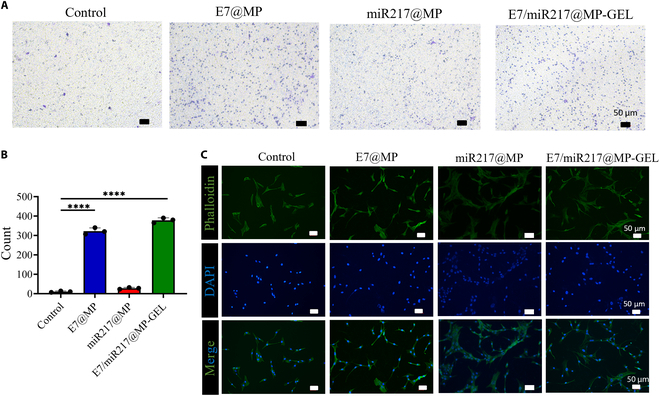
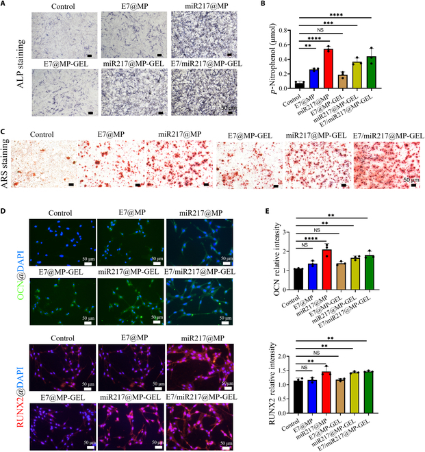
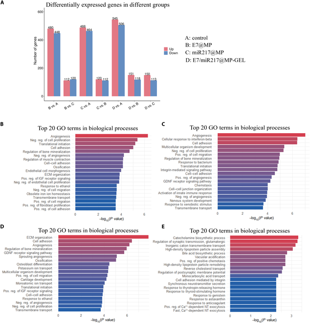
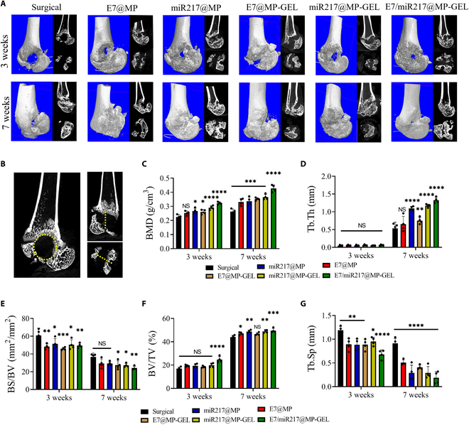
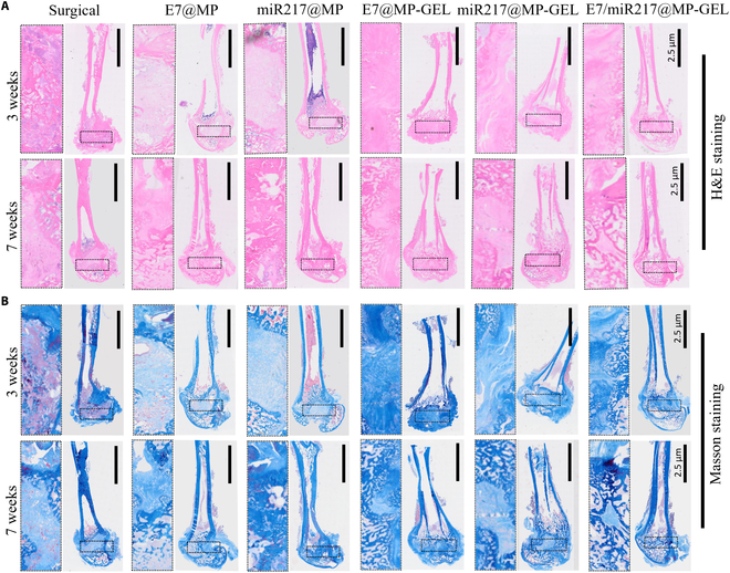
Large bone defects are still a persistent challenge in orthopedics. The availability limitations and associated complications of autologous and allogeneic bone have prompted an increasing reliance on tissue engineering and regenerative medicine. In this study, we developed an injectable scaffold combining an acellular extracellular periosteal matrix hydrogel with poly(d,l-lactate-co-glycol-acetate) microspheres loaded with the E7 peptide and miR217 (miR217/E7@MP-GEL). Characterization of the composites included morphological analysis by scanning electron microscopy, degradation and swelling tests, in vitro and in vivo biological evaluation, and the biological activity evaluation of mesenchymal stem cells (MSCs) through their effects on cell recruitment, proliferation, and osteogenic differentiation. The designed hydrogels demonstrated good physical and chemical properties that are cytocompatible and suitable for cell recruitment. In vitro studies confirmed the high biological activity of the release agent, which markedly enhanced the proliferation and osteogenic differentiation of MSCs. In vivo application to a rat model of a femur defect exhibited a significant increase in bone volume and density over 7 weeks, resulting in enhanced bone regeneration. Acellular periosteum-based hydrogels combined with the E7 peptide and miR217-loaded poly(d,l-lactate-co-glycol-acetate) microspheres can promote effective bone regeneration through the recruitment, proliferation, and osteogenic differentiation of MSCs, which provides a promising approach for the treatment of large bone defects.


Conflict of interest statement

Competing interests: The authors declare that they have no competing interests.

Figures

Fig. 1.
Fig. 1.
Sketch of the preparation and application of E7/miR217@MP-GEL composites. (A) Preparation of E7@MP and miR217@MP using poly(d,l-lactate-co-glycol-acetate) (PLGA) and polyvinyl alcohol (PVA). (B) Hydrogel of the periosteal extracellular matrix and preparation of E7/miR217@MP-GEL composites. (C) In situ injection of composites to promote bone regeneration (this scheme was created by BioRender).
Fig. 2.
Fig. 2.
Characteristics of the periosteum extracellular matrix hydrogel. (A) Periosteum decellularization before and after appearance comparison. (B) The appearance of periosteum powder. (C) Hydrogel formation before and after. (D) Hematoxylin and eosin (H&E) staining, 4′,6-diamidino-2-phenylindole (DAPI) staining, Masson staining, and Safranin O solid green staining (left to right) of periosteum tissue before and after decellularization. (E) Changes in DNA and glycosaminoglycans in periosteum tissue before and after decellularization. (F) Scanning electron microscopy (SEM) images of periosteal hydrogels. From left to right: ×5,000; ×10,000; ×15,000. (G) Swelling rate of periosteal hydrogels in phosphate-buffered saline (PBS). (H) Degradation rates of periosteal hydrogels in PBS at 23 and 37 °C. (I and J) Rheological properties of periosteal hydrogels: variation in energy storage modulus G′ and loss modulus G″ of the samples at a temperature of 37 °C with an increase in strain from 0.1% to 100%. Time sweeps of the storage modulus G′ and loss modulus G″ of samples at 37 °C with strains varying at 0.1% and 50% from each other. ***P < 0.005. NS, not significant.
Fig. 3.
Fig. 3.
Characterization of PLGA@MP prepared with different PVA and PLGA concentrations. (A) Appearance of PLGA@MP under microscope and SEM. (B) Rate of release at 37 °C of microspheres obtained by a PVA concentration of 1.5% (w/v) prepared with PLGA concentrations of 20, 40, and 60 mg/ml. (C) Rate of release at 37 °C of microspheres obtained from a PLGA concentration of 60 mg/ml prepared with PVA concentrations of 1%, 1.5% and 2% (w/v). (D) Microscopic and scanning electron microscopic appearance of methylene blue-simulated drugs after encapsulation by microspheres. (E) Release rates of microspheres encapsulated with methylene blue after preparation using PLGA and PVA concentration ratios of 60 mg/ml:1% (left) and 20 mg/ml:2% (right) in PBS solutions at 37 and 60 °C,. (F and G) After the microspheres were encapsulated with methylene blue, changes in their appearance were observed using microscopy and SEM in PBS solution at 37 and 60 °C.
Fig. 4.
Fig. 4.
Appearance and release performance of E7@MP-GEL and miR217@MP-GEL. (A and B) Microscopic appearance of E7@MP and miR217@MP. (C) Statistical plots of the appearance of fluorescein isothiocyanate (FITC)–E7@MP under fluorescence microscopy and the average fluorescence intensity of the region of interest (ROI). (D) Release rates of E7@MP and miR217@MP in PBS solution at 37 °C for 24 h and 6 d. (E and F) Appearance of E7@MP-GEL (top) and miR217@MP-GEL (bottom) in EP tubes and under SEM. (G) Release rates of FITC–E7@MP and FITC–E7@MP-GEL in PBS solution at 37 °C. Release rates of miR217@MP and miR217@MP-GEL in PBS solution at 37 °C. (H) Release rates of E7@MP-GEL and miR217@MP-GEL in PBS solution at 37 °C for 6 d.
Fig. 5.
Fig. 5.
Cytocompatibility of E7/miR217@MP-GEL. (A) Cell viability of E7@MP, miR217@MP, and E7/miR217@MP-GEL after 3 and 7 d of co-culture with bone marrow mesenchymal stem cells (BMSCs). (B and C) Results and statistical analysis of cell proliferation after 3 and 7 d of co-culture with BMSCs. (D and E) Results and statistical analysis of number of live and dead cells after 3 and 7 d of co-culture with BMSCs. (F and G) Results and statistical analysis of the apoptosis of BMSCs after 3 d of co-culture with BMSCs. (H) Results and statistical analysis of changes in the appearance after co-culture with erythrocytes and hemolysis. PI, propidium iodide; EdU, 5-ethynyl-2′-deoxyuridine. *P < 0.05, **P < 0.01, ***P < 0.005, and ****P < 0.001. NS, not significant.
Fig. 6.
Fig. 6.
E7/miR217@MP-GEL recruits BMSCs in vitro and promotes adhesion. (A and B) Results of Transwell experiments and statistical analysis of E7@MP, miR217@MP, and E7/miR217@MP-GEL after 3 d of co-culture with BMSCs. (C) Changes in cell appearance after 3 d of co-culture with BMSCs in each group; phalloidin is green, and DAPI is blue. ****P < 0.001.
Fig. 7.
Fig. 7.
E7/miR217@MP-GEL promotes osteogenic differentiation of BMSCs. (A and B) alkaline phosphatase (ALP) staining results and ALP content analysis of E7@MP, miR217@MP, E7@MP-GEL, miR217@MP-GEL, and E7/miR217@MP-GEL after 7 d of co-culture with BMSCs. (C) Alizarin red staining after 21 d of co-culture with BMSCs in each group. (D and E) Results and statistical analysis of osteocalcin (OCN) and Runt-related transcription factor 2 (RUNX2) immunofluorescence in cells after 7 d of co-culture with BMSCs in each group. ARS, alizarin red. **P < 0.01, ***P < 0.005, and ****P < 0.001. NS, not significant.
Fig. 8.
Fig. 8.
E7/miR217@MP-GEL affects the transcriptome of BMSCs. BMSCs were co-cultured with E7@MP, miR217@MP, and E7/miR217@MP-GEL for 7 d, and the transcriptome was analyzed using RNA sequencing. (A) Genes differentially expressed between E7@MP, miR217@MP, and E7/miR217@MP-GEL vs. control phases. (B) Biological process enrichment analysis of differentially expressed genes between the E7/miR217@MP-GEL and control groups (n = 3). (C and D) Differentially expressed genes between E7@MP and miR217@MP vs. control (n = 3). (E) Differentially expressed genes between E7@MP and miR217@MP (n = 3). GO, Gene Ontology; ECM, extracellular matrix; IGF, insulin-like growth factor; Neg. reg., negative regulation; Pos. reg., positive regulation; GDNF, glial cell line-derived neurotrophic factor; NT, neurotransmitter.
Fig. 9.
Fig. 9.
E7/miR217@MP-GEL treatment in a Sprague Dawley (SD) rat femoral defect model. (A) Changes in the computed tomography (CT) of femoral defect sites at weeks 3 and 7 by E7@MP, miR217@MP, E7@MP-GEL, miR217@MP-GEL, and E7/miR217@MP-GEL in an SD rat femoral defect model. (B) Schematic representation of ROI for CT analysis. (C to G) Quantification of microCT parameters, namely, bone mineral density (BMD), trabecular thickness (Tb.Th), bone surface (BS)/bone volume (BV), BV/tissue volume (TV), and bone separation coefficient (Tb.Sp). *P < 0.05, **P < 0.01, ***P < 0.005, and ****P < 0.001. NS, not significant.
Fig. 10.
Fig. 10.
Tissue staining of E7/miR217@MP-GEL at the site of femoral defects in SD rats. (A and B) H&E staining and Masson staining of E7@MP, miR217@MP, E7@MP-GEL, miR217@MP-GEL, and E7/miR217@MP-GEL at the femoral defect site at weeks 3 and 7.

Similar articles

References

    1. Wang J, Yin Q, Gu S, Wu Y, Rui Y. Induced membrane technique in the treatment of infectious bone defect: A clinical analysis. Orthop Traumatol Surg Res. 2019;105(3):535–539. - PubMed
    1. Wang X, Huang Y, Liu D, Zeng T, Wang J, Al Hasan MJ, Liu W, Wang D. The Masquelet induced membrane technique with PRP-FG-nHA/PA66 scaffold can heal a rat large femoral bone defect. BMC Musculoskelet Disord. 2024;25(1): Article 455. - PMC - PubMed
    1. Iseki T, Rothrauff BB, Kihara S, Overholt KJ, Taha T, Lin H, Alexander PG, Tuan RS. Enhanced osteochondral repair by leukocyte-depleted platelet-rich plasma in combination with adipose-derived mesenchymal stromal cells encapsulated in a three-dimensional photocrosslinked injectable hydrogel in a rabbit model. Stem Cell Res Ther. 2024;15(1): Article 159. - PMC - PubMed
    1. Ho-Shui-Ling A, Bolander J, Rustom LE, Johnson AW, Luyten FP, Picart C. Bone regeneration strategies: Engineered scaffolds, bioactive molecules and stem cells current stage and future perspectives. Biomaterials. 2018;180:143–162. - PMC - PubMed
    1. Urban IA, Montero E, Monje A, Sanz-Sánchez I. Effectiveness of vertical ridge augmentation interventions: A systematic review and meta-analysis. J Clin Periodontol. 2019;46(Suppl 21):319–339. - PubMed

LinkOut - more resources