. 2022 May 27;5(5):6995-7008.
doi: 10.1021/acsanm.2c00958. Epub 2022 May 16.

Increasing the Efficacy of Gold Nanorod Uptake in Stem Cell-Derived Therapeutic Cells: Implications for Stem Cell Labeling and Optical Coherence Tomography Imaging

Affiliations

Increasing the Efficacy of Gold Nanorod Uptake in Stem Cell-Derived Therapeutic Cells: Implications for Stem Cell Labeling and Optical Coherence Tomography Imaging

Grant W Marquart et al. ACS Appl Nano Mater. .

Abstract

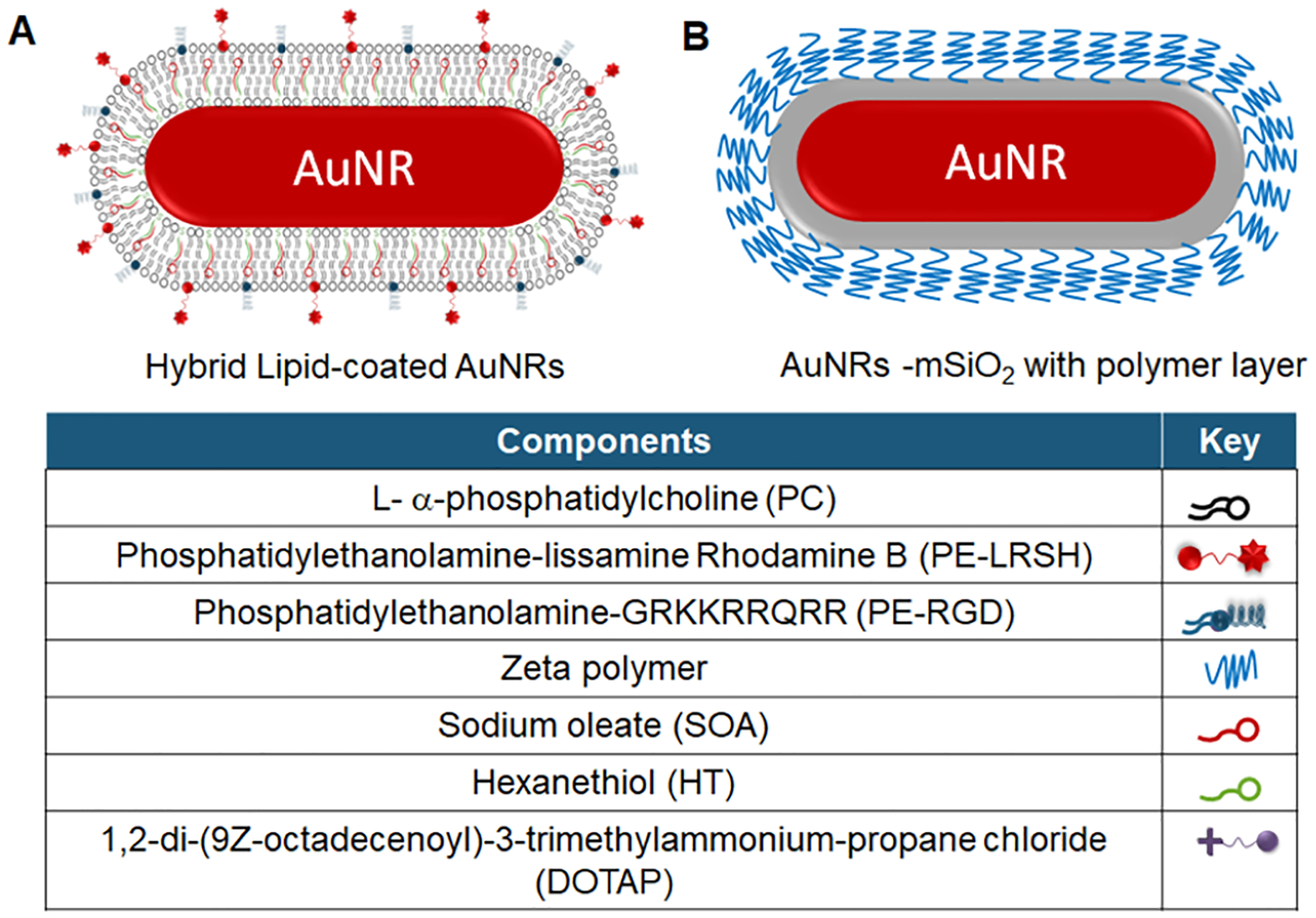
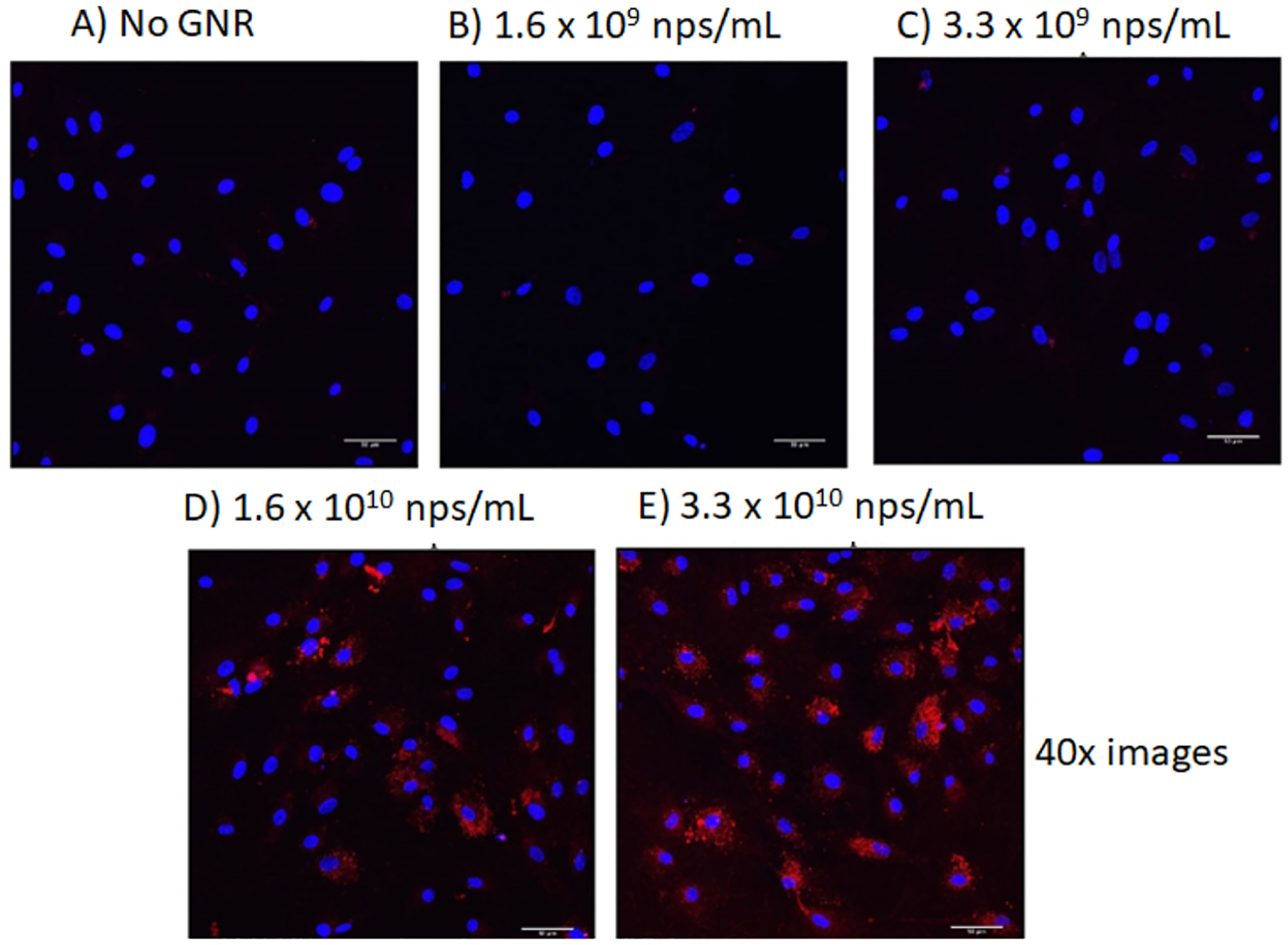
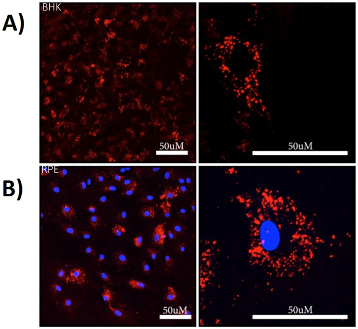
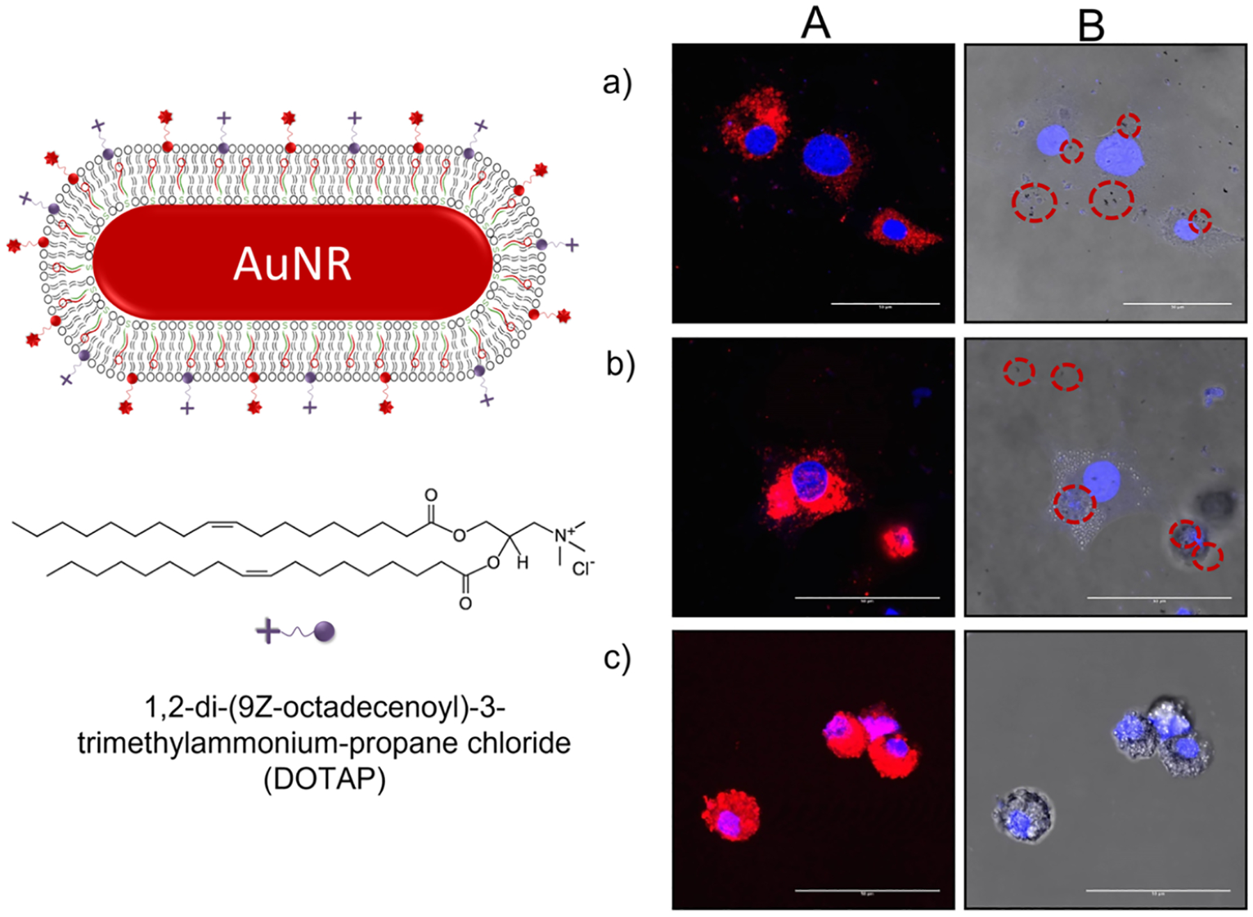
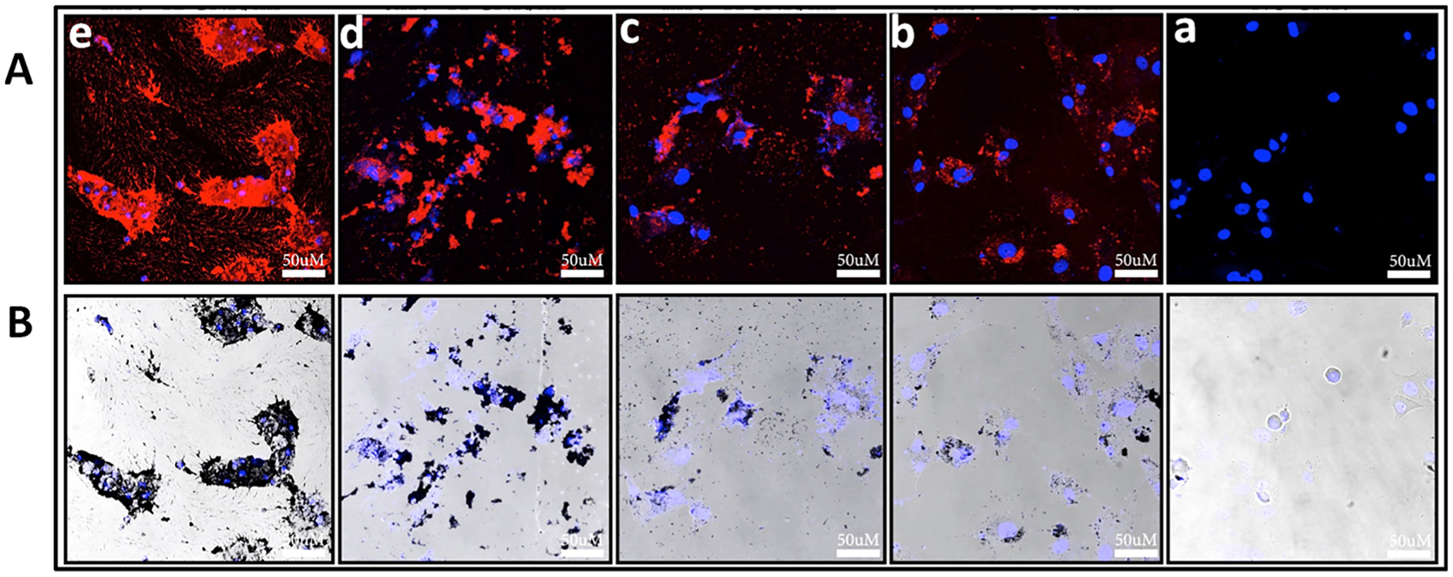
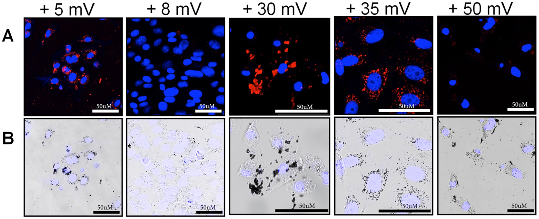
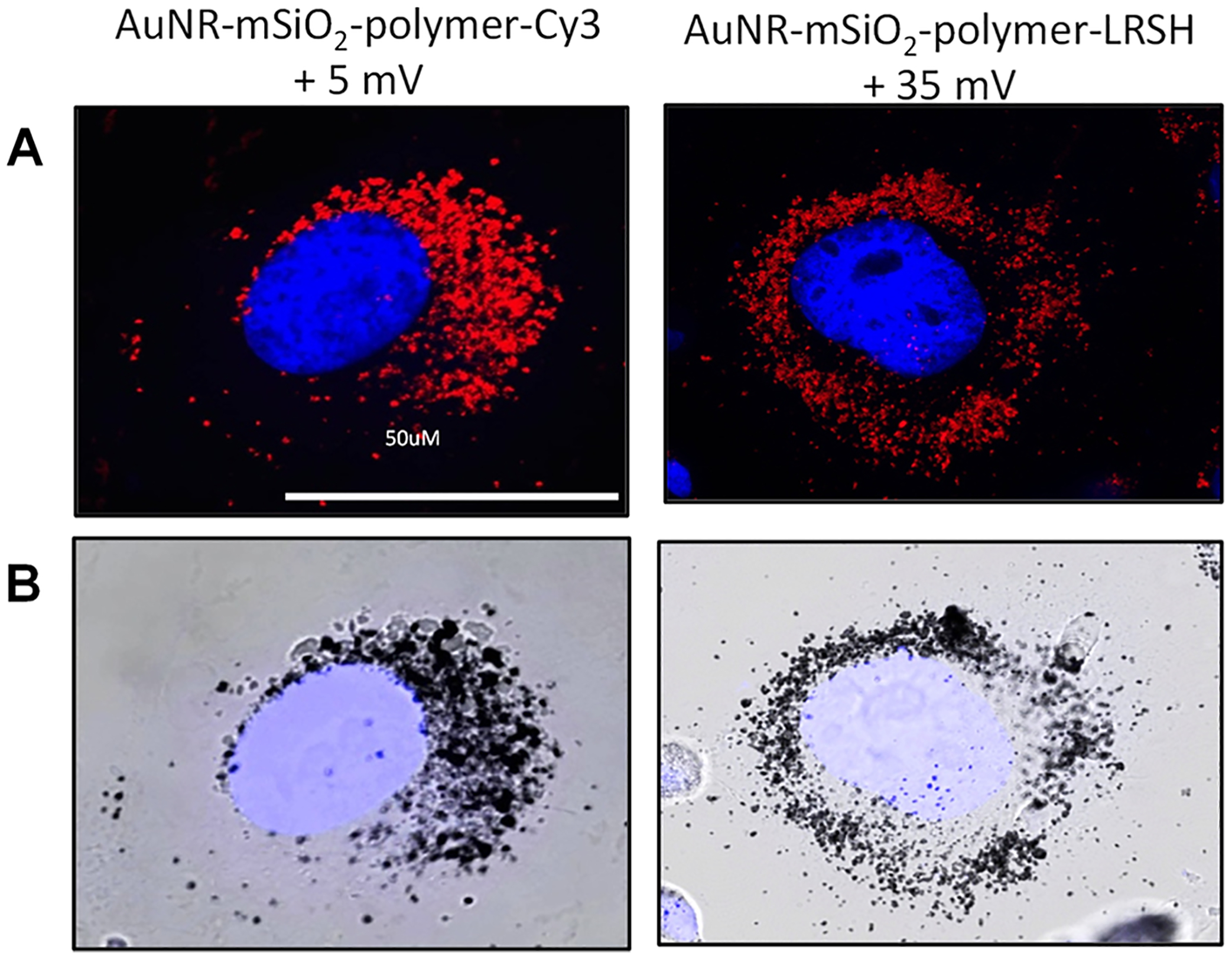
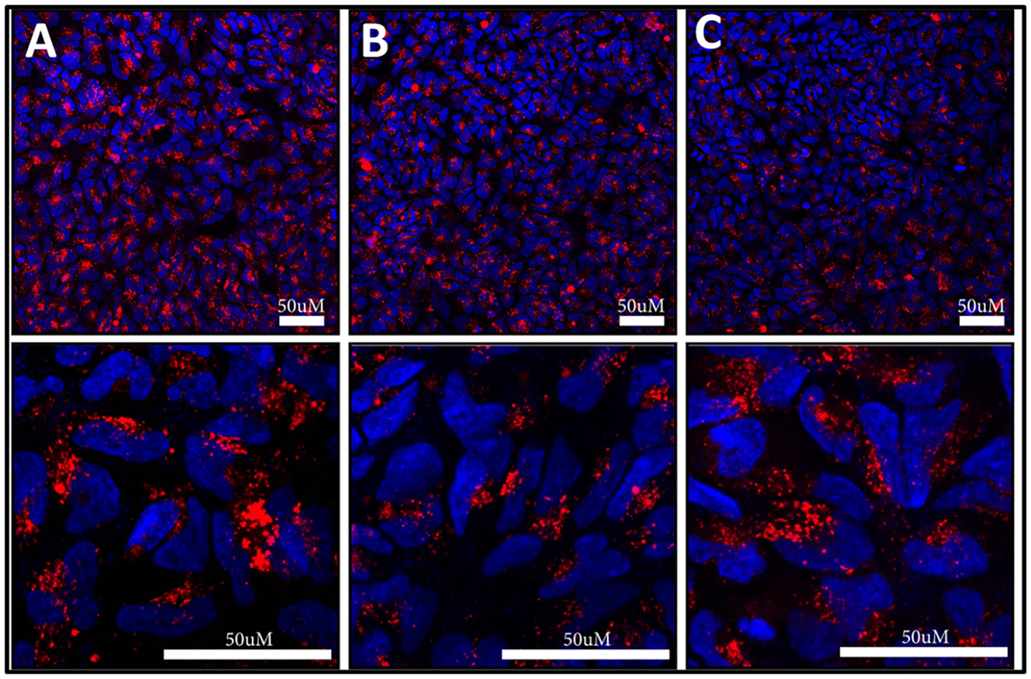
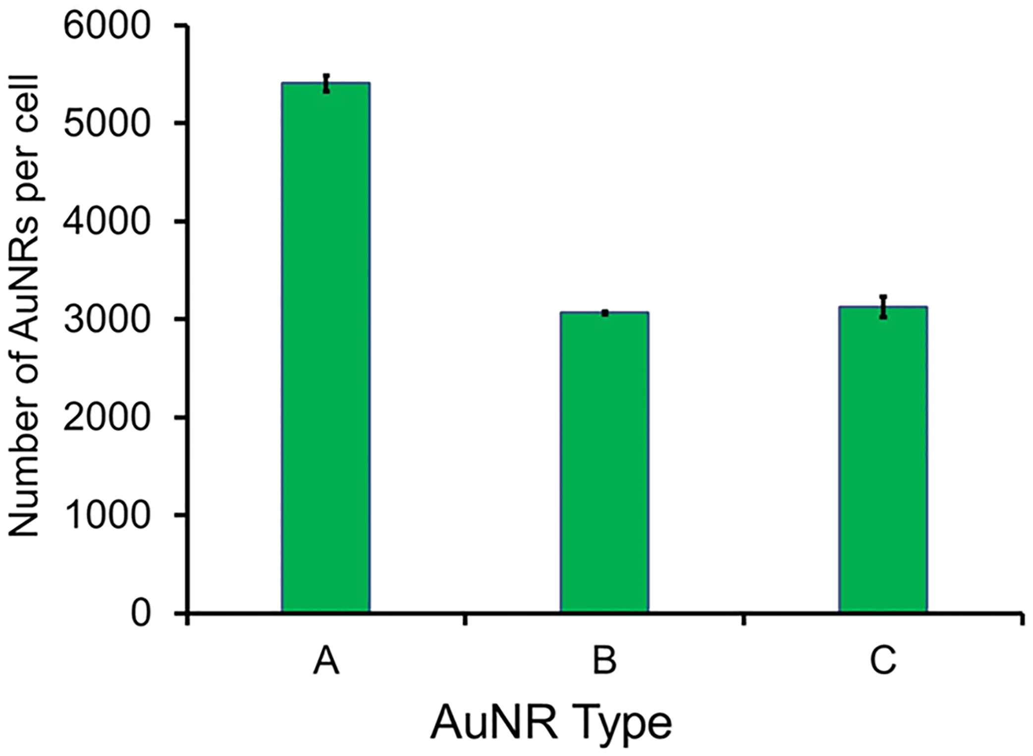
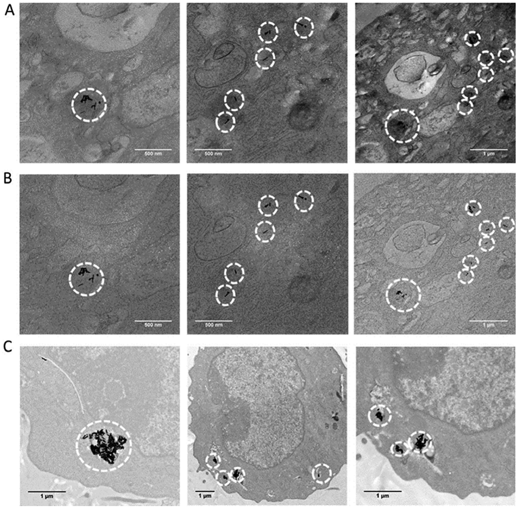
The advancement of safe nanomaterials for use as optical coherence tomography (OCT) imaging and stem cell-labeling agents to longitudinally visually track therapeutic derived retinal stem cells to study their migration, survival rate, and efficacy is challenged by instability, intracellular aggregation, low uptake, and cytotoxicity. Here, we describe a series of hybrid lipid-coated gold nanorods (AuNRs) that could solve these issues. These nanomaterials were made via a layer-by-layer assembly approach, and their stability in biological media, mechanism, efficiency of uptake, and toxicity were compared with a commercially available set of AuNRs with a 5 nm mesoporous silica (mSiO2)-polymer coating. These nanomaterials can serve as stem cell labeling and OCT imaging agents because they absorb in the near-infrared (NIR) region away from biological tissues. Although both subtypes of AuNRs were taken up by retinal pigment epithelial, neural progenitor, and baby hamster kidney cells, slightly negatively charged hybrid lipid-coated AuNRs had minimal aggregation in biological media and within the cytoplasm of cells (~3000 AuNRs/cell) as well as minimal impact on cell health. Hybrid lipid-coated AuNRs modified with cell-penetrating peptides had the least toxicological impact, with >92% cell viability. In contrast, the more "sticky" AuNRs with a 5 nm mSiO2-polymer coating showed significant aggregation in biological media and within the cytoplasm with lower-than-expected uptake of AuNRs (~5400 of AuNRs/cell) given their highly positive surface charge (35+ mV). Collectively, we have demonstrated that hybrid lipid-coated AuNRs, which absorb in the NIR-II region away from biological tissues, with tuned surface chemistry can label therapeutic derived stem cells with minimal aggregation and impact on cell health as well as enhance uptake for OCT imaging applications.

Keywords: cell uptake studies; cell-penetrating peptides; lipid-coated gold nanorods; optical coherence tomography imaging; stem cells; toxicity.


Conflict of interest statement

The authors declare no competing financial interest.

Figures

Scheme 1.
Scheme 1.
Cartoon of Gold Nanorods with a (A) Hybrid Lipid Membrane or a (B) 5 nm mSiO2 and Polymer Layer
Figure 1.
Figure 1.
Representative UV–vis spectra of 1 mL AuNR-PC/PE-LRSH/DOTAP (6:1:3) (i) before and (ii) after 1 h and (iii) 24 h of incubation with 20 μL of 307 mM KCN. All samples were in 10 mM sodium phosphate buffer at pH 8.0.
Figure 2.
Figure 2.
Confocal laser scanning microscopy images at 40× magnification of RPE cells (A) before and (B) after the addition of AuNR-SOA-PC/PE-LRSH-HT at an applied concentration of 1.6 × 10⁹ AuNRs/mL, (C) 3.3 × 10⁹ nanoparticles AuNRs/mL, (D) 1.6 × 10¹⁰ AuNRs/mL, and (E) 3.3 × 10¹⁰ AuNRs/mL. Blue fluorescence is from the DAPI-stained nuclei, and red is from the LRSH-labeled AuNRs. The scale bars indicate 50 μm. Cell concentration is 150,000 cells/mL.
Figure 3.
Figure 3.
Confocal laser scanning microscopy images (left 40× and right 63× magnification) of (A) BHK and (B) RPE cells after incubation with an applied concentration of 5 × 10¹⁰ AuNRs/mL of AuNR-SOA-PC/PE-LRSH-HT for 24 h in DMEM with 150,000 cells/mL. Blue fluorescence is from DAPI-stained nuclei, and red is from PE-LRSH-labeled AuNRs. The scale bars equal 50 μm resolution.
Figure 4.
Figure 4.
(A) Confocal laser scanning microscopy images at 40× magnification with a 4× digital zoom and (B) bright-field images of AuNR-SOA-PC/PE-LRSH-HT incubated at (a) 4.1 × 10¹⁰ nps/mL, (b) 8.3 × 10¹⁰ nps/mL, and (c) 4.1 × 10¹¹ nps/mL incubated with 150,000 RPE cells per mL for 24 h in DMEM. Blue fluorescence is the DAPI-stained nuclei, and red is PE-LRSH-labeled AuNRs. The scale bars indicate 50 μm. Red circles indicate where large aggregates of AuNRs are seen in the bright field.
Figure 5.
Figure 5.
Representative (A) confocal laser scanning microscopy images and (B) bright-field images at 40× magnification of BHK cells (a) without AuNRs and BHK cells incubated with increasing concentrations of AuNR-mSiO2-polymer-Cy3 at (b) 8.0 × 10¹⁰ nps/mL, (b) 4.0 × 10¹¹ nps/mL, (c) 8.0 × 10¹¹ nps/mL and (d) 4.0 × 10¹² nps/mL. All AuNR samples were incubated with 150,000 cells/mL for 24 h in DMEM. Blue fluorescence is the DAPI-stained nuclei, and red is Cy3-labeled polymer-coated mSiO2-layered AuNRs. The scale bars indicate 50 μm.
Figure 6.
Figure 6.
Representative (A) confocal laser scanning microscopy images and (B) bright-field images at 40× magnification with a 4× digital zoom of BHK cells incubated with 5.0 × 10¹⁰ AuNRs/mL of AuNR-mSiO2-polymer-LRSH with increasing the positive charge (right to left). All AuNR-mSiO2-polymer-LRSH samples were incubated with 150,000 cells/mL for 24 h in DMEM. Blue fluorescence is the DAPI-stained nuclei, and red is LRSH-labeled polymer-coated mSiO2-layered AuNRs. The scale bars indicate 50 μm.
Figure 7.
Figure 7.
Representative (A) confocal fluorescence microscopy and (B) bright-field images at 63× magnification of BHK cells incubated with 5.0 × 10¹⁰ AuNRs/mL of the AuNR-mSiO2-polymer-Cy3 (55 × 10 nm) with +5 mV zeta potential and the AuNR-mSiO2-polymer-LRSH (96 × 25 nm) with a +35 mV zeta potential. All mSiO2-polymer-AuNR samples were incubated with 150,000 cells/mL for 24 h in DMEM. Blue fluorescence is the DAPI-stained nuclei, and red is Cy3 or LRSH dye-labeled polymer-coated mSiO2-layered AuNRs. The scale bars indicate 50 μm.
Figure 8.
Figure 8.
Confocal laser scanning microscopy images of NP cells incubated with 5 × 10¹⁰ AuNRs/mL of (A) AuNR-SOA-PC/PE-LRSH/DOTAP-HT, (B) AuNR-SOA-PC/PE-LRSH/PE-RGD-HT, and (C) AuNR-SOA-PC/PE-LRSH/PE-RGD/DOTAP-HT in DMEM for 24 h. Blue fluorescence is the DAPI-stained nuclei, and red is PE-LRSH-labeled AuNRs. The top panel is at 40× magnification, and the bottom panel is at 40× magnification with a 4× digital zoom. The scale bars indicate 50 μm.
Figure 9.
Figure 9.
Quantification of AuNR uptake in BHK cells by ICP-MS. 5 × 10 AuNRs/mL of (A) AuNRs-mSiO2-polymer-LRSH at +35 mV, (B) AuNR-SOA-PC/PE-LRSH/DOTAP-HT, and (C) AuNR-SOA-PC/PE-RGD/PE-LRSH-HT were incubated with 106 BHK cells/mL for 24 h in DMEM at 37 °C. Data are reported as the average number of AuNRs/cell from triplicate trials.
Figure 10.
Figure 10.
TEM imaging of BHK cells after 24 h incubated with AuNR-PC/PE-RGD/PE-LRSH-HT (8:1:1) (#9 Table S1) stained with 1% osmium tetroxide (A) with objective aperture and (B) without objective aperture. TEM images reveal accumulation of AuNRs as black granules in the cytosol (red arrows), and (C) AuNRs-mSiO2-polymer-LRSH nanoparticles #5 unstained. The scale bars indicate 500 nm (2 left columns) and 1 μM (far right column and bottom).

Similar articles

References

    1. Stone EM A very effective treatment for neovascular macular degeneration. N. Engl. J. Med 2006, 355, 1493–1495. - PubMed
    1. Zhou JY; Zhang Z; Qian GS Mesenchymal stem cells to treat diabetic neuropathy: a long and strenuous way from bench to the clinic. Cell Death Discovery 2016, 2, 16055. - PMC - PubMed
    1. Limoli PG; Limoli C; Vingolo EM; Franzone F; Nebbioso M Mesenchymal stem and non-stem cell surgery, rescue, and regeneration in glaucomatous optic neuropathy. Stem Cell Res. Ther 2021, 12, 275. - PMC - PubMed
    1. Scarfe L; Brillant N; Kumar JD; Ali N; Alrumayh A; Amali M; Barbellion S; Jones V; Niemeijer M; Potdevin S; Roussignol G; Vaganov A; Barbaric I; Barrow M; Burton NC; Connell J; Dazzi F; Edsbagge J; French NS; Holder J; Hutchinson C; Jones DR; Kalber T; Lovatt C; Lythgoe MF; Patel S; Patrick PS; Piner J; Reinhardt J; Ricci E; Sidaway J; Stacey GN; Starkey Lewis PJ; Sullivan G; Taylor A; Wilm B; Poptani H; Murray P; Goldring CEP; Park BK Preclinical imaging methods for assessing the safety and efficacy of regenerative medicine therapies. npj Regener. Med 2017, 2, 28. - PMC - PubMed
    1. Huang D; Swanson EA; Lin CP; Schuman JS; Stinson WG; Chang W; Hee MR; Flotte T; Gregory K; Puliafito CA; Fujimoto JG Optical coherence tomography. Science 1991, 254, 1178–1181. - PMC - PubMed

LinkOut - more resources