. 2025 Jan;5(1):e70091.
doi: 10.1002/cpz1.70091.

Generation of Retinal Ganglion Cells from Reprogrammed Keratocytes of Non-Glaucoma and Glaucoma Donors

Affiliations

Generation of Retinal Ganglion Cells from Reprogrammed Keratocytes of Non-Glaucoma and Glaucoma Donors

Shahna S Hameed et al. Curr Protoc. 2025 Jan.

Abstract

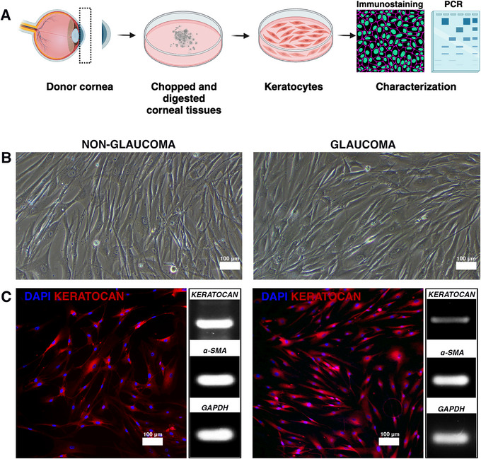
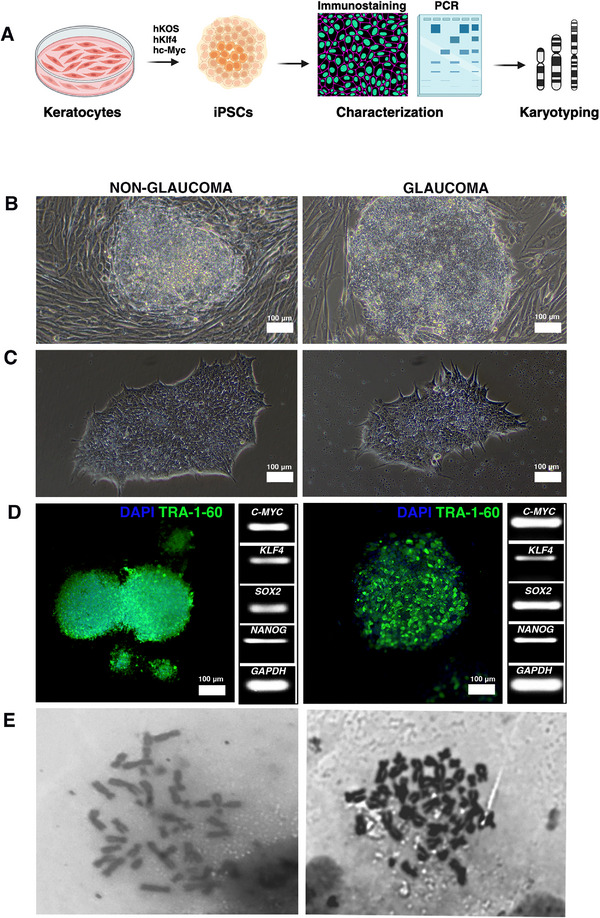
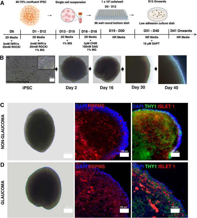
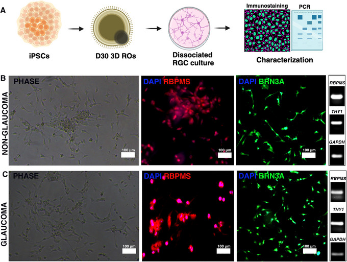
Human induced pluripotent stem cell (hiPSC)-based disease modeling can be successfully recapitulated to mimic disease characteristics across various human pathologies. Glaucoma, a progressive optic neuropathy, primarily affects the retinal ganglion cells (RGCs). While multiple groups have successfully generated RGCs from non-diseased hiPSCs, producing RGCs from glaucomatous human samples holds significant promise for understanding disease pathology by revealing patient-specific disease signatures. Given that keratocytes originate from the neural crest and previous reports suggest that ocular fibroblasts from glaucomatous donors carry pathogenic signatures, it is highly plausible that these signatures imprinted within the keratocytes will also be present in the derived RGCs. Thus, we aimed to generate RGCs from both glaucomatous and non-glaucomatous donor keratocytes and validate disease-specific signatures in 3D retinal organoids and in isolated RGCs. Our protocol describes the generation of iPSCs from keratocytes of both glaucomatous and non-glaucomatous donors, followed by their differentiation into retinal organoids. Subsequent isolation and culturing of RGCs were performed. Disease signatures in the RGCs were validated in both 3D retinal organoids (ROs) and 2D RGC cultures, and glaucomatous RGCs in 3D and 2D cultures demonstrated increased cleaved CASP3 and significant RGC loss, indicating disease imprints in the hiPSC-derived RGCs. This model offers a venue and high throughput platform for studying glaucomatous disease pathology and holds significant potential for drug discovery using RGCs derived from human donors. © 2025 The Author(s). Current Protocols published by Wiley Periodicals LLC. Basic Protocol 1: Culturing of keratocytes from human cadaveric donors Basic Protocol 2: Reprogramming donor keratocytes into iPSCs Basic Protocol 3: Evaluation of chromosomal loss during reprogramming in iPSCs by karyotyping Basic Protocol 4: Generation of 3D ROs Basic Protocol 5: Dissociation and culturing of RGCs from 3D ROs Support Protocol 1: Immunostaining for phenotypic characterization of cells Support Protocol 2: Sectioning of 3D ROs and immunostaining Support Protocol 3: Western blotting for cleaved CASP3 and THY1.

Keywords: glaucoma; induced pluripotent stem cells; keratocytes; retinal ganglion cells; retinal organoids.


Conflict of interest statement

The authors declare no conflict of interest.

Figures

Figure 1
Figure 1
Isolation and culture of keratocytes from non‐glaucomatous and glaucomatous donor corneas. (A). Schematic of the experimental paradigm. (B). Phase contrast images of cultured keratocytes from non‐glaucomatous and glaucomatous donors. (C). Immunostaining and PCR characterization of the keratocytes using markers keratocan and α‐SMA. GAPDH served as a housekeeping gene for PCR. α‐SMA: Alpha smooth muscle actin, PCR: Polymerase chain reaction, GAPDH: glyceraldehyde‐3‐phosphate dehydrogenase, Magnification: 20×.
Figure 2
Figure 2
Reprogramming of donor keratocytes into induced pluripotent stem cells. (A). Schematic of the experimental paradigm. (B). Generation of iPSC clones. (C). Propagation of individual clones in a rh‐laminin‐coated plate. (D). Immunostaining of the iPSCs using marker TRA‐1‐60 and PCR characterization with C‐MYC, KLF4, SOX2, NANOG, and GAPDH serving as a housekeeping control gene. (E). Phase contrast images of karyotyping depicting no chromosomal loss while reprogramming. iPSC: induced pluripotent stem cells, TRA‐1‐60: T cell receptor alpha locus, C‐MYC: cellular Myc, KLF4: Kruppel‐like factor 4, SOX2: SRY‐box 2, NANOG: Nanog homeobox, GAPDH: glyceraldehyde‐3‐phosphate dehydrogenase, Magnification: 20×.
Figure 3
Figure 3
Differentiation of induced pluripotent stem cells into retinal organoids. (A). Schematic of the experimental paradigm. (B). The stepwise differentiation of iPSCs into ROs demonstrates neuro‐retinal formation on day 2, which becomes more pronounced by day 16 and continues to thicken with lamination by day 30 and day 40. Phase contrast and immunostaining images of day 30 (C) non‐glaucomatous and (D) glaucomatous organoids, labeled with RGC markers RBPMS, ISLET‐1, and THY1, and counterstained with nuclear marker DAPI. iPSC: induced pluripotent stem cells, RGC: Retinal ganglion cells, ROs: Retinal organoids, RBPMS: RNA binding protein with multiple splicing, ISLET‐1: Insulin gene enhancer protein 1, THY1: thymus cell antigen 1, DAPI: 4',6‐diamidino‐2‐phenylindole, Magnification: 20×.
Figure 4
Figure 4
Generating retinal ganglion cells from retinal organoids. (A) Schematic of the experimental paradigm. (B) Non‐glaucomatous and (C) glaucomatous depicting phase contrast images of dissociated RGCs and their characterization by immunostaining (RBPMS, BRN3A, DAPI) and PCR (RBPMS, THY1). GAPDH is used as a housekeeping gene for PCR. RGC: Retinal ganglion cells, ROs: Retinal organoids, RBPMS: RNA binding protein with multiple splicing, BRN3A: brain‐specific homeobox/POU domain protein 3A, THY1: thymus cell antigen 1, GAPDH: glyceraldehyde‐3‐phosphate dehydrogenase, DAPI: 4',6‐diamidino‐2‐phenylindole, Magnification: 20×.
Figure 5
Figure 5
Assessment of neurodegenerative phenotype in the retinal organoids and dissociated retinal ganglion cells. (A). Non‐glaucomatous and (B) glaucomatous day 30 ROs showing expression of RGC marker RBPMS and apoptotic marker cleaved CASP3. The dissociated culture of RGCs from (C) non‐glaucomatous and (D) glaucomatous day 30 ROs expressing RGC marker RBPMS and apoptotic marker cleaved CASP3. (E) Western blotting depicting decreased expression of RGC marker THY1 with increased expression of cleaved CASP3 in glaucomatous ROs as compared to non‐glaucomatous ROs. Densitometric quantification shows significantly increased expression of (F) cleaved CASP3 (*p < 0.05) and decreased expression of (G) THY1 (*p < 0.05) in glaucomatous ROs as compared to non‐glaucomatous ROs. β‐actin was used as a housekeeping gene. RGC: Retinal ganglion cells, ROs: Retinal organoids, CASP3: Caspase 3, RBPMS: RNA binding protein with multiple splicing, THY1: thymus cell antigen. Magnification: 20×.

Similar articles

References

    1. Agarwal, D. , Dash, N. , Mazo, K. W. , Chopra, M. , Avila, M. P. , Patel, A. , Wong, R. M. , Jia, C. , Do, H. , Cheng, J. , Chiang, C. , Jurlina, S. L. , Roshan, M. , Perry, M. W. , Rho, J. M. , Broyer, R. , Lee, C. D. , Weinreb, R. N. , Gavrilovici, C. , … Wahlin, K. J. (2023). Human retinal ganglion cell neurons generated by synchronous BMP inhibition and transcription factor mediated reprogramming. NPJ Regenerative Medicine, 8(1), 55. 10.1038/s41536-023-00327-x - DOI - PMC - PubMed
    1. Bikkuzin, T. , Shi, Y. , Sun, B. , Guo, Y. , Jin, X. , Han, Z. , Pavlov, V. , & Zhang, H. (2019). Human induced pluripotent stem cell line HMUi001‐A derived from corneal stromal cells. Stem Cell Research, 37, 101409. 10.1016/j.scr.2019.101409 - DOI - PubMed
    1. Binte, M. Y. N. Z. , Riau, A. K. , Yam, G. H. F. , Binte Halim, N. S. H. , & Mehta, J. S. (2022). Isolation and propagation of human corneal stromal keratocytes for tissue engineering and cell therapy. Cells, 11(1). 10.3390/cells11010178 - DOI - PMC - PubMed
    1. Boal, A. M. , McGrady, N. R. , Holden, J. M. , Risner, M. L. , & Calkins, D. J. (2023). Retinal ganglion cells adapt to ionic stress in experimental glaucoma. Frontiers in Neuroscience, 17, 1142668. 10.3389/fnins.2023.1142668 - DOI - PMC - PubMed
    1. Bolinches‐Amoros, A. , Lukovic, D. , Castro, A. A. , Leon, M. , Kamenarova, K. , Kaneva, R. , Jendelova, P. , Blanco‐Kelly, F. , Ayuso, C. , Cortón, M. , & Erceg, S. (2018). Generation of a human iPSC line from a patient with congenital glaucoma caused by mutation in CYP1B1 gene. Stem Cell Research, 28, 96–99. 10.1016/j.scr.2018.01.004 - DOI - PubMed

MeSH terms