Review
. 2025 May 1;110(5):1061-1073.
doi: 10.3324/haematol.2023.284633. Epub 2025 Jan 9.

The modern use of hydroxyurea for children with sickle cell anemia

Affiliations
Review

The modern use of hydroxyurea for children with sickle cell anemia

Charles T Quinn et al. Haematologica. .

Abstract

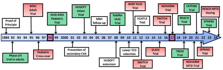
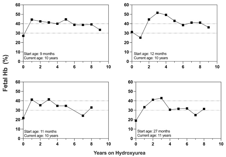
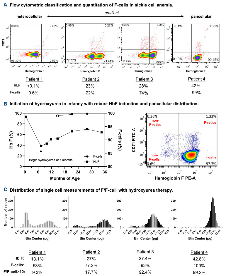
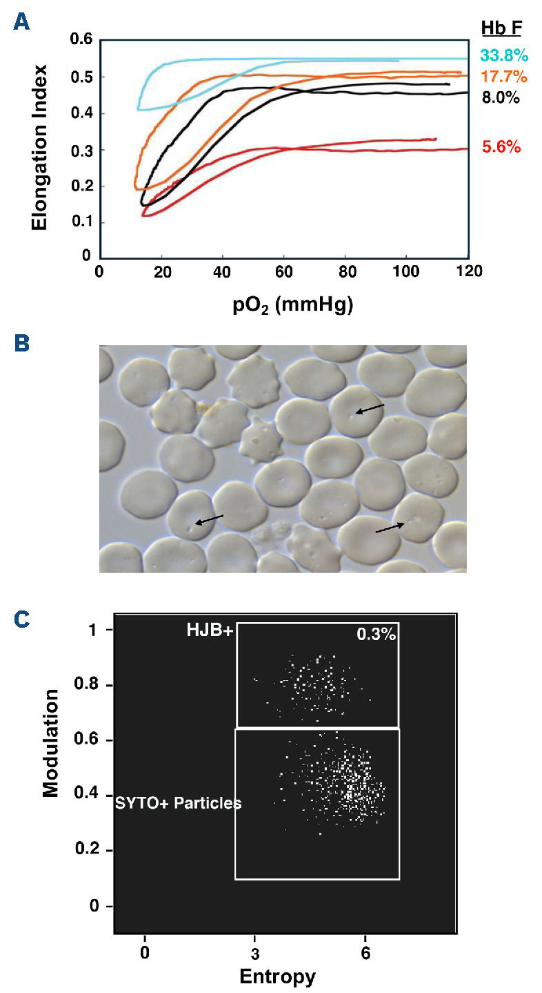
Over the past 40 years, the introduction and refinement of hydroxyurea therapy has led to remarkable progress in the care of individuals with sickle cell anemia (SCA). From initial small proof-of-principle studies to multicenter phase III controlled clinical trials and then numerous open-label studies, the consistent benefits of once-daily oral hydroxyurea have been demonstrated across the lifespan. Elevated fetal hemoglobin (HbF) serves as the most important treatment response, as HbF delays sickle hemoglobin polymerization and reduces erythrocyte sickling. Increased amounts of HbF, especially when distributed across the majority of erythrocytes, improve clinical outcomes by reducing hemolytic anemia and preventing vaso-occlusion, thereby ameliorating both acute and chronic - and overt and covert - complications. Additional benefits of hydroxyurea beyond HbF induction include lower neutrophil and platelet counts, reduced inflammation, and improved rheology. Toxicities of hydroxyurea in SCA are typically mild and predictable; modest cytopenia is expected and actually therapeutic, while occasional gastrointestinal and cutaneous manifestations are well-tolerated. Long-term risks of hydroxyurea for SCA are mainly theoretical but require ongoing surveillance. Accordingly, hydroxyurea should be initiated as part of standard of care, ideally in the first year of life. Proper dosing of hydroxyurea is critical, aiming through stepwise dose escalation to achieve modest but safe myelosuppression, with periodic adjustments for weight gain. Precision dosing using pharmacokinetics may facilitate optimal dosing without frequent dose adjustments. Although transformative and even curative therapies for SCA are emerging, hydroxyurea is the only available and accessible disease-modifying treatment that can address the global burden of disease, especially in low-resource settings within sub-Saharan Africa.


Figures

Figure 1.
Figure 1.
Timeline of hydroxyurea trials. This 40-year timeline illustrates the expanding portfolio of rigorous clinical trials that have consistently demonstrated the efficacy and effectiveness of hydroxyurea treatment for sickle cell anemia across the lifespan. Green-shaded boxes represent phase I/II trials, while red-shaded boxes represent phase III trials. Purple boxes denote the year of regulatory approval for adults (1998) and children (2017).
Figure 2.
Figure 2.
Sustained high values of fetal hemoglobin in children treated from infancy. Four representative examples are provided from over 40 children who initiated hydroxyurea before 3 years of age (9-27 months) who have had sustained treatment responses of fetal hemoglobin (HbF) >30% for 8-9 years. Reference lines are shown for HbF values of 30% and 40%. Note that some patients have occasional struggles with medication adherence, indicated by transient declines in HbF (e.g., the patients shown in the bottom two graphs at 7 years of therapy), which our medical and psychosocial teams can successfully address.
Figure 3.
Figure 3.
F-cell analysis for monitoring hydroxyurea therapy. (A) The gradient of F-cell (red blood cells containing fetal hemoglobin [HbF]) expression across four individuals from heterocellular to pancellular. Patient 1 does not have sickle cell anemia (SCA); patients 2-4 have SCA and different levels of HbF induction with hydroxyurea therapy. Note the sizable difference in F-cell fraction between patients 2 and 3 even though both have similar amounts of HbF. Patient 4 is a young child who initiated phamacokinetically-optimized hydroxyurea therapy by 9 months of age. Each graph shows singlet red blood cell events classified by HbF expression (x-axis) and CD71 expression (y-axis). The two right quadrants of each graph indicate F-cells. The top two quadrants of each graph indicate immature reticulocytes. The total F-cell fraction is the sum of the right upper quadrant (F-retics: circulating reticulocytes containing HbF) and right lower quadrant (mature F-cells). (B) Data for an infant who began hydroxyurea at 7 months of age. The left graph shows HbF values (left y-axis) and F-cell fraction (right y-axis) before and during hydroxyurea therapy. The open circle (o) indicates the timing of the complete F-cell study shown in the right graph, demonstrating pancellular expression of HbF that recapitulates the HbF expression in compound heterozygosity for HbS and gene-deletion hereditary persistence of HbF (HPFH). (C) Histograms of single-cell estimates of mean HbF per F-cell (F/F-cell) (pg) in the F-cell populations of four individuals. Patients 1-3 have SCA, are treated with hydroxyurea, and have increasing amounts of HbF induction. Patient 4 has compound heterozygosity for HbS and gene-deletion HP-FH. Patients 3 and 4 have >90% of F-cells with >10 pg HbF, indicating that their red blood cells are fully protected from sickling. F-cells: red blood cells containing HbF; F-retics: circulating reticulocytes containing HbF; F/F-cell: mean HbF per F-cell.
Figure 4.
Figure 4.
Advanced laboratory testing for monitoring of hydroxyurea therapy. (A) Oxygen-gradient ektacytometry depicts the relationship between red blood cell (RBC) deformability (elongation index), as an index of onset of sickling, and the partial pressure of O2 (pO2). Patients who have higher amounts of fetal hemoglobin (HbF) expression (identified on the right of the graph) have a progressively delayed onset of sickling that occurs at successively lower pO2. (B) Phase-contrast microscopy demonstrates pocked or pitted RBC, which correspond to submembrane inclusions that are normally removed by the spleen (examples shown by black arrows). The RBC pit count is a semiquantitative measurement of splenic function. (C) A novel flow cytometric quantitative method is illustrated; it is designed to detect and enumerate Howell-Jolly bodies (HJB), which are nuclear remnants that are normally removed by the spleen and increased in number in hyposplenic states. Here, RBC are stained with double-stranded DNA-avid dye, and morphological characteristics (size, shape) of the intra-RBC DNA inclusions are used to identify HJB.

Similar articles

Cited by

References

    1. Platt OS, Orkin SH, Dover G, Beardsley GP, Miller B, Nathan DG. Hydroxyurea enhances fetal hemoglobin production in sickle cell anemia. J Clin Invest. 1984;74(2):652-656. - PMC - PubMed
    1. Yawn BP, Buchanan GR, Afenyi-Annan AN, et al. . Management of sickle cell disease: summary of the 2014 evidence-based report by expert panel members. JAMA. 2014;312(1):1033-1048. - PubMed
    1. Watson J. The significance of the paucity of sickle cells in newborn Negro infants. Am J Med Sci. 1948;215(4):419-423. - PubMed
    1. Perrine RP, Pembrey ME, John P, Perrine S, Shoup F. Natural history of sickle cell anemia in Saudi Arabs. A study of 270 subjects. Ann Intern Med. 1978;88(1):1-6. - PubMed
    1. Jacob GF, Raper AB. Hereditary persistence of foetal haemoglobin production, and its interaction with the sickle cell trait. Br J Haematol. 1958;4(2):138-149. - PubMed

MeSH terms