. 2025 Feb 5;16(3):355-364.
doi: 10.1021/acschemneuro.4c00461. Epub 2025 Jan 9.

Biological Amyloids Chemically Damage DNA

Affiliations

Biological Amyloids Chemically Damage DNA

Istvan Horvath et al. ACS Chem Neurosci. .

Abstract

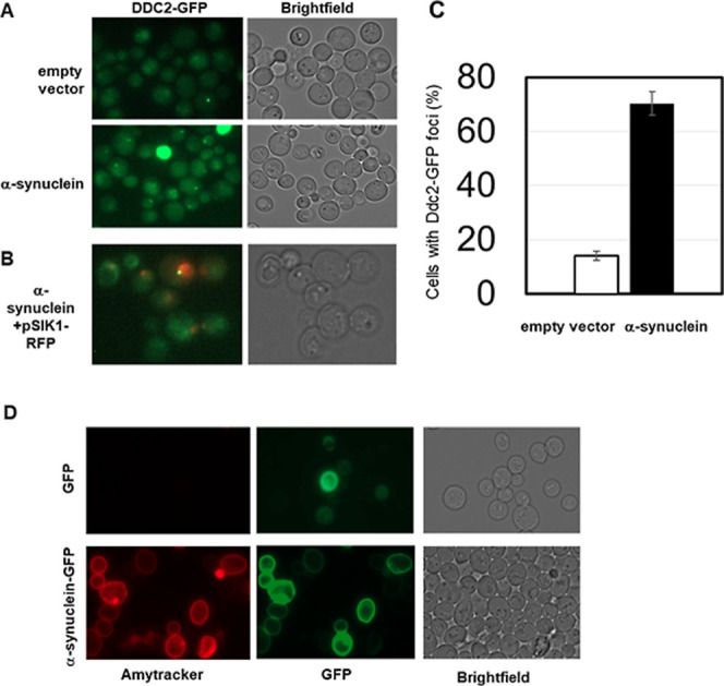
Amyloid fibrils are protein polymers noncovalently assembled through β-strands arranged in a cross-β structure. Biological amyloids were considered chemically inert until we and others recently demonstrated their ability to catalyze chemical reactions in vitro. To further explore the functional repertoire of amyloids, we here probe if fibrils of α-synuclein (αS) display chemical reactivity toward DNA. We demonstrate that αS amyloids bind DNA at micromolar concentrations in vitro. Using the activity of DNA repair enzymes as proxy for damage, we unravel that DNA-amyloid interactions promote chemical modifications, such as single-strand nicks, to the DNA. Double-strand breaks are also evident based on nanochannel analysis of individual long DNA molecules. The amyloid fold is essential for the activity as no DNA chemical modification is detected with αS monomers. In a yeast cell model, there is increased DNA damage when αS is overexpressed. Chemical perturbation of DNA adds another chemical reaction to the set of activities emerging for biological amyloids. Since αS amyloids are also found in the nuclei of neuronal cells of Parkinson's disease (PD) patients, and increased DNA damage is a hallmark of PD, we propose that αS amyloids contribute to PD by direct chemical perturbation of DNA.

Keywords: DNA damage; Parkinson’s disease; alpha-synuclein; amyloids; catalytic activity; nanochannels.


Conflict of interest statement

The authors declare no competing financial interest.

Figures

Figure 1
Figure 1
(A) Binding of monomeric (squares) and amyloid (circles) αS to immobilized DNA as measured by SPR, solid line shows hyperbolic fit. (B) AFM image of λ-DNA on mica surface. (C) AFM image of mixture of DNA and αS amyloids; blue arrows highlight where DNA appears to emerge after following along the amyloid long axis. Z-range for AFM images is 5 nm. (D) Box plot of height distribution of αS amyloids in the presence (average: 7.3 ± 1.0 nm) and absence (average: 6.1 ± 0.7 nm) of λ-DNA (P ≪ 0.0001). Inset shows an example cross section of λ-DNA (blue), αS amyloid alone (black) and αS amyloid with DNA (red).
Figure 2
Figure 2
(A) Scheme of DNA damage detection. λ-DNA incubation with αS amyloids or αS monomers was followed by enzymatic repair and thereafter incorporation of fluorescent nucleotides at the damage sites. (B) Fluorescence microscopy image of labeled λ-DNA after incubation with αS monomers or amyloids and stretched on a functionalized glass coverslip. The DNA backbone was stained with YOYO-1 (green) and red dots are fluorescent nucleotides incorporated at damage sites. Scale bar = 10 μm. (C) DNA damage detection using a repair enzyme cocktail. Error bars indicate standard deviation calculated from biological replicates. (D) Detection of DNA damage using single repair enzymes. Error bars indicate standard deviation calculated from technical duplicates. P-values; ns, not significant; ***P ≤ 0.0002; ****P < 0.0001.
Figure 3
Figure 3
(A) Schematic of the nanofluidic device. (B) Fluorescence images of λ-DNA molecules after incubation (and removal) with 0 μM (control, only DNA), 2.5, 4 and 10 μM αS amyloids in the nanochannels. (C) Distribution of lengths of λ-DNA molecules in the nanochannels. Median length of DNA molecules (arrows) and percentage of molecules with lengths of 4 μm or less are indicated in each panel.
Figure 4
Figure 4
Analysis of DNA damage in actively growing yeast cells. Exponentially growing cells expressing the double-stranded DNA break sensor protein Ddc2 fused to GFP were imaged by fluorescence microscopy. (A) Cells were transformed with either the empty multicopy vector control plasmid (pYX242) or αS expressed from a strong, constitutive promotor. (B) To verify nuclear localization of Ddc2-GFP foci, cells were also transformed with a plasmid expressing a Sik1/Nop56-RFP fusion protein and imaged by fluorescence microscopy. (C) Quantification of the fraction of control and αS expressing cells displaying Ddc2-GFP foci. On average 14.1 ± 1.8 (5.6% SD) of control cells contained foci whereas 70.3 ± 4.3 (16.1% SD) of αS expressing cells contained foci. A two-sided and two-tailed t-test (n = 10 vs n = 14) indicates a statistically significant difference with P < 4.7 × 10⁻¹⁰. (D) Cells expressing GFP tagged αS or GFP only (green) from a strong constitutive promoter were stained with Amytracker (red) to assess presence of amyloids.
Figure 5
Figure 5
(A) Illustration of possible amyloid-DNA interaction. High-resolution structure of wild-type αS amyloid (6h6b) with 5 layers of monomers in two protofilaments is shown next to a piece of B-form DNA (3bse) positioned at the suggested interaction site near the protofilament interface (see text). The surface of the αS amyloid is colored according to electrostatics (blue, positive; red, negative); in the DNA, phosphorus is orange and oxygen is red. The positions where N- and C-termini disordered segments will extend from the ordered amyloid core are indicated. (B) Chemical structures of substrates (PNPA, PNPP, ATP, DNA; the latter two, this work) reported to be cleaved by αS amyloids so far. PNPA, p-nitrophenyl acetate (ester bond); PNPP, p-nitrophenyl phosphate (phosphoester bond). Phosphodiester bonds, proposed cleavage sites in DNA, are marked with red arrows in the DNA chemical structure. We note that other bonds in the DNA backbone may also be targets for the amyloid reactivity.

References

    1. Chiti F.; Dobson C. M. Protein Misfolding, Amyloid Formation, and Human Disease: A Summary of Progress Over the Last Decade. Annu. Rev. Biochem. 2017, 86, 27–68. 10.1146/annurev-biochem-061516-045115. - DOI - PubMed
    1. Sawaya M. R.; Hughes M. P.; Rodriguez J. A.; Riek R.; Eisenberg D. S. The expanding amyloid family: Structure, stability, function, and pathogenesis. Cell 2021, 184 (19), 4857–4873. 10.1016/j.cell.2021.08.013. - DOI - PMC - PubMed
    1. Evans M. L.; Chapman M. R. Curli biogenesis: order out of disorder. Biochim. Biophys. Acta 2014, 1843 (8), 1551–1558. 10.1016/j.bbamcr.2013.09.010. - DOI - PMC - PubMed
    1. Otzen D. Functional amyloid. Prion 2010, 4 (4), 256–264. 10.4161/pri.4.4.13676. - DOI - PMC - PubMed
    1. Fink A. L. The aggregation and fibrillation of alpha-synuclein. Acc. Chem. Res. 2006, 39 (9), 628–634. 10.1021/ar050073t. - DOI - PubMed

LinkOut - more resources