. 2025 Jan 22;73(3):2019-2032.
doi: 10.1021/acs.jafc.4c09392. Epub 2025 Jan 9.

Enhanced Production of Rebaudioside D and Rebaudioside M through V155T Substitution in the Glycosyltransferase UGT91D2 from Stevia rebaudiana

Affiliations

Enhanced Production of Rebaudioside D and Rebaudioside M through V155T Substitution in the Glycosyltransferase UGT91D2 from Stevia rebaudiana

Tsubasa Shoji et al. J Agric Food Chem. .

Abstract

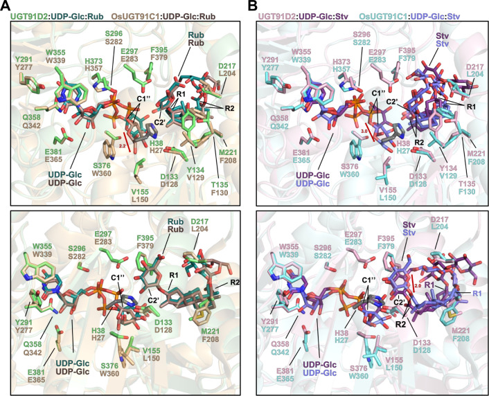
Steviol glycosides (SGs) are noncaloric natural sweeteners found in the leaves of stevia (Stevia rebaudiana). These diterpene glycosides are biosynthesized by attaching varying numbers of monosaccharides, primarily glucose, to steviol aglycone. Rebaudioside (Reb) D and Reb M are highly glucosylated SGs that are valued for their superior sweetness and organoleptic properties, yet they are present in limited quantities in stevia leaves. This study aims to improve the substrate preference and catalytic efficiency of UDP-sugar-dependent glycosyltransferase UGT91D2 from stevia, which acts as a bottleneck in the biosynthesis of Reb D and Reb M. We modeled the structure of UGT91D2 and substituted two amino acid residues, Y134 and V155, which are located near the glycosyl acceptor and donor, respectively. Expression of the UGT91D2V155T in budding yeast significantly enhanced the production of Reb D and Reb M. Furthermore, transient expression in Nicotiana benthamiana revealed that the V155T substitution improved the glucosylation activity of UGT91D2, suggesting that this substitution enhances UDP-glucose binding and reduces side reactions involving nonglucose donors. By coexpressing multiple stevia UGT genes in N. benthamiana, we successfully produced highly glucosylated SGs from steviol. Our results provide insights into the substrate specificity of UGT91D2 and contribute to the engineering of SG biosynthesis.

Keywords: UDP-glucose; UDP-sugar-dependent glycosyltransferase; stevia; steviol glycoside.


Conflict of interest statement

The authors declare the following competing financial interest(s): Y.T., M.K., J.T., and T.H. are/were employees of Suntory Global Innovation Center Ltd. The other authors declare that they have no competing interests.

Figures

Figure 1
Figure 1
Steviol glycosides (SGs) and their formation from steviol (St) through glycosylation in stevia. (A) A diagram illustrating the glycosylation steps in SG biosynthesis. Steps catalyzed by UGT85C2, UGT74G1, UGT91D2, and UGT76G1 are indicated by black, gray, red, and blue arrows, respectively. The R1 (C13 hydroxyl group) and R2 (C19 carboxyl group) are highlighted in red next to the St structure. Glucose (Glc), rhamnose (Rh), and xylose (Xy) moieties are represented according to their respective linkages. Glycosylation steps occurring on the R1 and R2 sides are depicted in horizontal and vertical directions, respectively. (B) Structures of minor SGs. The analogs differ from the indicated SGs by the addition of one monosaccharide moiety, either in terms of position or type. Dul, dulcoside; Reb, rebaudioside; Rub, rubusoside; Sb, steviolbioside; S13G, steviol 13-O-glucoside; S19G, steviol 19-O-glucoside; Stv, stevioside; UGT, UDP-sugar-dependent glycosyltransferase.
Figure 2
Figure 2
Structures of UGT91D2 and OsUGT91C1 docked with substrates. The same structures are viewed from different angles in the upper and lower panels, with a focus on the active site. Color codes are as follows: oxygen (red), nitrogen (blue), sulfur (yellow), phosphorus (orange), and carbon backbones of the catalytic H27 in OsUGT91C1 and H38 in UGT91D2 (dark gray). (A) Superimposed views of UGT91D2:UDP-Glc:Rub and OsUGT91C1:UDP-Glc:Rub. (B) Superimposed views of UGT91D2:UDP-Glc:Stv and OsUGT91C1:UDP-Glc:Stv. The shifts of the Glc moiety of UDP-Glc in the upper panels (measured at C5′’) and the shifts of Stv in the lower panels (also measured at C5′’) between UGT91D2 and OsUGT91 structures are indicated by red arrows, along with the measured distances (Å).
Figure 3
Figure 3
UGT-mediated glycosylation of St in budding yeast. The average levels of SGs are shown along with standard deviations, based on 4–7 biological replicates analyzed by LC-MS/MS. Significant differences compared to the wild-type (WT) controls were assessed using a Student’s t-test, with significance indicated above each bar: **p < 0.01, *p < 0.05. ns indicates not significant. The graphs were arranged in the same order as in Figure 1A. Conversions mediated by UGT91D2 are marked with light gray arrows connecting the graphs of substrates to their corresponding products.
Figure 4
Figure 4
Conversion of SG substrates by UGT91D1, UGT91D2, or UGT91D2V155T in N. benthamiana leaves. UGT91D1, UGT91D2, or UGT91D2V155T was transiently expressed in N. benthamiana leaves to convert S13G (A), Rub (B), Stv (C), or Reb A (D), each of which was infiltrated into the leaves, into their corresponding products. The pBI121 vector served as a control (C). Conversions mediated by UGT91D2 are indicated with light gray arrows connecting the substrate and product graphs. Narrow arrows with dashed lines represent side reactions. Error bars indicate the standard deviation based on 8–12 biological replicates. Significant differences relative to the control (C) (indicated above each bar) and between UGT91D2 and UGT91D2V155T were assessed using a Student’s t-test: **p < 0.01, *p < 0.05. ns indicates not significant. DW denotes dry weight.
Figure 5
Figure 5
Formation of SGs from St in N. benthamianaleaves. The contents of St and major SGs are displayed. UGT85C2, UGT74G1, and UGT76G1 are collectively referred to as 3UGT. Error bars represent the standard deviation from 14 or 15 biological replicates. Significant differences compared to the control (C) (indicated above each bar) and among the samples connected by lines were assessed using a Student’s t-test: **p < 0.01, *p < 0.05. ns indicates not significant. The graphs are arranged in the same order as in Figure 1A. Conversions mediated by UGT91D2 are indicated with light gray arrows connecting the substrate and product graphs. DW denotes dry weight.
Figure 6
Figure 6
UGT-mediated production of minor SGs in N. benthamiana. UGT85C2, UGT74G1, and UGT76G1 are collectively referred to as 3UGT. Analogs of the minor SGs are indicated in brackets. Error bars represent standard deviation based on 14 or 15 biological replicates. Significant differences compared to the control (shown above each bar) and among the samples connected by lines were assessed using a Student’s t-test: **p < 0.01, *p < 0.05. ns indicates not significant.
Figure 7
Figure 7
UGT91D2V155T model structure docked with substrates. Structures around the active site are displayed with distances in Å indicated beside relevant dashed lines. Color codes are as follows: oxygen (red), nitrogen (blue), sulfur (yellow), phosphorus (orange), and carbon backbones of the catalytic H38 (dark gray). (A) UGT91D2V155T model structure docked with UDP-Glc and Rub (UGT91D2V155T:UDP-Glc:Rub). (B) UGT91D2V155T model structure docked with UDP-Glc and Stv (UGT91D2V155T:UDP-Glc:Stv).

Similar articles

References

    1. WHO, Guideline: Sugars Intake for Adults and children. 2015, https://www.who.int/pubulication/i/item/9789241549028. - PubMed
    1. Kinghorn A. D.Stevia: The Genus Stevia; Routledge: London, 2002.
    1. Ceunen S.; Geuns J. M. Steviol glycosides: chemical diversity, metabolism, and function. J. Nat. Prod. 2013, 76 (6), 1201–1228. 10.1021/np400203b. - DOI - PubMed
    1. Gerwig G. J.; Te Poele E. M.; Dijkhuizen L.; Kamerling J. P. Stevia glycosides: chemical and enzymatic modifications of their carbohydrate moieties to improve the sweet-tasting quality. Adv. Carbohydr. Chem. Biochem. 2016, 73, 1–72. 10.1016/bs.accb.2016.05.001. - DOI - PubMed
    1. Yang T.; Zhang J.; Ke D.; Yang W.; Tang M.; Jiang J.; Cheng G.; Li J.; Cheng W.; Wei Y.; Li Q.; Naismith J. H.; Zhu X. Hydrophobic recognition allows the glycosyltransferase UGT76G1 to catalyze its substrate in two orientations. Nat. Commun. 2019, 10 (1), 3214.10.1038/s41467-019-11154-4. - DOI - PMC - PubMed

MeSH terms

LinkOut - more resources