. 2025 Feb 4;24(2):163-175.
doi: 10.1158/1535-7163.MCT-24-0588.

OBI-992, a Novel TROP2-Targeted Antibody-Drug Conjugate, Demonstrates Antitumor Activity in Multiple Cancer Models

Affiliations

OBI-992, a Novel TROP2-Targeted Antibody-Drug Conjugate, Demonstrates Antitumor Activity in Multiple Cancer Models

Wan-Fen Li et al. Mol Cancer Ther. .

Abstract

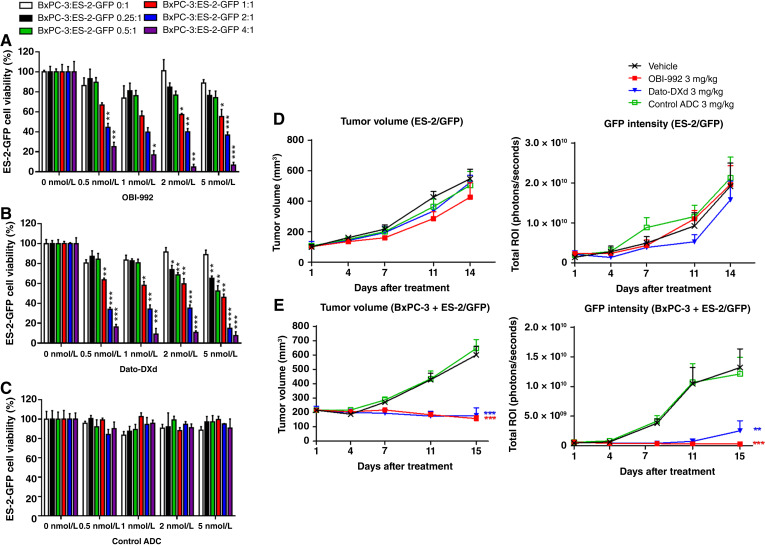
Trophoblast cell surface antigen 2 (TROP2) is highly expressed in multiple cancers relative to normal tissues, supporting its role as a target for cancer therapy. OBI-992 is an antibody-drug conjugate (ADC) derived from a novel TROP2-targeted antibody linked to the topoisomerase 1 (TOP1) inhibitor exatecan via an enzyme-cleavable hydrophilic linker, with a drug-antibody ratio of 4. This study evaluated and compared the antitumor activity of OBI-992 with that of benchmark TROP2-targeted ADCs datopotamab deruxtecan (Dato-DXd) and sacituzumab govitecan (SG) in cell line-derived xenograft (CDX) and patient-derived xenograft (PDX) models. OBI-992 treatment exhibited statistically significant antitumor activity versus controls at doses of 3 and 10 mg/kg in various CDX and PDX models, demonstrating comparable or better antitumor activity with benchmark ADCs. In a large-tumor model, longer survival times were observed in OBI-992-treated mice compared with Dato-DXd-treated mice. OBI-992 treatment induced marked bystander killing of TROP2-negative cells in the presence of nearby TROP2-positive cells in both in vitro and in vivo studies. In lung adenocarcinoma CDX models with overexpression of either P-glycoprotein (P-gp) or breast cancer resistance protein (BCRP) to mimic ATP-binding cassette transporter-mediated multidrug resistance, OBI-992 treatment maintained antitumor activity when Dato-DXd treatment became less effective. The combination of OBI-992 at suboptimal doses with either poly (ADP-ribose) polymerase (PARP) inhibitors or an immune check point inhibitor produced synergistic antitumor effects in mouse models. Taken together, these translational results support further development of OBI-992 as a cancer therapy.


Conflict of interest statement

W.-F. Li reports a patent for PCT/US2024/021729 pending and a patent 113111546 pending. T.-Y. Huang reports a patent for PCT/US2024/021729 pending and a patent 113111546 pending. M.-T. Lai reports a patent for PCT/US2024/021729 pending and a patent for 113111546/Taiwan pending. No disclosures were reported by the other authors.

Figures

None
Graphical abstract
Figure 1.
Figure 1.
TROP2 expression in various cancer tissues. A, The expression levels of TROP2 in tissue samples from 19 cancer types were analyzed by IHC. Dots indicate TROP2 H-score for each sample, and solid bars indicate the median H-score of each cancer type. B, TROP2 prevalence rates (x-axis) were calculated as the percentage of samples with TROP2 H-score ≥100. The ranking order of cancer types was based on the TROP2 prevalence rate. C, Plot of percentage of cells from tumor (blue) and corresponding normal tissues (gray) with TROP2 membrane expression determined by IHC. Each dot represents an individual sample. Solid bars indicate the median percent of TROP2-expressing cells for each tissue type. The numbers of specimens used for each cancer type are indicated in the plots. Representative IHC images of TROP2 high (3+), moderate (2+), and low (1+) expression are shown in Supplementary Fig. S3. Adeno, adenocarcinoma; BC, breast carcinoma; Ca., cancer/carcinoma; CRC, colorectal cancer; H&N, head and neck carcinoma; HER2, human epidermal growth factor receptor 2; HR, hormone receptor; HRPC, hormone-resistant prostate cancer; IHC, immunohistochemistry; NSCLC, non-small cell lung cancer; RCC, renal cell renal carcinoma; SCC, squamous carcinoma; SCLC, small cell lung carcinoma; TNBC, triple-negative breast carcinoma.
Figure 2.
Figure 2.
In vivo antitumor activity of OBI-992 on multiple CDX models. Mice were intravenously treated with indicated doses of drugs or vehicle control. A–E, Changes to tumor volume are plotted against number of days after an initial treatment dose for (A) BxPC-3 (n = 5 per group), (B) NCI-N87 (n = 5 per group), (C) NCI-H1975 (n = 6 per group), and (D) MDA-MB-231 CDX mice (n = 5 per group) as well as for (E) BxPC3 large-tumor CDX mice (n = 6 per group). TGI was calculated for each treatment group and is denoted on the right side of tumor growth plots. F and G, Survival curves were produced for BxPC-3 large-tumor xenograft mice treated with (F) 10 mg/kg or (G) 3 mg/kg OBI-992 or Dato-DXd. *, P < 0.05; **, P < 0.01; ***, P < 0.001 for treatment groups versus vehicle control, unless otherwise noted with a bracket. Dots and error bars indicate the mean ± SEM. All animals survived to scheduled sacrifice with two exceptions: one control animal in C was sacrificed on day 11 due to ulcered tumor, and two OBI-992 (10 mg/kg)-treated mice in D were sacrificed on day 17 due to body weight loss >15%. In large-tumor model (E, F, G), animals were sacrificed if the tumor size exceeded 1,400 mm3 or with ulcered tumor diameter larger than 15 mm. No animals died directly due to tumor burden. BIW × 4, twice weekly for four doses; Dato-DXd, datopotamab deruxtecan; QW × 3, once every week for three doses; SG, sacituzumab govitecan; TGI, tumor growth inhibition.
Figure 3.
Figure 3.
In vivo antitumor activity of OBI-992 on multiple PDX models. Mice were intravenously treated with indicated doses of drugs. Changes to tumor volume are plotted against number of days after an initial treatment dose for (A) BR9464 breast cancer, (B) GA0091 gastric cancer, (C) LU-01-1370 lung cancer, (D) PC-07-0003 pancreatic cancer, (E) ES-06-0010 esophageal cancer, (F) OV-10-0068 ovarian cancer, (G) LU-01-1004 and (H) LU-01-0236 lung cancer, and (I) GA6866 gastric cancer PDX mice. TGI was calculated for each treatment group and is denoted on the right side of tumor growth plots. *, P < 0.05; **, P < 0.01; ***, P < 0.001 for treatment groups versus vehicle control, unless otherwise noted with a bracket. Dots and error bars indicate the mean ± SEM. A–F, n = 3 and (G–I) n = 6. All animals survived to the scheduled endpoint. Dato-DXd, datopotamab deruxtecan; SEM, standard error of the mean; TGI, tumor growth inhibition.
Figure 4.
Figure 4.
Bystander killing effect with OBI-992 treatment. A–C, TROP2-positive BxPC3 cells were co-cultured with TROP2-negative ES-2/GFP cells at indicated ratios. The cultures were treated with serially diluted (A) OBI-992, (B) Dato-DXd, or (C) control ADC. Viability of ES-2/GFP cells was measured by the intensity of GFP fluorescence relative to the corresponding non-treated group (0 nmol/L). Dots and error bars indicate the mean ± SEM (n = 3 independent experiments). D and E, CDX models of TROP2-negative ES-2/GFP cells (D) alone or (E) mixed with TROP2-positive BxPC-3 cells were treated with vehicle, 3 mg/kg OBI-992, 3 mg/kg Dato-DXd, or control ADC as a single dose. Tumor volume and GFP intensity are plotted against the number of days after treatment. ***, P < 0.001 for treatment groups versus vehicle control. Dots and error bars indicate the mean ± SEM. D, n = 5 and (E) n = 10. All animals survived to the scheduled endpoint. ADC, antibody–drug conjugate; ROI, region of interest; SEM, standard error of the mean.
Figure 5.
Figure 5.
OBI-992 maintained antitumor activity in both P-gp and BCRP overexpression tumor models. A, ABCB1 (encodes P-gp) and (B) ABCG2 (encodes BCRP) mRNA levels in HCC827, HCC827/ABCB1, and HCC827/ABCG2 cells were measured by qRT-PCR. Relative transcript levels are normalized to levels in HCC827 cells. Dots and error bars indicate the mean ± SEM (n = 3). C–H, Mice were intravenously treated with indicated doses of drugs. OBI-992 or Dato-DXd was administered as a single dose on day 1, whereas SG was administered twice a week for four doses. Changes to tumor volume are plotted against number of days after an initial treatment dose for (C and D) HCC827 (n = 5 per group), (E and F) HCC827/ABCB1 (n = 6 per group), and (G and H) HCC827/ABCG2 CDX mice (n = 6 per group). TGI was calculated for each treatment group and is denoted on the right side of tumor growth plots. *, P < 0.05; **, P < 0.01; ***, P < 0.001 for treatment group versus vehicle control. Dots and error bars indicate the mean ± SEM (n = 5–6). All animals survived to the scheduled endpoint. BCRP, breast cancer resistance protein; Dato-DXd, datopotamab deruxtecan; P-gp, P-glycoprotein; SEM, standard error of the mean; SG, sacituzumab govitecan; TGI, tumor growth inhibition.
Figure 6.
Figure 6.
Synergistic antitumor effect of OBI-992 in combination with PARP inhibitors or anti-PD-1 antibody. A and B, In vitro cytotoxicity assay of HRD-positive Capan-1 and HRD-negative BxPC3 cells treated with OBI-992 and/or (A) olaparib or (B) talazoparib at decreasing doses. The cell viability presented was normalized to a vehicle control. Dots and error bars indicate the mean ± SEM (n = 3 technical replicates). The synergistic biologic activity of OBI-992 in combination with olaparib or talazoparib was examined using CDI as described in “Methods and Materials”; combination groups that showed synergistic effect (CDI <1) were labeled with an asterisk. The calculated CDI values were described in Supplementary Table S8. C and D, Capan-1 CDX mice were treated with the indicated suboptimal doses of OBI-992 via intravenous injection and/or (C) olaparib or (D) talazoparib by oral gavage. All animals survived to scheduled endpoint. E, Colon cancer MC38/hTROP2 syngeneic mice (immunocompetent B6 mice; n = 6 per group) were treated with the indicated suboptimal doses of OBI-992 and/or anti-PD-1 via intravenous injection. The experiment was terminated on day 11 due to rapid tumor growth in the syngeneic model. No tumor rejection was observed in animals. C–E, Changes to tumor volume are plotted against the number of days after an initial treatment dose. TGI was calculated for each treatment group and is denoted on the right side of tumor growth plots. *, P < 0.05; **, P < 0.01; ***, P < 0.001 for the comparisons indicated by brackets. Dots and error bars indicate the mean ± SEM (n = 6). Anti-PD-1, anti-programmed cell death protein 1 antibody; IC50, half-maximal inhibitory concentration; SEM, standard error of the mean; TGI, tumor growth inhibition.

References

    1. Fu Z, Li S, Han S, Shi C, Zhang Y. Antibody drug conjugate: the “biological missile” for targeted cancer therapy. Signal Transduct Target Ther 2022;7:93. - PMC - PubMed
    1. Shastry M, Gupta A, Chandarlapaty S, Young M, Powles T, Hamilton E. Rise of antibody-drug conjugates: the present and future. Am Soc Clin Oncol Educ Book 2023;43:e390094. - PubMed
    1. Drago JZ, Modi S, Chandarlapaty S. Unlocking the potential of antibody-drug conjugates for cancer therapy. Nat Rev Clin Oncol 2021;18:327–44. - PMC - PubMed
    1. Shvartsur A, Bonavida B. Trop2 and its overexpression in cancers: regulation and clinical/therapeutic implications. Genes Cancer 2015;6:84–105. - PMC - PubMed
    1. Cubas R, Zhang S, Li M, Chen C, Yao Q. Trop2 expression contributes to tumor pathogenesis by activating the ERK MAPK pathway. Mol Cancer 2010;9:253. - PMC - PubMed

MeSH terms

LinkOut - more resources