Review
. 2025 Jun 21;27(5):1165-1179.
doi: 10.1093/neuonc/noae276.

A hitchhiker's guide to cerebrospinal fluid biomarkers for neuro-oncology

Affiliations
Review

A hitchhiker's guide to cerebrospinal fluid biomarkers for neuro-oncology

Cecile Riviere-Cazaux et al. Neuro Oncol. .

Erratum in

Abstract

Cerebrospinal fluid (CSF) has emerged as a valuable liquid biopsy source for glioma biomarker discovery and validation. CSF produced within the ventricles circulates through the subarachnoid space, where the composition of glioma-derived analytes is influenced by the proximity and anatomical location of sampling relative to tumor, in addition to underlying tumor biology. The substantial gradients observed between lumbar and intracranial CSF compartments for tumor-derived analytes underscore the importance of sampling site selection. Moreover, radiographic features, such as tumor-CSF contact and blood-brain barrier disruption, are critical covariates that may affect biomarker detection and the abundance of plasma-derived analytes in CSF, respectively. Longitudinal intracranial CSF sampling, enabled by access devices like Ommaya reservoirs, may offer a window into treatment response and disease progression, though variability in analyte yield, sample volumes, and the dynamic effects of surgical resection pose challenges. This review critically evaluates the anatomic, radiographic, and longitudinal factors, or "time-space continuum," that impact glioma CSF biomarker abundance. Practical considerations for longitudinal CSF biobanking, including access device placement and collection, are also reviewed. Key takeaways and recommendations for CSF glioma biomarker discovery and validation are provided as a "hitchhiker's guide" based on our collective experience, along with resources for investigators aiming to develop CSF biobanking at their institutions.

Keywords: biomarker; cerebrospinal fluid; glioma; monitoring; neuro-oncology.


Conflict of interest statement

The authors have no competing interests to declare that are directly related to the work in this review.

Figures

Figure 1.
Figure 1.
Biomarker distribution in CSF based on contact with CSF spaces. The relative concentration of biomarkers in CSF is depicted for tumors with (A) pial contact, (B) ventricular contact, (C) no contact (fully intraparenchymal tumors), and (D) leptomeningeal disease.
Figure 2.
Figure 2.
D-2-hydroxyglutarate (D-2-HG) as a candidate biomarker of IDH-mutant gliomas. (A) D-2-hydroxyglutarate (D-2-HG) concentrations were compared in paired lumbar and intracranial CSF samples from patients with IDH-mutant gliomas (n = 14 pairs), as well as in (B) IDH-wild-type tumors (glioma and other central nervous system tumors; n = 12 pairs), via a Wilcoxon matched-pairs signed-rank test. (C) D-2-HG concentrations were compared in lumbar CSF from patients with IDH-mutant gliomas versus IDH-wild-type tumors via Mann-Whitney test. The normality of the distribution was tested using a D’Agostino-Pearson normality test. Lines indicate the mean with standard deviation.
Figure 3.
Figure 3.
Location of CSF acquisition impacts glioma biomarker discovery. (A) FGF1 abundance is shown in intracranial and lumbar CSF from patients with gliomas (paired; n = 16), as well as from patients with NPH (unpaired lumbar, n = 6, and intracranial, n = 9). Wilcoxon signed-rank test was performed on paired glioma intracranial versus lumbar samples; all other tests performed were Mann-Whitney U tests. (B) The abundances of five of the most differentially abundant proteins between intracranial glioma versus NPH CSF are shown; lumbar glioma versus NPH CSF results are also shown (Mann-Whitney U tests). The glioma and NPH LP and intracranial sample sizes are as in (A). (C) CSF was obtained intra-operatively from a patient with a grade 2 IDH-mutant astrocytoma from the Sylvian fissure, temporal horn of the lateral ventricle, and ambient cistern, as well as the lumbar cistern via an LP. Post-resection CSF was obtained on POD 2 via an Ommaya reservoir implanted at resection. D-2-HG was quantified in each sample. (D) A Mann-Whitney U test was performed on the abundance of calcyphosin-like protein in ventricular (n = 20) versus subarachnoid (n = 23) CSF from patients with GBM. Lines indicate the median and 95% confidence intervals for each group. ACSF2 = acyl-CoA synthetase family member 2, APOBEC3G = apolipoprotein B mRNA editing enzyme catalytic subunit 3G, HPGDS = hematopoietic prostaglandin D synthase; IFI16 = gamma-interferon-inducible protein 16; CLIC2 = chloride intracellular channel protein 2.
Figure 4.
Figure 4.
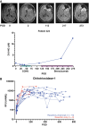
Longitudinal intracranial glioma CSF: impacts of progression and resection. (A) CSF was obtained intra-operatively from the lateral ventricle during resection for a recurrent grade 4 IDH-mutant astrocytoma, during which an Ommaya reservoir was placed for longitudinal CSF access. CSF was then collected at multiple points during treatment with lomustine (CCNU) and bevacizumab, with disease progression occurring near POD73 and 217. (B) The abundance of chitrotriosidase-1 was evaluated in longitudinal CSF samples from patients with gliomas beginning at the time of resection (POD0), 13 of whom underwent chemoradiation and 3 of whom only underwent resection.
Figure 5.
Figure 5.
Ommaya reservoir catheters in the resection cavity. The location of the ventricular catheters attached to Ommaya reservoirs over time is shown in open (left) versus collapsed (right) resection cavities from two patients on T2-weighted MRI scans. The catheter tip is circled.

References

    1. Stupp R, Mason WP, van den Bent MJ, et al. ; European Organisation for Research and Treatment of Cancer Brain Tumor and Radiotherapy Groups. Radiotherapy plus concomitant and adjuvant temozolomide for glioblastoma. N Engl J Med. 2005;352(10):987–996. - PubMed
    1. Wen PY, Bent M, Youssef G, et al. RANO 2.0: update to the response assessment in Neuro-Oncology criteria for high- and low-grade gliomas in adults. J Clin Oncol. 2023;41(33):5187–5199. - PMC - PubMed
    1. Thust SC, van den Bent MJ, Smits M.. Pseudoprogression of brain tumors. J Magn Reson Imaging. 2018;48(3):571–589. - PMC - PubMed
    1. Gilbert MR, Dignam JJ, Armstrong TS, et al. A randomized trial of bevacizumab for newly diagnosed glioblastoma. N Engl J Med. 2014;370(8):699–708. - PMC - PubMed
    1. Barthel FP, Johnson KC, Varn FS, et al. ; GLASS Consortium. Longitudinal molecular trajectories of diffuse glioma in adults. Nature. 2019;576(7785):112–120. - PMC - PubMed

Substances