

Automated quantification of anterior chamber cells using swept-source anterior segment optical coherence tomography
- PMID: 39786628
- PMCID: PMC11717729
- DOI: 10.1186/s12348-025-00456-y
Automated quantification of anterior chamber cells using swept-source anterior segment optical coherence tomography
Abstract
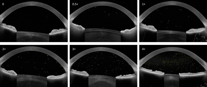
Purpose: To validate automated counts of presumed anterior chamber (AC) cells in eyes with histories of uveitis involving the anterior segment using swept-source (SS) anterior segment optical coherence tomography (AS-OCT) against manual counts and compare automated counts against Standardized Uveitis Nomenclature (SUN) criteria.
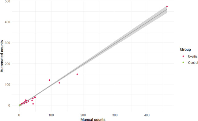
Methods: Eyes were imaged with the ANTERION SS AS-OCT device (Heidelberg Engineering). A fully automated custom algorithm quantified the number of hyper-reflective foci (HRF) in line-scan images. Automated and manual counts were compared using interclass correlation (ICC) and Pearson correlation coefficient. Automated counts were compared to SUN grades using a mixed-effects linear regression model.
Results: 90 eyes (54 participants) were included; 67 eyes (41 participants) had histories of uveitis, while 23 eyes (13 healthy participants) served as controls. ICC comparing automated to manual counts was 0.99 and the Pearson correlation coefficient was 0.98. Eyes at each SUN grade with corresponding median HRF (interquartile range [IQR]) were: Grade 0, 42 eyes, 2 HRF (0,4); 0.5+, 10 eyes, 10 HRF (8,15); 1+, 9 eyes, 22 HRF (15,33); 2+, 3 eyes, 27 HRF; 3+, 2 eyes, 128 HRF; 4+, 1 eye, 474 HRF. For every 1-step increase in grade, automated count increased by 38 (p < 0.001) or 293% (Pearson correlation coefficient 0.80, p < 0.001). Automated counts differed significantly between clinically inactive eyes (2 HRF [0,4]) and controls (0 HRF [0,1], p = 0.02).
Conclusions: Our algorithm accurately counts HRF when compared to manual counts, with strong correlation to SUN clinical grades. SS AS-OCT offers the advantage of imaging of the entire AC and may allow detection of subclinical inflammation in eyes that appear clinically inactive.
Keywords: Anterior chamber inflammation; Image analysis; Optical coherence tomography (OCT); Standardization of Uveitis nomenclature (SUN); Uveitis.
© 2025. The Author(s).
Conflict of interest statement
Declarations. Ethics approval and consent to participate: The study was approved by the UCLA Health Institutional Review Board (IRB#19-001732). Consent for publication: Written informed consent was obtained from all study participants, unless they were < 18 years of age, in which case written informed consent was obtained from a parent of the study participant and an additional assent document was signed by study participants 7 years of age and older. Competing interests: The authors declare no competing interests.
Figures

References
Grants and funding
LinkOut - more resources
Full Text Sources
Research Materials
