. 2025 Apr 1;142(4):692-703.
doi: 10.1097/ALN.0000000000005360. Epub 2025 Jan 8.

Impaired Macroscopic Cerebrospinal Fluid Flow by Sevoflurane in Humans during and after Anesthesia

Affiliations

Impaired Macroscopic Cerebrospinal Fluid Flow by Sevoflurane in Humans during and after Anesthesia

Juliana Zimmermann et al. Anesthesiology. .

Abstract

Background: According to the model of the glymphatic system, the directed flow of cerebrospinal fluid (CSF) is a driver of waste clearance from the brain. In sleep, glymphatic transport is enhanced, but it is unclear how it is affected by anesthesia. Animal research indicates partially opposing effects of distinct anesthetics, but corresponding results in humans are lacking. Thus, this study aims to investigate the effect of sevoflurane anesthesia on CSF flow in humans, both during and after anesthesia.

Methods: Using data from a functional magnetic resonance imaging experiment in 16 healthy human subjects before, during, and 45 min after sevoflurane monoanesthesia of 2 volume percent (vol%), the authors related gray matter blood oxygenation level-dependent signals to CSF flow, indexed by functional magnetic resonance imaging signal fluctuations, across the basal cisternae. Specifically, CSF flow was measured by CSF functional magnetic resonance imaging signal amplitudes, global gray matter functional connectivity by the median of interregional gray matter functional magnetic resonance imaging Spearman rank correlations, and global gray matter-CSF basal cisternae coupling by Spearman rank correlations of functional magnetic resonance imaging signals.

Results: Anesthesia decreased cisternal CSF peak-to-trough amplitude (median difference, 1.00; 95% CI, 0.17 to 1.83; P = .013) and disrupted the global cortical blood oxygenation level-dependent and functional magnetic resonance imaging-based connectivity (median difference, 1.5; 95% CI, 0.67 to 2.33; P < 0.001) and global gray matter-CSF coupling (median difference, 1.19; 95% CI, 0.36 to 2.02; P = 0.002). Remarkably, the impairments of global connectivity (median difference, 0.94; 95% CI, 0.11 to 1.77; P = 0.022) and global gray matter-CSF coupling (median difference, 1.06; 95% CI, 0.23 to 1.89; P = 0.008) persisted after re-emergence from anesthesia.

Conclusions: Collectively, the authors' data show that sevoflurane impairs macroscopic CSF flow via a disruption of coherent global gray matter activity. This effect persists, at least for a short time, after regaining consciousness. Future studies need to elucidate whether this contributes to the emergence of postoperative neurocognitive symptoms, especially in older patients or those with dementia.


Conflict of interest statement

The authors declare no competing interests.

Figures

Fig. 1.
Fig. 1.
Decreased cerebrospinal fluid (CSF) signal peak-to-trough amplitude as a marker of flow during anesthesia. (A) Top, Representative sagittal T1-weighted image of a subject and positioning of the imaging volume as well as the bottom slice (yellow). Bottom, Bottom slice of the imaging volume containing the CSF voxel mask (yellow). (B) Representative normalized CSF signal traces of a representative subject during preanesthesia wakefulness (wake_pre, top), anesthesia with 2% sevoflurane (sevo, middle), and postanesthesia wakefulness (wake_post, bottom). (C) Box plot of the peak-to-trough amplitude of the slice 1 CSF signal depicted in B for all n = 16 subjects during wake_pre (left), 2% sevoflurane (middle), and wake_post. Friedman test with post hoc comparison using Tukey honestly significant difference procedure. *P < 0.05; n.s., not significant.
Fig. 2.
Fig. 2.
Decreased absolute functional connectivity during anesthesia and after emergence. (A) Surface projection of the 247 subregions used for the correlation analysis. (B) Correlation matrix (Spearman correlation) of the subregions defined in A for one representative subject during the preanesthesia wakefulness (wake_pre), 2% sevoflurane, and wake_post stages. (C) Box plot of the median absolute global functional connectivity value (Spearman rank) for all n = 16 subjects during preanesthesia wakefulness (wake_pre, left), 2% sevoflurane (sevo, middle), and wake_post conditions. Friedman test with post hoc comparison via Tukey honestly significant difference procedure. **P < 0.01; *P < 0.05; n.s., not significant.
Fig. 3.
Fig. 3.
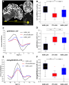
Impaired global gray matter–cerebrospinal fluid (CSF) coupling during and after anesthesia. (A) Left, Sagittal blood oxygenation level–dependent (BOLD) image of a representative subject (left) with the global gray matter mask superimposed in white and the slice 1 CSF mask in yellow. Right, Axial slices from the same subject. (B) Box plot of the Spearman correlation coefficients between global gray matter (gGM)–BOLD and CSF for all n = 16 subjects during preanesthesia wakefulness (wake_pre) (left), 2% sevoflurane (middle), and wake_post conditions. Friedman test with post hoc comparison using Tukey honestly significant difference procedure. (C) Cross-correlation of the averaged gGM-BOLD and CSF signals under wake_pre (black), 2% sevoflurane (red), and wake_post (blue) conditions. (D) same as B for the amplitude of the lag at +4 s, the timepoint with the strongest anticorrelation. (E) Cross-correlation of the averaged –dt/t of gGM-BOLD and CSF signals under wake_pre (black), 2% sevoflurane (red), and wake_post (blue) conditions. (F) Amplitude of the cross-correlation shown in E for the timepoint of –2 s, the timepoint with the highest correlation. *** P < 0.001; **P < 0.01; *P < 0.05; n.s., not significant. CSF, cerebrospinal fluid.
Fig. 4.
Fig. 4.
The correlation of cortical connectivity or CSF flow with global gray matter (gGM)–CSF coupling is disrupted by sevoflurane. (A) Relationship between median absolute global functional connectivity (FC) and gGM–CSF coupling for preanesthesia wakefulness (wake_pre, black, left), during sevoflurane (red, middle), and after sevoflurane (wake_post, blue, right). Each circle corresponds to an individual subject. r and P values are derived from Spearman rank correlation testing. (B) Same as A for the relationship of CSF amplitude and gGM-CSF coupling.

References

    1. Hladky SB, Barrand MA: The glymphatic hypothesis: The theory and the evidence. Fluids Barriers CNS 2022; 19:9. - PMC - PubMed
    1. Iliff JJ, Wang M, Liao Y, et al. : A paravascular pathway facilitates CSF flow through the brain parenchyma and the clearance of interstitial solutes, including amyloid beta. Sci Transl Med 2012; 4:147ra11 - PMC - PubMed
    1. Hablitz LM, Nedergaard M: The glymphatic system: A novel component of fundamental neurobiology. J Neurosci 2021; 41:7698–711 - PMC - PubMed
    1. Fultz NE, Bonmassar G, Setsompop K, et al. : Coupled electrophysiological, hemodynamic, and cerebrospinal fluid oscillations in human sleep. Science 2019; 366:628–31 - PMC - PubMed
    1. Hablitz LM, Pla V, Giannetto M, et al. : Circadian control of brain glymphatic and lymphatic fluid flow. Nat Commun 2020; 11:4411. - PMC - PubMed