. 2025 Jan 9;20(1):e0313255.
doi: 10.1371/journal.pone.0313255. eCollection 2025.

Knockout of the ING5 epigenetic regulator confirms roles in stem cell maintenance and tumor suppression in vivo

Affiliations

Knockout of the ING5 epigenetic regulator confirms roles in stem cell maintenance and tumor suppression in vivo

Buthaina Al Shueili et al. PLoS One. .

Abstract

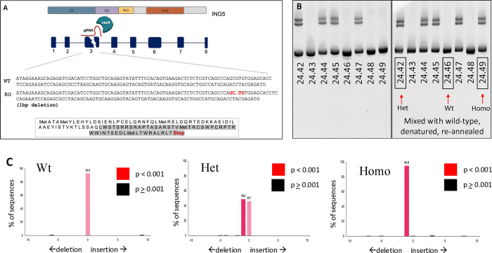
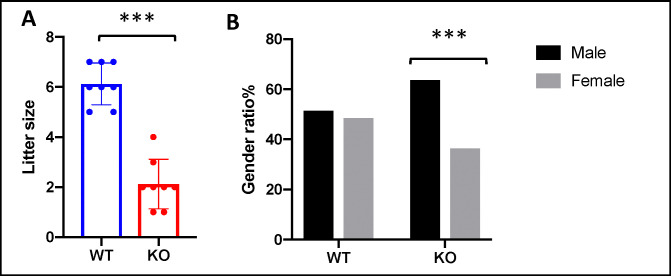
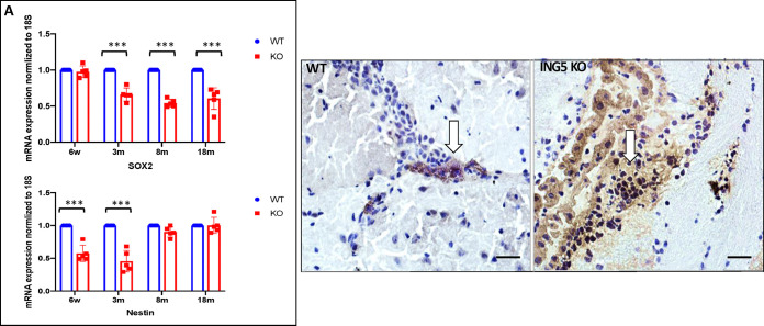
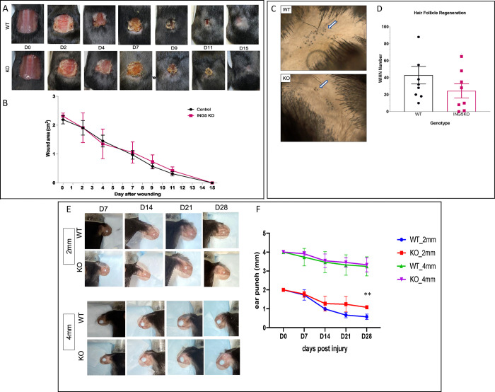
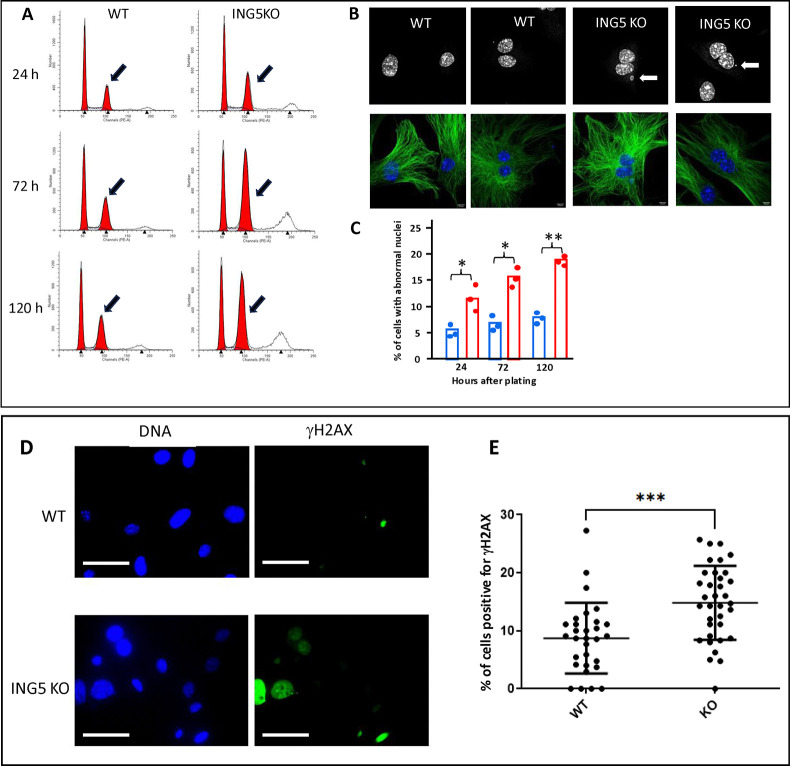
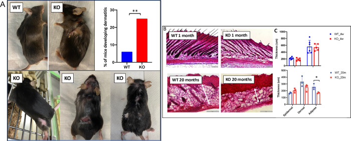
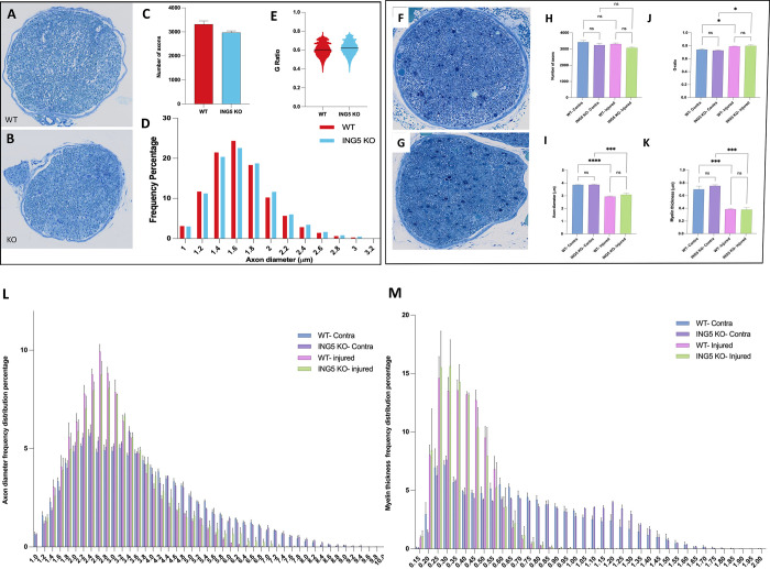
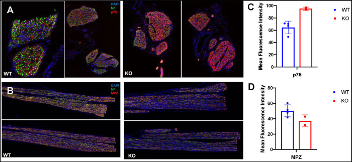
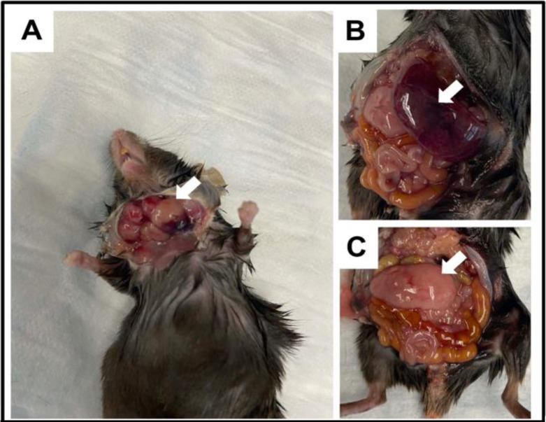
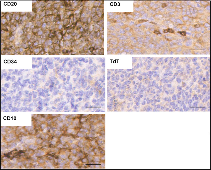
INhibitor of Growth (ING1-5) proteins are epigenetic readers that target histone acetyltransferase (HAT) or histone deacetylase (HDAC) complexes to the H3K4Me3 mark of active transcription. ING5 targets Moz/Morf and HBO1 HAT complexes that alter acetylation of H3 and H4 core histones, affecting gene expression. Previous experiments in vitro indicated that ING5 functions to maintain stem cell character in normal and in cancer stem cells. Here we find that CRISPR/Cas9 ING5 knockout (KO) mice are sub-fertile but show no decrease in lifespan or ability to heal wounds despite indications of depleted stem cell pools in several tissues. ING5 KO mouse embryo fibroblasts accumulate in G2 of the cell cycle, have high levels of abnormal nuclei and show high basal levels of the γH2AX indicator of DNA damage. KO animals also develop severe dermatitis at a 5-fold higher rate that wild-type littermates. Consistent with ING5 serving a tumor suppressive role, ING5 KO mice developed germinal centre diffuse large B-cell lymphomas at a rate 6-fold higher than control mice at 18 months of age. These data suggest that ING5 functions in vivo to maintain stem cell character in multiple organs, that reduction of stem cell populations is not limiting for murine lifespan and that like a subset of other ING family members, ING5 functions as a tumor suppressor in hematopoietic cells in vivo.


Conflict of interest statement

All authors declare no competing interests for this study.

Figures

Fig 1
Fig 1. Generation of CRISPR/Cas9 ING5 knockout (KO) mice.
A) The CRISPR/Cas9 targeting strategy used for generating ING5 knockout (KO) mice. Exons are indicated in blue boxes, and introns are noted by black lines. The targeting site for each of the guide RNAs (gRNAs) used is in exon3 (a unique site that distinguishes ING5 from ING4). The nucleotide and amino acid (in black) sequences of exon3 in wild-type (WT) and KO alleles are shown. The Gray area represents exon3 amino acids with a premature stop codon. B) For genotyping line 24 (8bp deletion), DNA was isolated from mouse ear samples and amplified using ING5 primers. PCR products analyzed on 10% polyacrylamide gels showing a single band are wild-type while heterozygous samples give additional retarded band(s). For homozygous KO samples, wildtype DNA is mixed with the samples, denatured, and re-annealed. If the sample is homozygous mutant, two bands are observed. C) For genotyping of the one bp deletion line, DNA was isolated from mouse ear samples, amplified using ING5 primers and PCR products were purified for sequencing and then analyzed using TIDE. Wild-type samples show bars with zero mutation, heterozygous samples are identified with two bars; one with zero mutation and the other with a 1bp deletion and homozygous samples give one bar with a 1bp deletion.
Fig 2
Fig 2. ING5 knockout progeny and demography.
A) Litter sizes from WT and KO matings. Individual data points represent 1 mating that resulted in a litter. Graph shows mean ±SEM, ***p<0.0005. B) Percentage of males versus females in WT and KO mice. Total numbers of WT animals examined was 192 males and 181 females and in KO animals 128 males and 73 females. ***p<0.005.
Fig 3
Fig 3. ING5 KO mice show reduction in neural stem cell markers and an increase in differentiated progenitors in brain.
A) mRNA levels of the SOX2 and nestin neural stem cell markers in ING5 KO and WT brain tissues at 1.5, 3, 8 and 18 months. The graph shows mean ±SEM. (n = 5), ***p<0.005. B) Indirect immunostaining of 3-month-old WT and KO mice for the differentiation marker doublecortin (DC) in the brain subventricular zone. Arrows identify cells with brown positive staining and nuclei staining blue. The percentage of positive cells in WT in 4 fields was 8.8 ± 1% and in the KO 31.3 ± 24% (n = 3). DC expression is a measure of neural stem cells differentiating into neural precursor cells (NPCs) and forming newly differentiated neurons. Scale bars = 50mM.
Fig 4
Fig 4. ING5 KO mice repair skin wounds efficiently but produce fewer hair follicles after wounding.
A) Wound closure for full-thickness square wounds (≥1.5cm diameter) in 28 day old mice. Wounds were monitored every 48h until full healing. B) Quantification of the wound area using Image J. The graph shows mean ±SEM, (n = 8). The scale bar = 5 cm. C) Skin section from healed wounds showing newly generated hair follicles in WT and KO mice. The arrows identify follicles. D) Quantification of wound-induced hair follicle neogenesis (WIHN) in WT and KO mice (n = 8). E) Ear punchs (2mm and 4mm) for 8 week old WT and KO mice in the centre of the right and left ear, respectively. Wounds were monitored every 7 days for 28 days. F) Quantification of the ear punch area. The graph shows mean ±SEM. (n = 3). **P<0.01.
Fig 5
Fig 5. ING5 KO fibroblasts rapidly accumulate in G2 of the cell cycle.
A) Representative flow cytometry plots for cell cycle analysis of WT and ING5-KO mouse embryo fibroblasts. Cells were harvested 24, 72 and 120 h after plating, and were harvested, fixed and stained with propidium iodide. Fifteen thousand cells were collected per sample. The arrows highlight peaks containing 4N amounts of DNA corresponding to cells containing 2 nuclei. B) Representative images of nuclei in WT and ING5 KO MEFs. Nuclei are stained with Hoechst and alpha-tubulin is shown in green. Arrows indicate micronuclei seen more frequently in ING5 KO cells. C) Quantification of the number of abnormal nuclei observed in WT and ING5 KO MEFs. Each bar graph represents 3 fields of 120 cells each counted in a blind experimental protocol. *p<0.05; **p<0.02. D) Representative images of WT and ING5 KO primary mouse embryo fibroblasts stained for DNA with Hoechst and with an antibody for ɣH2AX. Images were taken with a 40x objective and the bar = 50 μM. E) Quantification of cells positive for ɣH2AX in WT and ING5 KO primary mouse embryo fibroblasts. Cells that contained one or more foci were deemed positive. Cells with pan-nuclear staining of ɣH2AX were excluded from the analysis. 80–110 cells were counted per sample with 3 biological replicates for each cell strain (240–330 total cells per cell strain). Graph shows mean +/- SD ***p<0.005; n = 3.
Fig 6
Fig 6. ING5 KO mice frequently develop dermatitis with ageing.
A) WT and ING5 KO mice in upper panels are 18 months old. KO mice in lower panels show differing severities of dermatitis. The graph indicates the percentage of mice developing dermatitis by 18 months of age (n = 90 WT and 105 KO). **p<0.01. B) H&E staining of skin sections of WT and KO in young (1 month) and old (20 month) mice. C) Quantification of epidermal and dermal thickness in WT and KO young and old mice. The graph shows mean ±SEM (n = 5). Scale bar = 200 μM. *p<0.05.
Fig 7
Fig 7. Axon regrowth and myelination is not compromised in ING5 knockouts.
Neonatal wild type A) or knockout B) pups had sciatic nerves removed and semi-thin sections were stained with Toluidine blue to visualize axons and myelin. Axon number C), axon diameter D) and G-ratio E) were similar in wild type and knockout mice (n = 3). Eight week-old wild type F) or knockout G) mice had sciatic nerves removed four weeks after injury and semi-thin sections were stained with toluidine blue to visualize axons and myelin. H) Axon number, I) Axon thickness, J) G-ratio and K) myelin thickness were similar in wild type and knockout mice. Axon diameter and myelin thickness was also similarly reduced in WT and ING5 KO mice after injury (n = 3). The ranges of axon diameter L) and myelin thickness M) is shown for animals in the injured and contralateral (uninjured) sciatic nerves. *P<0.05, ***<0.005.
Fig 8
Fig 8. ING5 KO mice show delayed expression of the MPZ Schwann cell marker.
Eight-week-old mice had their sciatic nerve crushed and harvested four weeks after injury. Harvested nerves were examined for regrowth and healing of axons by staining for the axonal neurofilament (NF) marker and for expression of MPZ marking myelinating Schwann cells or p75 marking non-myelinating Schwann cells. A) transverse and B) longitudinal sections of sciatic nerve proximal to crush points stained for the indicated markers in wild-type or ING5 KO animals. Mean fluorescence intensity for p75 C) and D) MPZ from 4 WT or 2 ING5 KO animals each, indicates that by four weeks ING5 KO animals had slightly reduced staining for markers of myelinating axons.
Fig 9
Fig 9. ING5 KO mice develop enlarged lymph nodes, spleens and pancreata.
A subset of ING5 KO mice (17 of 129 at 18 months of age vs. 3 of 132 wild-type littermates) show enlarged lymph nodes (arrow in panel A), enlarged spleen (arrow in panel B) and enlarged pancreas (panel C with spleen removed). The mouse shown is 15 months old.
Fig 10
Fig 10. Staining ING5 KO tumors.
Strong staining for CD20 and weak intermittent staining for CD3 indicate B-cell and not T-cell origin. A lack of CD34 or TdT staining indicates the tumor formed from a more mature cell lineage while CD10 staining is consistent with ING5 KO tumors representing germinal center diffuse large B-cell lymphomas. Scale bars = 50μm.

Similar articles

Cited by

References

    1. Jaenisch R., Young R., Stem cells, the molecular circuitry of pluripotency and nuclear reprogramming, Cell. 132 (2008) 567–582. doi: 10.1016/j.cell.2008.01.015 - DOI - PMC - PubMed
    1. Takahashi K., Yamanaka S., Induction of Pluripotent Stem Cells from Mouse Embryonic and Adult Fibroblast Cultures by Defined Factors, Cell. 126 (2006) 663–676. doi: 10.1016/j.cell.2006.07.024 - DOI - PubMed
    1. Morgan H.D., Santos F., Green K., Dean W., Reik W., Epigenetic reprogramming in mammals, Human Molecular Genetics. 14 (2005) R47–R58. doi: 10.1093/hmg/ddi114 - DOI - PubMed
    1. Reik W., Stability and flexibility of epigenetic gene regulation in mammalian development, Nature. 447 (2007) 425–432. doi: 10.1038/nature05918 - DOI - PubMed
    1. Allis C.D., Jenuwein T., The molecular hallmarks of epigenetic control, Nat Rev Genet. 17 (2016) 487–500. doi: 10.1038/nrg.2016.59 - DOI - PubMed

Substances

LinkOut - more resources