. 2025 Jan 7;53(1):gkae1309.
doi: 10.1093/nar/gkae1309.

Cu2+ mediates the oxidation of the transcription factor MscA to regulate the antioxidant defense of mycobacteria

Affiliations

Cu2+ mediates the oxidation of the transcription factor MscA to regulate the antioxidant defense of mycobacteria

Yuling Xiang et al. Nucleic Acids Res. .

Abstract

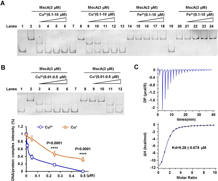
Copper (Cu), a trace element with redox activity, is both essential and toxic to living organisms. Its redox properties make it a cofactor for a variety of proteins, but it also causes oxidative stress, hence the need to maintain intracellular copper homeostasis. However, the role of copper in the regulation of antioxidant defense in bacteria remains unclear, and the involved transcription factors remain to be explored. In this study, we identified a novel transcription factor, MscA, that responded directly to Cu2+ to regulate the antioxidant defense of mycobacteria. Cu2+ directly bound to MscA to mediate oxidation and inhibit the DNA binding activity of MscA, subsequently downregulating the expression of antioxidant gene cluster to increase the accumulation of reactive oxygen species in mycobacteria, ultimately leading to oxidative damage to mycobacteria. Therefore, we firstly reported that the Cu2+ responsive transcription factor regulated the antioxidant defense in bacteria. This finding firstly and directly links the function of Cu2+ to the antioxidant defense of bacteria, and provides a new insight into bacterial antioxidant defense.


Figures

Graphical Abstract
Graphical Abstract
Figure 1.
Figure 1.
MscA directly and positively regulates the expression of antioxidant gene clusters to promote the antioxidant activity of M. smegmatis. (A) Assays for the effect of H2O2 stress on the growth of M. smegmatis. The growth of different mycobacterial strains, Msm/pMV261, ΔmscA/pMV261 and ΔmscA/pMV261-mscA, in 7H9 medium supplemented with 0.75 mM H2O2. (B) The schematic of the mscA operon and its regulatory region. (C) EMSAs of the DNA binding activity of MscA on mscAp promoter. The mscAp substrate was co-incubated with progressively increasing amounts of MscA (lanes 2–6). (D) β-Galactosidase activity assays. The effect of MscA on the gene expression was assayed by constructing mscAp-lacZ plasmid. The expression of mscAp-lacZ in the wild-type strain (Msm/WT) and mscA deletion mutant strain (ΔmscA) was examined. The data were presented as Miller units (right panel). The left column shows schematic representation of each plasmid used to generate recombinant strains. hsp60-lacZ , null promoter-lacZ and ms1839p were used as controls. (E) The growth of different mycobacterial strains, Msm/pMV261, Msm/pMV261-mscA, ΔmscAp/pMV261 and ΔmscAp/pMV261-mscA, in 7H9 medium supplemented with 1 mM H2O2. Error bars represent the variant range of the data derived from three biological replicates.
Figure 2.
Figure 2.
The regulation of antioxidant activity by MscA is conserved in mycobacteria. (A) The effect of allogenic mscAMmi on the gene expression was assayed by constructing mscAp-lacZ and mscAp- mscAMmi plasmids in the wild-type strain (Msm/WT). The activity of β-galactosidase in mscAp or mscAp-mscAMmi co-expressed recombinant strains was determined. (B) The growth of different mycobacterial strains, Msm/pJAM2, ΔmscA/pJAM2 and ΔmscA/pJAM2-mscAMmi, in 7H9 medium supplemented with 0.75 mM H2O2. (C) The left panel shows absence of different motifs of mscAp promoter schematic. Two DNA binding motifs, box1 and box2, located on the genome in M. smegmatis are indicated. The right panel shows EMSAs for the importance of two DNA binding motifs of MscA. Three mutant DNA substrates derived from the mscAp promoter, designated as mscAp1 (box2 deletion), mscAp2 (box1 deletion), mscAp3 (box1 and box2 deletion), were synthesized. These different DNA substrates were co-incubated with MscA (0–3 μM) and sampled on the gel for analysis. (D) The activity of β-galactosidase in Msm/mscAp-lacZ strain, Msm/mscAp1-lacZ strain, Msm/mscAp2-lacZ strain and Msm/mscAp3-lacZ strain was determined. Error bars represent the variant range of the data derived from three biological replicates.
Figure 3.
Figure 3.
Cu2+ specifically binds to MscA and inhibits its DNA binding activity. (A) EMSAs for the effect of Fe2+/Fe3+ or Cu+/Cu2+ on the DNA binding activity of MscA in vitro. The concentration of MscA was immobilized and the increasing amounts of Fe2+/Fe3+ or Cu+/Cu2+ (0.1–10 μM) were added into the reactions mixture and incubated for 15 min. Then, the DNA fragment was added into the reaction mixture and incubated for 10 min. (B) Comparison of concentration of Cu2+ and Cu+ on the DNA binding activity of MscA. The lower panel shows the quantitative comparison of the effects of Cu2+ and Cu+ on DNA/protein complexes. (C) ITC assays for the specific interaction between MscA and Cu2+. Raw titration data and integrated thermal measurements are shown in the top and bottom panels.
Figure 4.
Figure 4.
Cu2+ affects MscA oligomerization and inhibits its DNA binding activity. (A) The secondary structure of MscA protein was detected by circular dichroic chromatography. MscA was incubated with Cu2+ for 15 min and the secondary structure was detected by circular dichrometer. (B) SDS–PAGE assays for detecting the oligomerization state of MscA. The MscA and Cu2+ were incubated for 15 min. Then, the EDTA, DTT and urea were added into the reactions and incubated for 10 min, and the samples were added to the gel for detection. The lower panel shows quantitative analysis the monomer of MscA. (C) EMSAs for the effect of EDTA and DTT on the DNA binding activity of MscA. The concentration of MscA was immobilized and the amounts of Cu2+ (0.5 μM) were added into the reactions and incubated for 15 min, and then different concentrations of EDTA and DTT (0–2 mM) were added into the reactions and incubated for 15 min. The DNA substrate was added into the mixture and incubated for 10 min.
Figure 5.
Figure 5.
The key amino acid residue for Cu2+ oxidation of MscA is Cys40. (A) EMSAs for the effect of Cu2+ on DNA binding activity of MscA (C40A) mutant proteins. The concentrations of MscA and MscA (C40A) mutant proteins were immobilized, and the increasing amounts of Cu2+ (0.01–0.5 μM) were added into the reactions incubated for 15 min. The DNA fragment was added into the reactions mixture and incubated for 10 min. (B) ITC assays for the specific interaction between MscA (C40A) and Cu2+. Raw titration data and integrated thermal measurements are shown in the top and bottom panels. (C) The model of Cu2+ mediating the dimer formation of MscA.
Figure 6.
Figure 6.
Cu2+ inhibits the growth of mycobacteria under H2O2 stress by downregulating the expression of antioxidant genes. (A,B) β-Galactosidase activity assays. The effect of Cu2+ on gene expression was assayed by constructing mscAp-lacZ plasmid in the wild-type strain (Msm/WT) and mscA deletion mutant strain (ΔmscA). (C) β-Galactosidase activity assays. The effect of Cu2+ on gene expression was assayed by constructing mscAp-mscA-lacZ overexpression plasmid in the wild-type strain (Msm/WT). (D,E) The effects of Cu2+ on the growth of M. smegmatis under H2O2 stress. Both wild-type strain (Msm/WT) and mscA deletion mutant strain (ΔmscA) were grown in 7H9 medium supplemented with 0.3 mM (D) or 0.1 mM H2O2 (E), and different concentrations of Cu2+ were added. (F) Msm/WT strain, ΔmscA strain and ΔmscAp strain were grown in 7H9 medium and supplemented with different concentrations of Cu2+, and then fluorescent dye H2DCFDA was used to detect the bacterial intracellular ROS.
Figure 7.
Figure 7.
The model of which Cu2+ mediates the oxidation of the transcription factor MscA to regulate the antioxidant defense of mycobacteria.

Similar articles

References

    1. Andrei A., Ozturk Y., Khalfaoui-Hassani B., Rauch J., Marckmann D., Trasnea P.I., Daldal F., Koch H.G. Cu homeostasis in bacteria: the ins and outs. Membranes (Basel). 2020; 10:242. - PMC - PubMed
    1. Furukawa Y., Shintani A., Kokubo T. A dual role of cysteine residues in the maturation of prokaryotic Cu/Zn-superoxide dismutase. Metallomics. 2021; 13:mfab050. - PubMed
    1. Furukawa Y., Shintani A., Narikiyo S., Sue K., Akutsu M., Muraki N. Characterization of a novel cysteine-less Cu/Zn-superoxide dismutase in Paenibacillus lautus missing a conserved disulfide bond. J. Biol. Chem. 2023; 299:105040. - PMC - PubMed
    1. Frangipani E., Haas D. Copper acquisition by the SenC protein regulates aerobic respiration in Pseudomonas aeruginosaPAO1. FEMS MicrobioL Lett. 2009; 298:234–240. - PubMed
    1. Changela A., Chen K., Xue Y., Holschen J., Outten C.E., O’Halloran T.V., Mondragon A. Molecular basis of metal-ion selectivity and zeptomolar sensitivity by CueR. Science. 2003; 301:1383–1387. - PubMed

MeSH terms