. 2025 Jan 7;53(1):gkae1308.
doi: 10.1093/nar/gkae1308.

The role of ribosomal protein networks in ribosome dynamics

Affiliations

The role of ribosomal protein networks in ribosome dynamics

Youri Timsit et al. Nucleic Acids Res. .

Abstract

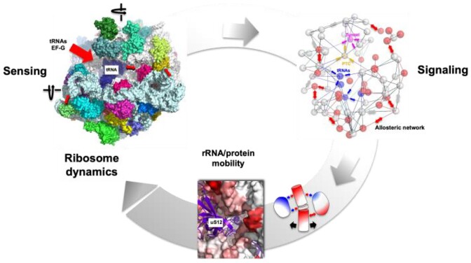
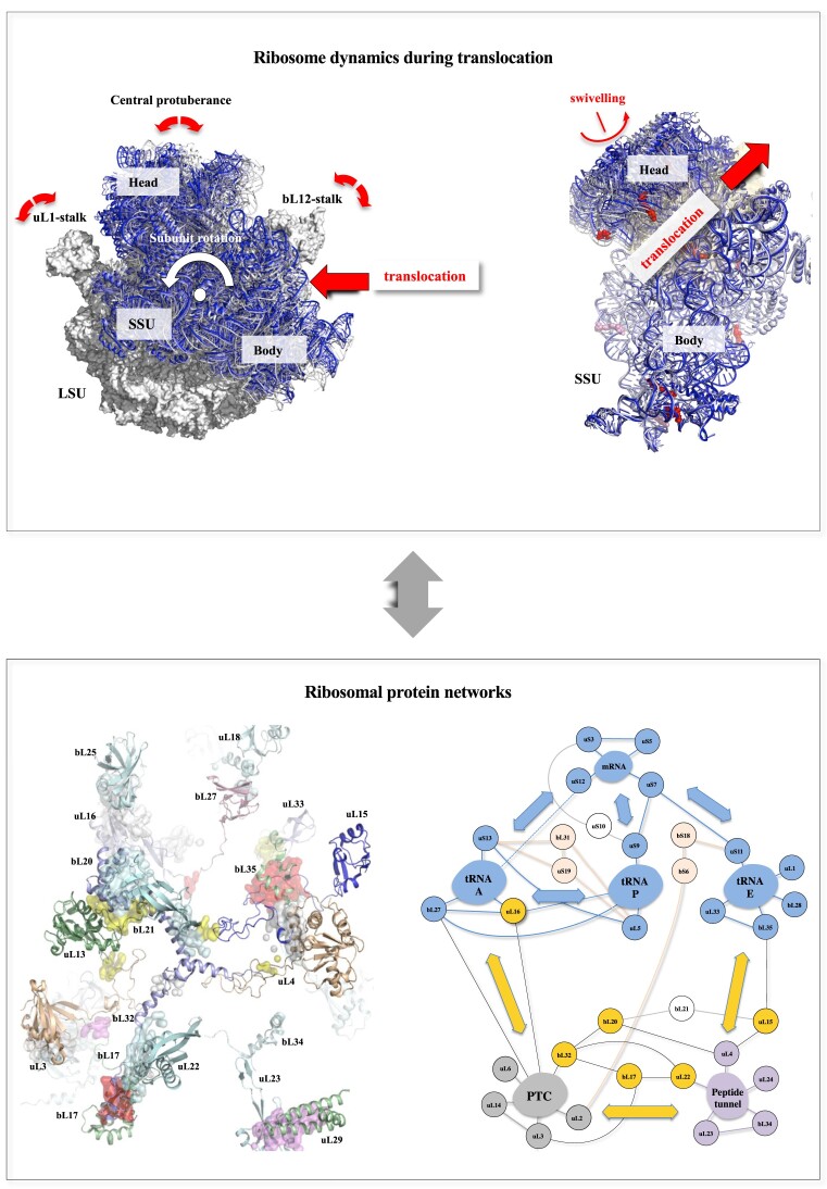
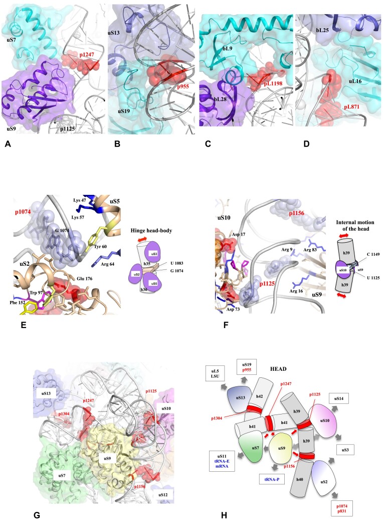
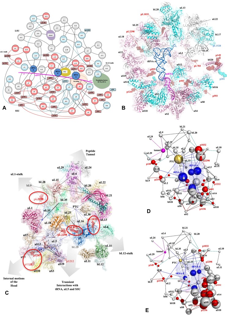
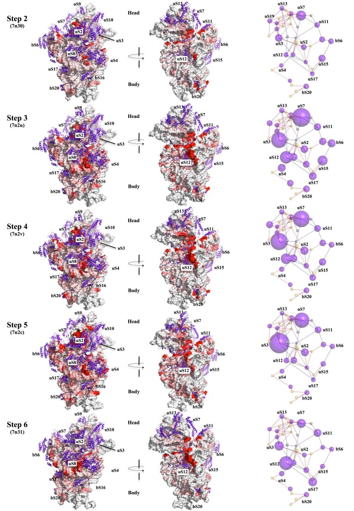
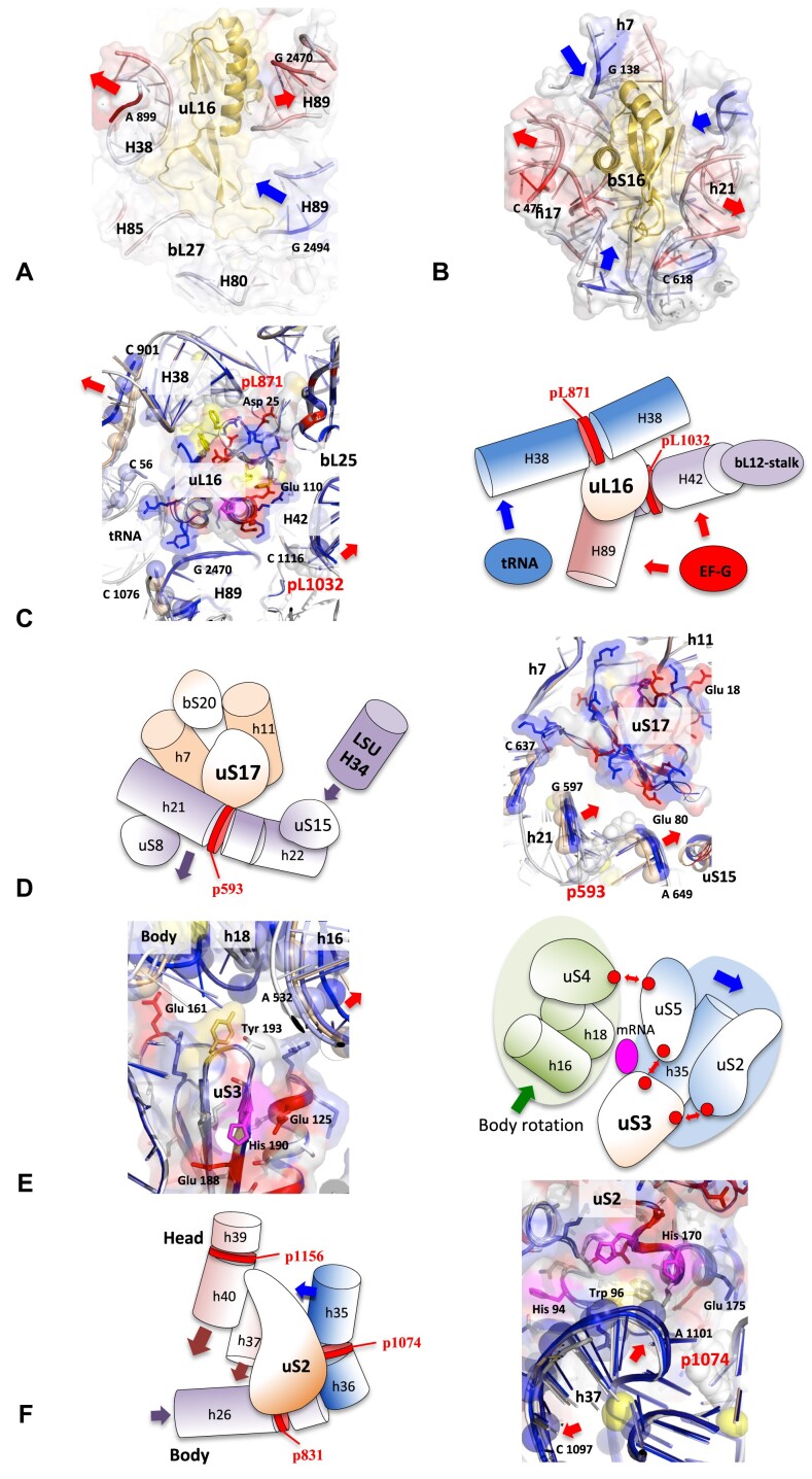
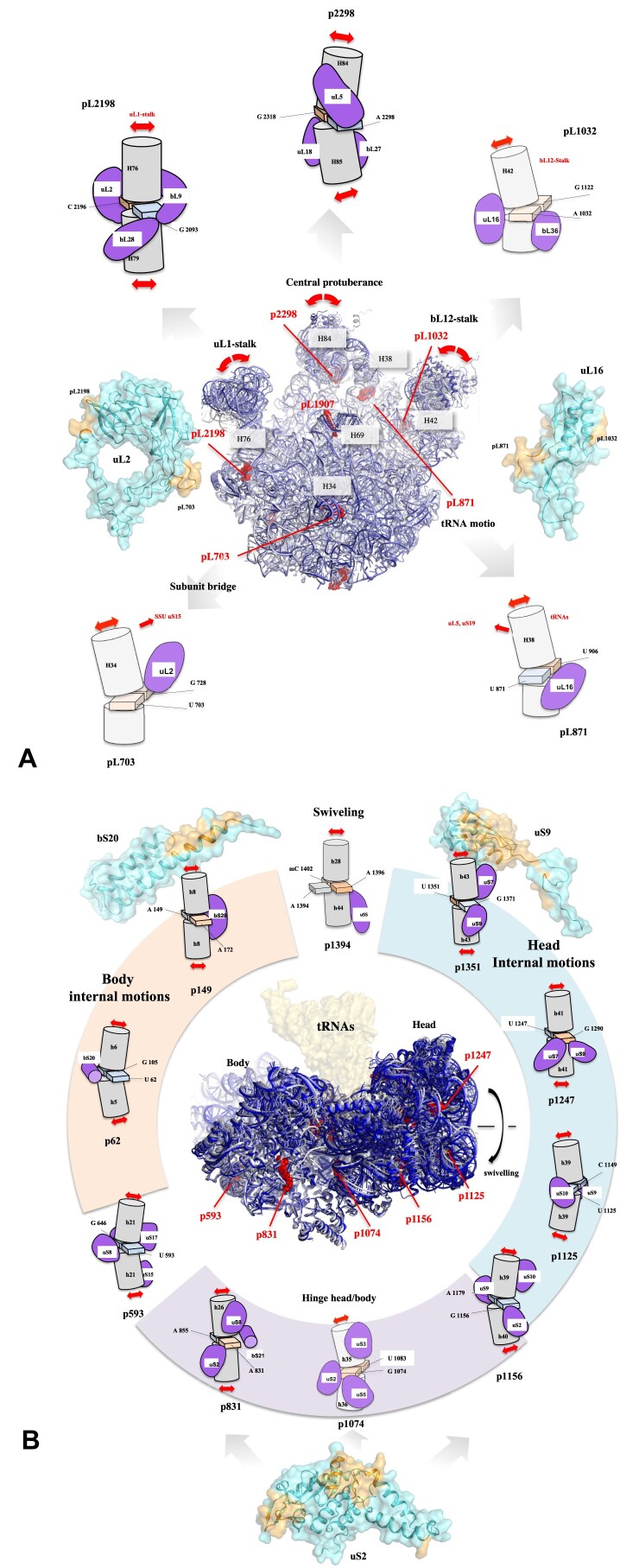
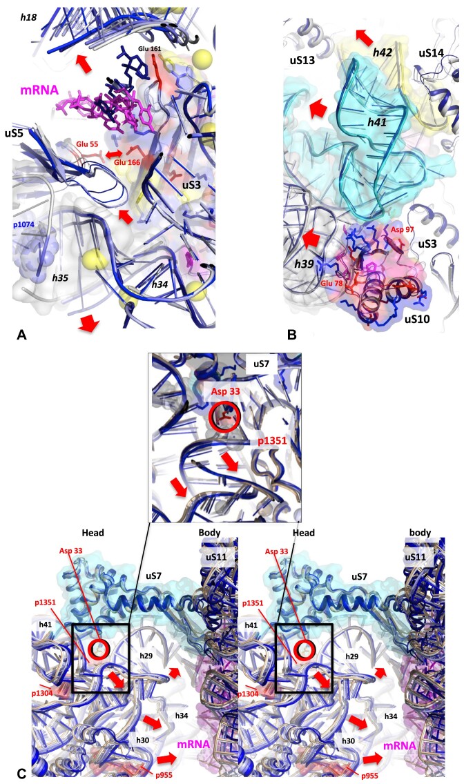
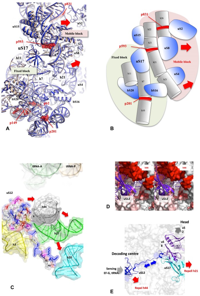
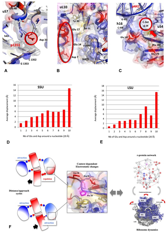
Accurate protein synthesis requires ribosomes to integrate signals from distant functional sites and execute complex dynamics. Despite advances in understanding ribosome structure and function, two key questions remain: how information is transmitted between these distant sites, and how ribosomal movements are synchronized? We recently highlighted the existence of ribosomal protein networks, likely evolved to participate in ribosome signaling. Here, we investigate the relationship between ribosomal protein networks and ribosome dynamics. Our findings show that major motion centers in the bacterial ribosome interact specifically with r-proteins, and that ribosomal RNA exhibits high mobility around each r-protein. This suggests that periodic electrostatic changes in the context of negatively charged residues (Glu and Asp) induce RNA-protein 'distance-approach' cycles, controlling key ribosomal movements during translocation. These charged residues play a critical role in modulating electrostatic repulsion between RNA and proteins, thus coordinating ribosomal dynamics. We propose that r-protein networks synchronize ribosomal dynamics through an 'electrostatic domino' effect, extending the concept of allostery to the regulation of movements within supramolecular assemblies.

Plain language summary

For accurate protein synthesis, ribosomes must coordinate signals and movements across their functional centers, but how this synchronization occurs remains unclear. Our study highlights the role of ribosomal protein networks in regulating ribosome dynamics. We show that major motion centers interact specifically with ribosomal proteins, and ribosomal RNA displays high mobility near negatively charged amino acids (Glu and Asp). Periodic electrostatic changes likely drive cycles of RNA–protein distance and approach, controlling key movements during translocation. Ribosomal protein networks may synchronize these dynamics through an ‘electrostatic domino effect,’ extending the concept of allostery to the regulation of molecular motions in supramolecular assemblies.


Figures

Graphical Abstract
Graphical Abstract
Figure 1.
Figure 1.
The relationship between ribosome dynamics during translocation and ribosomal protein networks. The aim of this study is to understand how r-protein network may contribute to coordinate ribosome dynamics. Top left panel: Summary of the main ribosomal motions during the translocation. The LSU in the classic unrotated state is represented with a white surface (PDB_id: 7n1p). The subunit rotation (the counter clockwise rotation of the 30S subunit relatively to the 50S subunit) is illustrated by a set of cartoons of the SSU during the six steps of the translocation described in the study of Rundlet et al. (2021) [48] with different colours. The colour code is maintained throughout the manuscript: white: step 1 (7n1p); light blue: step 2 (7n30); slate: step 3 (7n2u); blue: step 4 (7n2v); deep blue: step 5 (7n2c) and wheat: step 6 (7n31). Top right panel: Superimposition of the SSU observed in the six steps to highlight the swivelling motion. The direction of the translocation is represented by red arrows. Bottom left panel: Summary of the main properties of the r-protein networks. The r-proteins of the LSU of the bacterial ribosome of T. thermophilus (PDB_id: 4v8i) are represented by cartoons. The network interactions are represented by coloured surfaces. Bottom right panel: Schematic representation of the connectome of the bacterial ribosome where r-proteins are grouped into module around functional centers. The connections between the different functional modules are represented by arrows (adapted from Timsit et al., 2021 [102].
Figure 2.
Figure 2.
Properties of the interactions between CMs and r-proteins in the bacterial ribosome. Interactions between the CMs (red spheres) of the SSU and LSU of T. thermophilus ribosome (pdb id : 4v9h) and dimer of r-proteins (blue cartoons and surfaces): (A) uS7-uS9 and p1247. (B) uS13-uS19 and p955. (C) bL9-bL28 and pL1198. (D) uL16-bL25 and pL871. Asymmetry of charge distribution in CMs–r-protein interactions. (E) CM p1074 and uS2 and u5. (F) CM p1125 and uS9 and uS10. Ensemble of local CMs–r-protein interactions with the SSU head. (G) Interactions around the protein uS9 (H) Schematic representation of the network of interactions around uS9 (G). The CMs are represented in red and the r-proteins are represented by coloured cartoons and surfaces.
Figure 3.
Figure 3.
Network between r-proteins, CMs and functional centers of the bacterial ribosome. (A) schematic and (B) cartoon representations of the connectome linking the bacterial r-proteins to the CMs and the functional center. R-proteins that make direct interactions with CMs are represented in pink. R-proteins that interact indirectly with CMs are represented in cyan. (C) Network drawing (black lines) between the r-proteins and CMs, indicating the moving domains controlled by the CMs. (D) and (E) Network of r-proteins (grey spheres), functional centers (yellow spheres) and motion centers (red spheres) in the bacterial ribosome. The diameter of the spheres that correspond to the centers of mass of each component are proportional to the value of the maxima of centrality (D) Betweenness centrality (E) EigenVector centrality.
Figure 4.
Figure 4.
Spatio temporal map of rRNA–r-protein motions in the SSU. During the early translocation steps of E. coli ribosome. SSU map of 16S RNA–r-protein movements observed at step 2–6, relatively to step 1 (PDB_id: 7n1p). Surface representations of the 16 RNA coloured from white to red according to the measured phosphorus–phosphorus displacements of each nucleotide, from step 1 to step 6, around each r-protein (represented by violet cartoons). Right panel: the network of CMs (small yellow spheres) – r-proteins (violet spheres whose diameters are proportional to the sum of rRNA displacements around them (within a distance of 15 Å).
Figure 5.
Figure 5.
How does rRNA move around the r-proteins during translocation? RNA–protein distance approach cycles trapped at step 5 (7n1c) around uL16 (A) and bS16 (B). This is a graphic representation of the values reported in the tables of Supplementary Data 1. The r-proteins are represented as yellow cartoons surrounded by rRNA helices represented by ribbon and surfaces. The RNA is coloured according to whether it has undergone a shift towards (Max = blue) or away (Max = red) from the the Center of Mass of protein in step 5, in comparison to its initial position relative to the protein in step 1. Cartoons and schematic representation of the motions of the rRNA helices around representative r-proteins (C) uL16, rRNA motion induced by external factors: tRNA step 3, EF-G step 4. (D) uS17, the moving away of H21 from uS17 induced by LH34 (LSU). (E) uS3, uS3 and mRNA channel: A combination of rRNA–protein and protein–protein motions. (F) uS2, rRNA/r-protein motion away from the transit of external factors. White cartoons represent the structures of the r-proteins and its RNA layer at step 1 (7n1p). Light blue: step 2 (7n30); Slate: step 3 (7n2u); Blue: step 4 (7n2v); Deep blue: step 5 (7n2c) and Wheat: step 6 (7n31). The structures of the r-protein (at step 1) are represented by cartoons and surfaces. The phosphate groups of rRNA helices observed in steps 2–5 which have moved >1 Å from their position in step 1 are represented by spheres.
Figure 6.
Figure 6.
Global view of the CM–r-protein interactions in the context of the ribosome dynamics during translocation. The schematic figures represent the main CMs (represented as coloured squares) bound by the r-proteins and the corresponding motions during the translocation in the LSU (A) and the SSU (B). The r-proteins that mediate bridges between two or several CMs are represented by surfaces coloured in cyan. The interaction surfaces are represented in orange.
Figure 7.
Figure 7.
Influence of the local rRNA–protein mobility on the dynamics of the head during translocation. The sum of the localized rRNA–r-protein motions around each r-protein rearranges the internal structure of the head and modulates the sizes of the entry and exit mRNA channel in the head (A–C) during the head swivelling (occurring from steps 3–5). Motions of the r-proteins and rRNA helices around uS3 (A), uS10 (B) and uS7 (C) (each one is fixed during superimposition of the structures observed from steps 2–5). The red arrows highlight the motions during the translocation steps (same colour code as in Figure 1). See also Supplementary Figures S7 and S20–S23 for details and stereo views.
Figure 8.
Figure 8.
Influence of the local rRNA–protein mobility on the dynamics of the body during translocation. (A) The uS17 protein plays a pivotal role in orchestrating a significant reorganization of the body during the back-swivelling movement (step 6) in repelling h21 in front of p593. (B) The schematic view of the body represented in A illustrates the splitting of the body into mobile and moving blocks on both sides of uS17. (C) A further reorganization is initiated by uS12 on the opposite side of the body, resulting in the repulsion of h44 at step 6. (D) Stereo view focusing on uS12 in the SSU map (Figure 4, step 6). The 16S RNA surface around uS12 is coloured from white to red according to the phosphorus-phosphorus displacements at step 6. This view also shows how the long extension of uS12 passes right through the body to reach uS17 on the other side. (E) A model showing how uS12 and uS17 may be synchronized through a process of ‘electrostatic dominoes’ allostery (blue arrows), in their repulsion of the large helices h44 and h21 during back-swivelling.
Figure 9.
Figure 9.
r-protein motifs observed at the vicinity of rRNA moving regions. (A) uS7-p1351: The conserved Asp 33 interact directly with the moving base pair of p1351. (B) uL10-H43 at the base of the bL12-stalk: Cluster of negatively charged residues is observed just in front of the highly moving H43 helix. (C) uS4-h16: The moving residues of the 16S are located in front of a C-ter extremity of an alpha helix of uS4. The structures of the r-proteins (at step 1) are represented by cartoons and surfaces, the phosphate groups which have moved more than 1 Å from their position in step 1 are represented by spheres. (D–E) Averages of displacement of a nucleotide (P-P distance) of rRNA in the SSU (D) and in the LSU (E) in function of the number of Glu and Asp around it. This is a graphic representation of Supplementary Table S5A and B and Supplementary Data 2. (F) A model of a ‘modulated electrostatic lever arm’. The context-induced electrostatic changes of the r-protein surfaces modulate the interaction with the rRNA and control the cycles of repulsion-attraction between rRNA and protein in function of electrostatic signals transmitted through the network.

Similar articles

Cited by

References

    1. Melnikov S., Ben-Shem A., Garreau de Loubresse N., Jenner L., Yusupova G., Yusupov M. One core, two shells: bacterial and eukaryotic ribosomes. Nat. Struct. Mol. Biol. 2012; 19:560–567. - PubMed
    1. Bashan A., Yonath A. Correlating ribosome function with high-resolution structures. Trends Microbiol. 2008; 16:326–335. - PubMed
    1. Steitz T.A. A structural understanding of the dynamic ribosome machine. Nat. Rev. Mol. Cell Biol. 2008; 9:242–253. - PubMed
    1. Schmeing T.M., Ramakrishnan V. What recent ribosome structures have revealed about the mechanism of translation. Nature. 2009; 461:1234–1242. - PubMed
    1. Sheng K., Li N., Rabuck-Gibbons J.N., Dong X., Lyumkis D., Williamson J.R. Assembly landscape for the bacterial large ribosomal subunit. Nat. Commun. 2023; 14:5220. - PMC - PubMed

Grants and funding

LinkOut - more resources