Review
. 2025 Jan 10;10(1):9.
doi: 10.1038/s41392-024-02044-3.

Mitochondrial diseases: from molecular mechanisms to therapeutic advances

Affiliations
Review

Mitochondrial diseases: from molecular mechanisms to therapeutic advances

Haipeng Wen et al. Signal Transduct Target Ther. .

Abstract

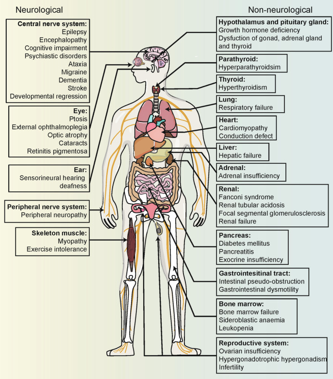
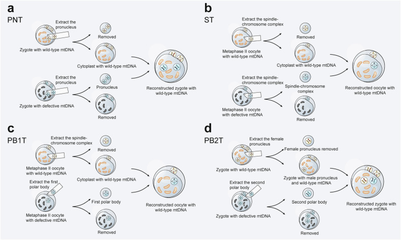
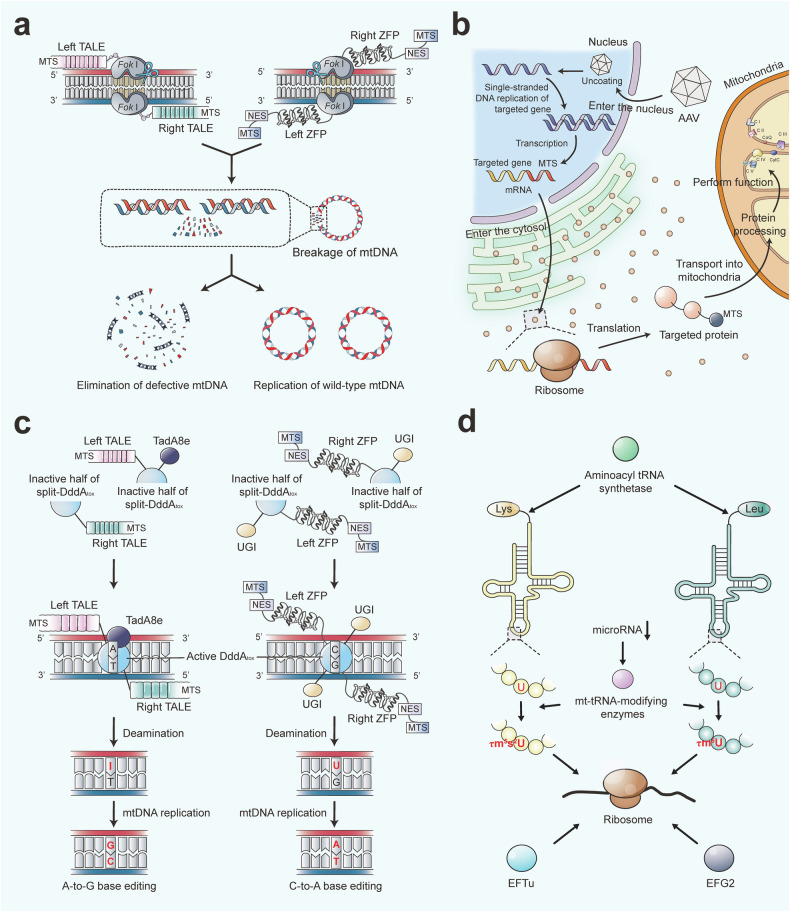
Mitochondria are essential for cellular function and viability, serving as central hubs of metabolism and signaling. They possess various metabolic and quality control mechanisms crucial for maintaining normal cellular activities. Mitochondrial genetic disorders can arise from a wide range of mutations in either mitochondrial or nuclear DNA, which encode mitochondrial proteins or other contents. These genetic defects can lead to a breakdown of mitochondrial function and metabolism, such as the collapse of oxidative phosphorylation, one of the mitochondria's most critical functions. Mitochondrial diseases, a common group of genetic disorders, are characterized by significant phenotypic and genetic heterogeneity. Clinical symptoms can manifest in various systems and organs throughout the body, with differing degrees and forms of severity. The complexity of the relationship between mitochondria and mitochondrial diseases results in an inadequate understanding of the genotype-phenotype correlation of these diseases, historically making diagnosis and treatment challenging and often leading to unsatisfactory clinical outcomes. However, recent advancements in research and technology have significantly improved our understanding and management of these conditions. Clinical translations of mitochondria-related therapies are actively progressing. This review focuses on the physiological mechanisms of mitochondria, the pathogenesis of mitochondrial diseases, and potential diagnostic and therapeutic applications. Additionally, this review discusses future perspectives on mitochondrial genetic diseases.


Conflict of interest statement

Competing interests: The authors declare no competing interest.

Figures

Fig. 1
Fig. 1
Timeline of Major Historical Events in the Study of Mitochondrial Diseases. From the initial discovery to current advancements, our understanding of the mechanisms underlying mitochondrial diseases has continually deepened. Over time, research explorations and progress have contributed to the development of diagnostic and treatment methods, ultimately providing insights into more efficient and accurate diagnostic and therapeutic strategies
Fig. 2
Fig. 2
Overview of Mitochondrial Metabolism. As the central hub of bioenergetics, the mitochondrion utilizes NADH and FADH2 produced by the TCA cycle to generate ATP through electron transfer and the H+ gradient across the respiratory chain complexes. Complexes I and III are the primary sources of mtROS, which cause oxidative damage or signaling transduction. The mtROS also can induce the opening of mPTP. Glucose and lipids (via β-oxidation) both contribute to the TCA cycle. Citrate can cross the mitochondrial membrane, allowing acetyl-CoA to be transported into the cytoplasm for various functions. TCA cycle tricarboxylic acid cycle; CACT carnitine-acylcarnitine translocase; CPT I/II carnitine palmitoyltransferase I/II; mtROS mitochondrial reactive oxygen species; ANT adenine nucleotide translocator; VDAC voltage-dependent anion channel; mPTP mitochondrial permeability transition pore
Fig. 3
Fig. 3
Mitochondrial Quality Control Network. Mitochondria employ both intercellular and intracellular quality control mechanisms to maintain homeostasis and redox balance. These mechanisms include mitochondrial biogenesis, fusion, fission, axonal transport, docking, mitophagy, the mitochondrial integrated stress response, and intercellular mitochondrial transfer. IMM inner mitochondrial membrane; OMM outer mitochondrial membrane; Δψm mitochondrial membrane potential; Ub ubiquitin; mt-ISR mitochondrial integrated stress response; mt-UPR mitochondrial unfolded protein response; MDV(s) mitochondria-derived vesicle(s)
Fig. 4
Fig. 4
Mitochondrial Apoptosis and Inflammation. Components within the IMS or matrix can trigger apoptosis or inflammation if leaked into the cytosol, primarily due to the mPTP and MOMP. Upon MOMP formation and mPTP opening, cytochrome C, SMAC, and mtDNA are released into the cytosol. Cytochrome C interacts with APAF1, activating caspase 9 to initiate the caspase cascade leading to apoptosis. SMAC accelerates this process by inhibiting XIAP. After binding with mtDNA, the cGAS enzyme produces cGAMP from ATP and GTP, activating the cGAS-STING signaling pathway and inducing type I interferon expression and NF-κB activation. The NLRP3 inflammasome can also bind with (oxidized) mtDNA to promote IL-1β and IL-18 cleavage. However, caspase 3 cleaves cGAS and IRF3 during apoptosis, inhibiting inflammation. MOMP mitochondrial outer membrane permeabilization; mPTP mitochondrial permeability transition pore; SMAC second mitochondrial-derived activator of caspases; APAF1 apoptosis protease activating factor 1; XIAP X-linked inhibitor of apoptosis protein; cGAS cyclic GMP-AMP synthase; STING stimulator of interferon genes; cGAMP cyclic guanosine monophosphate–adenosine monophosphate; TBK1 TANK binding kinase 1; IKK IκB kinase; IRF3 interferon regulatory factor 3
Fig. 5
Fig. 5
Multisystem Clinical Presentation of Mitochondrial Diseases. Due to the ubiquitous presence of mitochondria, mitochondrial diseases can present in any tissue of the body. Tissues and organs with high energy demands, such as skeletal muscle and brain, are particularly susceptible to oxidative phosphorylation defects, leading to common manifestations like myopathy and encephalopathy in mitochondrial diseases. The diverse and variable symptoms associated with these conditions increase the risk of misdiagnosis
Fig. 6
Fig. 6
Diagnostic Methodology for Mitochondrial Diseases. Diagnostic strategies have evolved from a biopsy-first approach to a genetic-first approach. Initial screenings should utilize blood, urine, and cerebrospinal fluid samples. Biomarker testing is essential in this process. For suspected mitochondrial diseases, mtDNA sequencing and analysis are the preferred methods, while nDNA sequencing should be considered in cases of mtDNA multiple deletions, depletion, or early-onset symptoms. RNA sequencing (transcriptomics) and respirometry also contribute to accurate diagnosis. Biopsy specimens, typically obtained from muscle or skin, remain valuable for confirming tissue-specific mtDNA mutations that may not be detected in blood or urine samples. Histopathological examination and respiratory chain enzyme analysis can be applied to these tissue samples, revealing abnormal mitochondrial structure, morphology, and function. Thus, biopsy retains significant diagnostic value. mtDNA mitochondrial DNA; nDNA nuclear DNA; ccf-mtDNA circulating cell-free mitochondrial DNA; Δψm mitochondrial membrane potential; SDH succinate dehydrogenase; COX cytochrome c oxidase; NADH-TR nicotinamide adenine dinucleotide tetrazolium reductase
Fig. 7
Fig. 7
Procedure for Mitochondrial Replacement Therapy. a The procedure for pronuclear transfer (PNT) involves extracting the pronucleus from a zygote of a healthy donor with wild-type mtDNA and from a patient with defective mtDNA. The cytoplast from the patient and the pronucleus from the healthy donor are then removed. Finally, the pronucleus from the patient and the cytoplast containing wild-type mtDNA from the healthy donor are fused, reconstructing a zygote with the patient’s pronucleus and wild-type mtDNA. b The procedure for spindle-chromosome complex transfer (ST) involves extracting the spindle-chromosome complex from a metaphase II oocyte of a healthy donor with wild-type mtDNA and from a patient with defective mtDNA. The cytoplast from the patient and the spindle-chromosome complex from the healthy donor are removed. Finally, the spindle-chromosome complex from the patient and the cytoplast containing wild-type mtDNA from the healthy donor are fused, reconstructing an oocyte with the patient’s spindle-chromosome complex and wild-type mtDNA. c The first polar body transfer (PB1T) procedure involves extracting the spindle-chromosome complex from a metaphase II oocyte of a healthy donor with wild-type mtDNA and the first polar body from a metaphase II oocyte of a patient with defective mtDNA. The cytoplast from the patient and the spindle-chromosome complex from the healthy donor are then removed. Finally, the first polar body from the patient and the cytoplast containing wild-type mtDNA from the healthy donor are fused, reconstructing an oocyte with the patient’s first polar body and wild-type mtDNA. d The second polar body transfer (PB2T) procedure involves extracting the female pronucleus from a zygote of a healthy donor with wild-type mtDNA and the second polar body from a zygote of a patient with defective mtDNA. The zygote from the patient and the female pronucleus from the healthy donor are then removed. Finally, the second polar body from the patient and the zygote containing wild-type mtDNA and the male pronucleus from the healthy donor are fused, reconstructing a zygote with the patient’s second polar body and wild-type mtDNA
Fig. 8
Fig. 8
Gene Therapy and Post-Transcriptional Modification Strategies for Mitochondrial Genetic Disorders. a MitoTALENs consist of TALE fused with FokI nucleases, while mtZFNs are composed of ZFP linked to FokI nucleases. MTS guides mtZFNs and mitoTALENs to the mitochondria. ZFP and TALE selectively bind to predetermined defective mtDNA target sequences, after which FokI dimerizes and cleaves the mtDNA adjacent to these binding sites, causing double-strand breaks that lead to the elimination of defective mtDNA. The remaining wild-type mtDNA can then replicate, altering the heteroplasmy ratio. b The targeted gene sequence, along with transcriptional regulatory elements and MTS, is packaged into an AAV vector, which is then delivered to the nucleus of the recipient cell. Inside the nucleus, the AAV uncoats and releases single-stranded DNA, which replicates to form double-stranded DNA. RNA polymerase then transcribes this DNA into mRNA. The mRNA exits the nucleus and is translated into the corresponding protein at the ribosomes in the cytoplasm. MTS guides these proteins to the mitochondria, where they undergo further processing and perform their respective functions. c DdCBEs are engineered by fusing MTS, split-DddAtox halves, UGIs, and either ZFPs or TALEs. DddAtox catalyzes the deamination of cytosine to uracil, while UGIs prevent uracil-DNA glycosylase from excising uracil, resulting in C-to-T editing during replication. Additionally, by linking MTS, split-DddAtox halves, TadA8e (an engineered adenine deaminase), and TALEs, TadA8e catalyzes the deamination of adenine to inosine, which pairs with cytosine during replication, thereby achieving targeted A-to-G editing. d Modifying the tRNA binding domain of nuclear-encoded aminoacyl tRNA synthetase or overexpressing aminoacyl tRNA synthetase can enhance aminoacylation efficiency and stabilize translation products. The expression of post-transcriptional negative regulators like microRNAs can inhibit the expression of mitochondrial RNA-modifying enzymes, thereby affecting mt-tRNA modifications. Using microRNA antagonists could potentially reverse disease phenotypes. Furthermore, overexpressing mt-tRNA-modifying enzymes can correct anticodon first nucleotide modification defects in mt-tRNA, improving ribosomal translation within mitochondria. Overexpression of mitochondrial translation elongation factors EFTu and EFG2 can also partially suppress amino acid misincorporation caused by mtDNA mutations during the translation elongation process. mtDNA mitochondrial DNA; mitoTALENs mitochondria-targeted transcription activator-like effector nucleases; mtZFNs mitochondria-targeted zinc-finger nucleases; MTS mitochondrial targeting sequence; NES nuclear export signal; DdCBEs DddAtox-derived cytosine base editors; ZFP zinc finger proteins; TALE transcription activator-like effector; UGIs uracil glycosylase inhibitors; TadA8e deoxyadenosine deaminase; AAV adeno-associated virus

Similar articles

Cited by

References

    1. Mitchell, P. Coupling of phosphorylation to electron and hydrogen transfer by a chemi-osmotic type of mechanism. Nature191, 144–148 (1961). - PubMed
    1. Gorman, G. S. et al. Mitochondrial diseases. Nat. Rev. Dis. Prim.2, 16080 (2016). - PubMed
    1. Schapira, A. H. Mitochondrial diseases. Lancet379, 1825–1834 (2012). - PubMed
    1. La Morgia, C., Maresca, A., Caporali, L., Valentino, M. L. & Carelli, V. Mitochondrial diseases in adults. J. Intern Med.287, 592–608 (2020). - PubMed
    1. Gropman, A. L. Diagnosis and treatment of childhood mitochondrial diseases. Curr. Neurol. Neurosci. Rep.1, 185–194 (2001). - PubMed

MeSH terms

LinkOut - more resources