Review
. 2025 Jan 9;11(1):4.
doi: 10.1038/s41378-024-00751-z.

Microfluidic platforms for monitoring cardiomyocyte electromechanical activity

Affiliations
Review

Microfluidic platforms for monitoring cardiomyocyte electromechanical activity

Wei Wang et al. Microsyst Nanoeng. .

Abstract

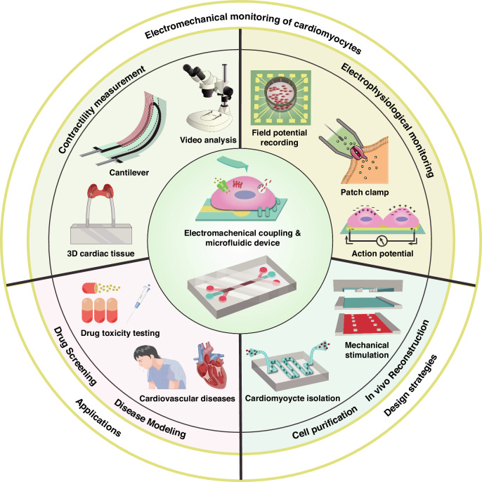
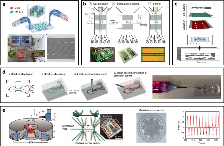
Cardiovascular diseases account for ~40% of global deaths annually. This situation has revealed the urgent need for the investigation and development of corresponding drugs for pathogenesis due to the complexity of research methods and detection techniques. An in vitro cardiomyocyte model is commonly used for cardiac drug screening and disease modeling since it can respond to microphysiological environmental variations through mechanoelectric feedback. Microfluidic platforms are capable of accurate fluid control and integration with analysis and detection techniques. Therefore, various microfluidic platforms (i.e., heart-on-a-chip) have been applied for the reconstruction of the physiological environment and detection of signals from cardiomyocytes. They have demonstrated advantages in mimicking the cardiovascular structure and function in vitro and in monitoring electromechanical signals. This review presents a summary of the methods and technologies used to monitor the contractility and electrophysiological signals of cardiomyocytes within microfluidic platforms. Then, applications in common cardiac drug screening and cardiovascular disease modeling are presented, followed by design strategies for enhancing physiology studies. Finally, we discuss prospects in the tissue engineering and sensing techniques of microfluidic platforms.


Conflict of interest statement

Conflict of interest: The authors declare no competing interests.

Figures

Fig. 1
Fig. 1. Schematic diagram of the microfluidic platforms used for physiological studies of cardiomyocytes.
The main contents comprise contractility and electrophysiological signal detection, its applications, and design strategies
Fig. 2
Fig. 2. Nonmicrofluidic and microfluidic platforms for measuring cardiomyocyte contractility.
a Force mapping method based on a “light nanoantenna” array with the piezo-phototronic effect. Reproduced with permission from ref.. b Micropost arrays for characterizing the contractile force, velocity, and power produced. Cell contractility was analyzed using microscopy with a high-speed camera. Reproduced with permission from ref.. c Magnetic hydrogel-based strain sensors for wireless-passive monitoring. The contraction and relaxation of cardiomyocytes on the hydrogel were evaluated via magnetic field detection. Reproduced with permission from ref.. d Working principle and photograph of the Ag/CNT-PDMS sensor. Cardiomyocyte beating results in changes in resistance between nanocracks. Reproduced with permission from ref.. e Fluorescence imaging and contractile stress analysis of 3D cardiac tissues. The GelMA hydrogel was used for cell encapsulation, and the PAm hydrogels in the top and bottom layers were used as “stress sensors” for quantifying the contractile stresses generated by the encapsulated cardiomyocytes. Reproduced with permission from ref.. f Outline of the microfluidic device with a schematic and picture of the channels used to monitor the beating frequency of cardiac bodies (CBs) via video imaging. Reproduced with permission from ref.. g MTF chip and assembly of a microfluidic device for the measurement of contractile stresses. Reproduced with permission from ref.. h Working principle and quantitative performance of the graphene hybrid anisotropic structural color film capable of reflecting cardiomyocyte behavior. Reproduced with permission from ref.. i A heart-on-a-chip microdevice (HMD) for visualizing the kinetics of cardiac microtissue pulsations by monitoring particle displacement. Reproduced with permission from ref.
Fig. 3
Fig. 3. Microfluidic platforms for monitoring electrophysiological signals.
a Microfluidic device integrated with electrical stimulation for measuring field potentials and distinguishing excitable cells from electrically non-excitable cells. Reproduced with permission from ref.. b Photograph and schematic of the organ-on-a-chip integrating both MEAs and TEER measurements. Dynamic detection of vascular permeability and cardiac function under the inflammatory stimulus tumor necrosis factor-alpha (TNF-α) or the cardiac-targeting drug isoproterenol. Reproduced with permission from ref.. c Multielectrode array decorated with 3D hollow nanotubes integrated with microfluidic channels for electrical recording and drug delivery. Reproduced with permission from ref.. d Heart-on-a-chip for the long-term dynamic culture of cardiomyocytes and field potential recording with Au electrodes. Reproduced with permission from ref.. e High-content drug screening (10 types of drugs, each with 5 concentrations to be assayed simultaneously) using high-resolution Ca2+ imaging. Reproduced with permission from ref.. f Laminar cardiac tissues formed through topographical cues and integration with commercial MEAs in a microfluidic device. Reproduced with permission from ref.. g Microfluidic device for action potential recording, where cell ionic currents are transduced into mirror charges. It detects the effects of nifedipine recorded as MAPs (mirror action potentials). Reproduced with permission from ref.
Fig. 4
Fig. 4. Microfluidics for drug screening and disease modeling.
a Microfibrous scaffolds for evaluating cardiovascular toxicity in the engineered endothelialized myocardium. Reproduced with permission from ref.. b Cardiac microphysiological system containing nutrient channels and cell channels. Reproduced with permission from ref.. c Heart-on-a-chip for studying the effects of acute hypoxia on cardiac function. Reproduced with permission from ref.. d Microfluidic myocardium-on-a-chip for CMs and ECs cocultured with capillary-like channels. Reproduced with permission from ref.. e Microfluidic device integrated with CMs and liver cancer cells to simulate the side effects of the anticancer drug doxorubicin (DXR). Reproduced with permission from ref.
Fig. 5
Fig. 5. Microfluidic platform design strategies for monitoring the physiological signals of cardiomyocytes.
a Microfluidic device for hiPSC-CM purification comprising a peristaltic pump, cell suspension reservoirs, and a serpentine channel. Reproduced with permission from ref.. b Microfluidic device for electrophysiological cell sorting in response to stimuli. Reproduced with permission from ref.. c Heart-on-a-chip providing mechanical and biochemical costimulation. Reproduced with permission from ref.. d Perfusable system connected with a heart-on-a-chip for measuring myocardial‒microvascular interactions. Reproduced with permission from ref.. e Microfluidic platform fabricated by DLW lithography and soft lithography for monitoring oscillatory forces generated by microtissues under a prescribed mechanical loading and pacing. Reproduced with permission from ref.

Similar articles

References

    1. World Health Organization. Invisible Numbers: The True Extent of Noncommunicable Diseases and What to Do About Them (World Health Organization, 2022).
    1. World Health Organization. World Health Statistics 2023: Monitoring Health for the SDGs, Sustainable Development Goals (World Health Organization, 2023).
    1. Congressional Budget Office. Research and Development in the Pharmaceutical Industry.https://www.cbo.gov/publication/57126 (2021).
    1. DiMasi, J. A., Grabowski, H. G. & Hansen, R. W. Innovation in the pharmaceutical industry: new estimates of R&D costs. J. Health Econ.47, 20–33 (2016). - PubMed
    1. Sun, D., Gao, W., Hu, H. & Zhou, S. Why 90% of clinical drug development fails and how to improve it? Acta Pharm. Sin. B12, 3049–3062 (2022). - PMC - PubMed

LinkOut - more resources