. 2025 Jan 10;17(1):7.
doi: 10.1038/s41368-024-00340-w.

Adhesive and injectable hydrogel microspheres for NRF2-mediated periodontal bone regeneration

Affiliations

Adhesive and injectable hydrogel microspheres for NRF2-mediated periodontal bone regeneration

Yu Wang et al. Int J Oral Sci. .

Abstract

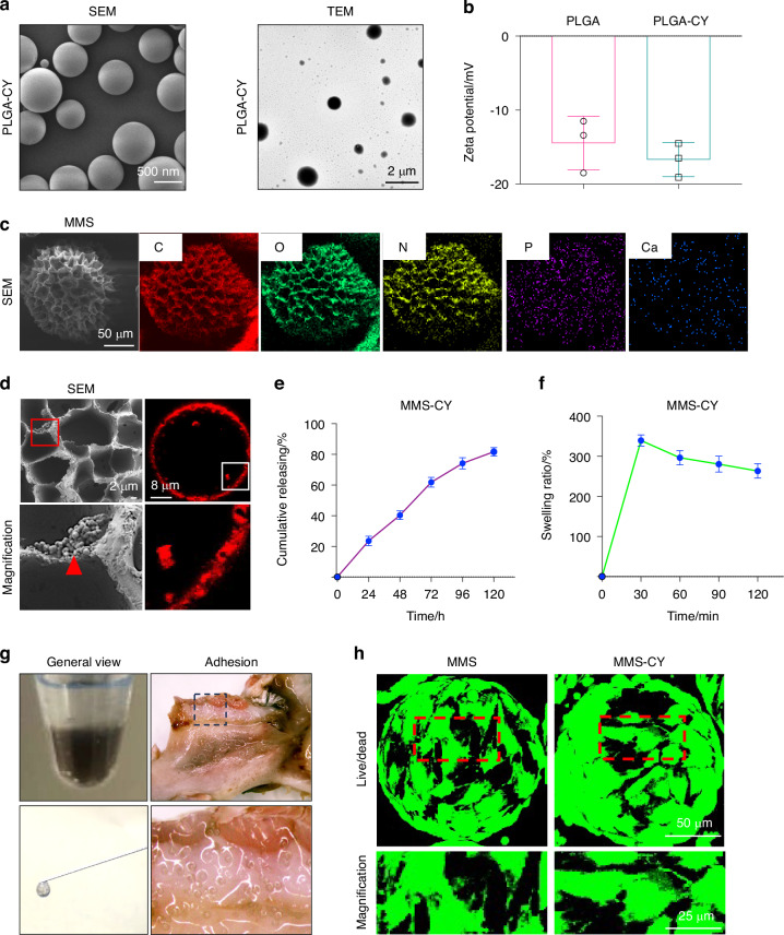
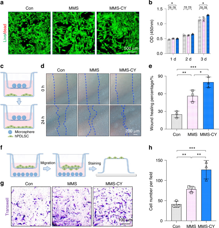
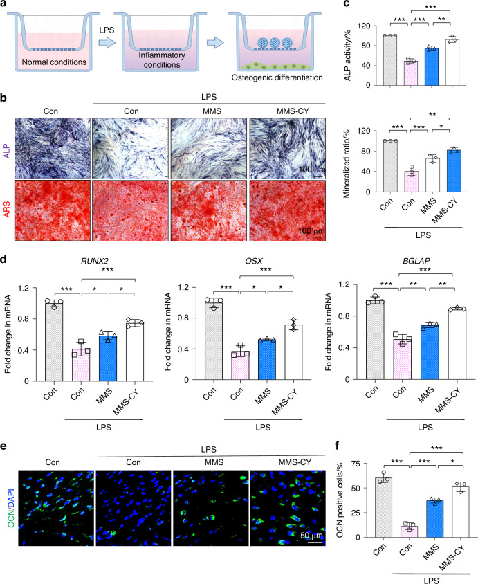
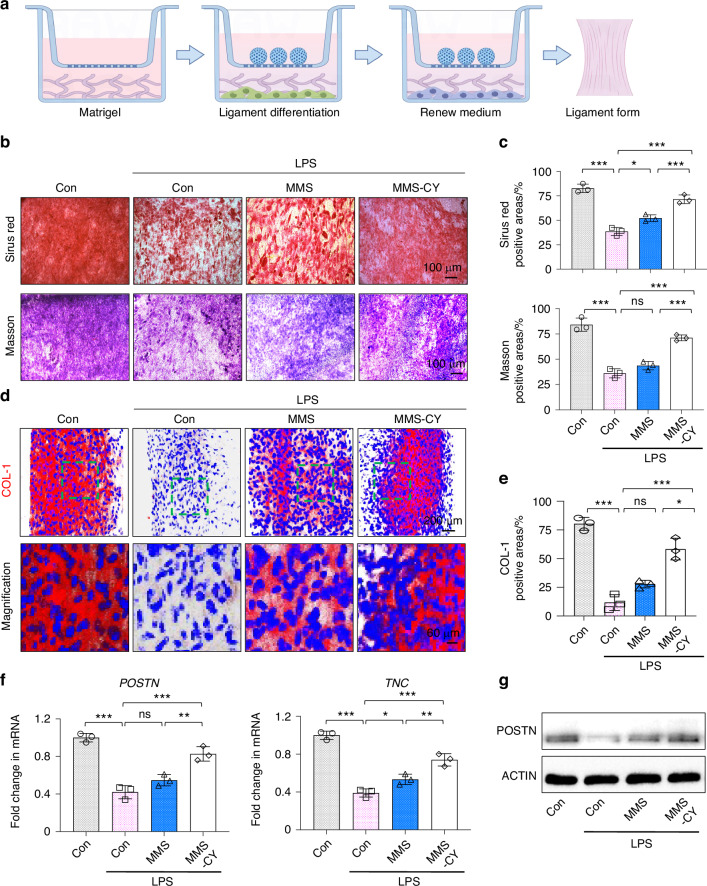
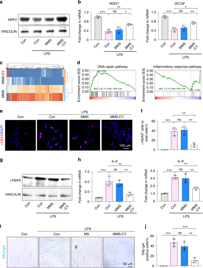
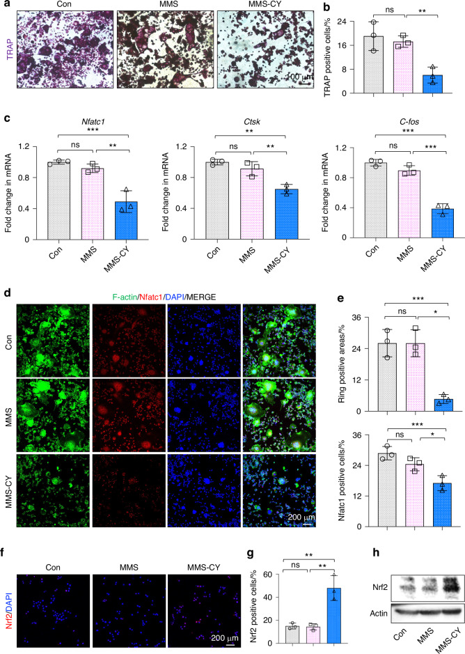
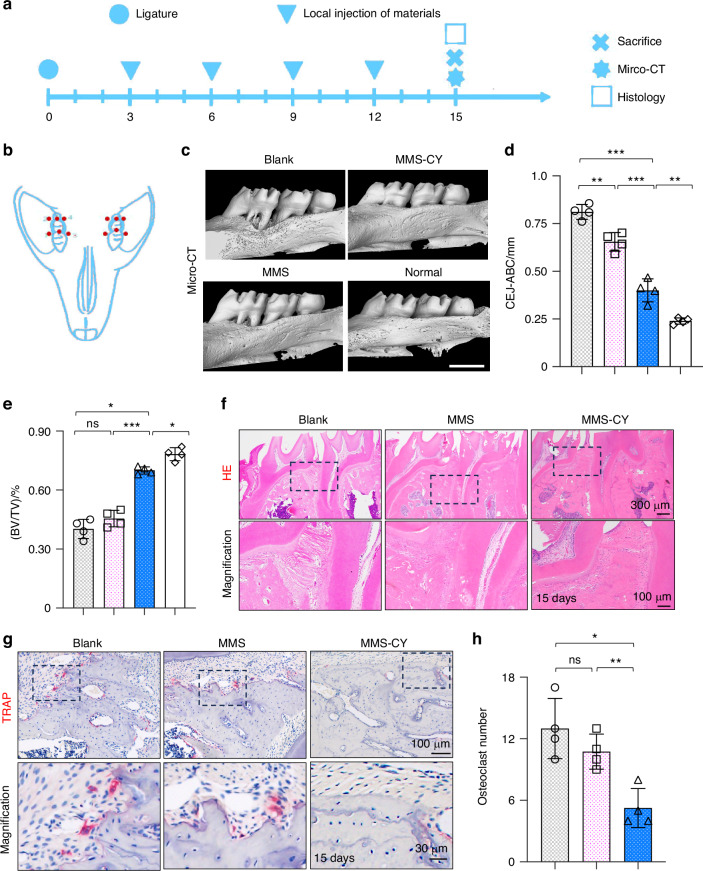
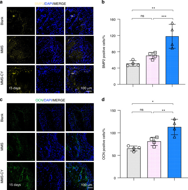
Regenerating periodontal bone defect surrounding periodontal tissue is crucial for orthodontic or dental implant treatment. The declined osteogenic ability of periodontal ligament stem cells (PDLSCs) induced by inflammation stimulus contributes to reduced capacity to regenerate periodontal bone, which brings about a huge challenge for treating periodontitis. Here, inspired by the adhesive property of mussels, we have created adhesive and mineralized hydrogel microspheres loaded with traditional compound cordycepin (MMS-CY). MMS-CY could adhere to the surface of alveolar bone, then promote the migration capacity of PDLSCs and thus recruit them to inflammatory periodontal tissues. Furthermore, MMS-CY rescued the impaired osteogenesis and ligament-forming capacity of PDLSCs, which were suppressed by the inflammation stimulus. Moreover, MMS-CY also displayed the excellent inhibitory effect on the osteoclastic activity. Mechanistically, MMS-CY inhibited the premature senescence induced by the inflammation stimulus through the nuclear factor erythroid 2-related factor (NRF2) pathway and reducing the DNA injury. Utilizing in vivo rat periodontitis model, MMS-CY was demonstrated to enhance the periodontal bone regeneration by improving osteogenesis and inhibiting the osteoclastic activity. Altogether, our study indicated that the multi-pronged approach is promising to promote the periodontal bone regeneration in periodontitis condition by reducing the inflammation-induced stem cell senescence and maintaining bone homeostasis.


Conflict of interest statement

Competing interests: The authors declare no competing interests.

Figures

Fig. 1
Fig. 1
Characterization of MMS-CY. a SEM and TEM images of PLGA-CY. b Zeta potential of PLGA and PLGA-CY. c Elemental mapping images of MMS-CY. d SEM images and immunofluorescence images of MMS-CY. e Release profiles of cordycepin from MMS-CY. f Swelling ratio of MMS-CY. g Overall views of injectable MMS-CY. h Live/dead assay staining of PDLSCs cultured on different kinds of microspheres
Fig. 2
Fig. 2
Assessment of MMS-CY biocompatibility and recruitment capacity. a Live/dead assay of MMS and MMS-CY-treated PDLSCs. b CCK-8 test of MMS and MMS-CY-treated PDLSCs (n = 3 biologically independent samples, by one-way ANOVA Turkey’s multiple comparison test: ns: not significant, *P < 0.05). c Schematic diagram of the scratch wound healing assay. d Microscope images of scratch lines at 0 and 24 h. e Semi-quantification of the percentage of wound healing (n = 3 biologically independent samples, by one-way ANOVA Turkey’s multiple comparison test: ***P < 0.001, **P < 0.01, *P < 0.05). f Schematic diagram of the chemotaxis experiment. g Microscope images of the migration of PDLSCs from the upper chamber. h Semi-quantification of the number of migrating cells. (n = 3 biologically independent samples, by one-way ANOVA Turkey’s multiple comparison test: ***P < 0.001, **P < 0.01). Data are represented as mean ± SD
Fig. 3
Fig. 3
MMS-CY improves the osteogenic capacity of PDLSCs under the inflammation stimulus. a Schematic diagram of MMS-CY restoring the impaired osteogenic differentiation capacity of PDLSCs under the inflammatory condition. b ALP and ARS staining of PDLSCs after coculture with MMS and MMS-CY for 7 days and 14 days respectively. c Quantification of the ALP activity on day 7 and the ratio of ARS positive areas in total areas on day 14 (n = 3 biologically independent samples, by one-way ANOVA Turkey’s multiple comparison test: ns: not significant, ***P < 0.001, **P < 0.01, *P < 0.05). d RT-qPCR of the osteogenic gene expression RUNX2, OSX and BGALP (n = 3 biologically independent samples, by one-way ANOVA Turkey’s multiple comparison test: ***P < 0.001, **P < 0.01, *P < 0.05). e Immunofluorescence staining of OCN of MMS and MMS-CY-treated PDLSCs under the inflammatory condition. f Semi-quantification of OCN (n = 3 biologically independent samples, by one-way ANOVA Turkey’s multiple comparison test: ***P < 0.001, *P < 0.05). Data are represented as mean ± SD
Fig. 4
Fig. 4
MMS-CY rescues the impaired ligament-forming capacity of PDLSCs under the inflammation stimulus. a Schematic diagram of MMS-CY restoring impaired ligament-forming capacity of PDLSCs under the inflammatory condition. b Sirius red and Masson’s trichrome staining of PDLSCs after the coculture with MMS and MMS-CY for 14 days respectively. c Quantification of Sirius red positive areas and Masson positive areas in total areas on day 14 (n = 3 biologically independent samples, by one-way ANOVA Turkey’s multiple comparison test: ns: not significant, ***P < 0.001, **P < 0.01, *P < 0.05). d Immunofluorescence staining of COL-1 of MMS and MMS-CY-treated PDLSCs under the inflammatory condition. e Semi-quantification of COL-1 (n = 3 biologically independent samples, by one-way ANOVA Turkey’s multiple comparison test: ns: not significant, ***P < 0.001, *P < 0.05). f RT-qPCR of the gene exression of POSTN and TNC (n = 3 biologically independent samples, by one-way ANOVA Turkey’s multiple comparison test: ns: not significant, ***P < 0.001, **P < 0.01, *P < 0.05). g Western blot of POSTN in MMS and MMS-CY-treated PDLSCs under the inflammatory condition. Data are represented as mean ± SD
Fig. 5
Fig. 5
MMS-CY restores the impaired function of PDLSCs by reducing the DNA injury. a Western blot of NRF2 in MMS and MMS-CY-treated PDLSCs under the inflammatory condition. b RT-qPCR of the gene expression of NQO1 and GCLM. (n = 3 biologically independent samples, by one-way ANOVA Turkey’s multiple comparison test: ns: not significant, ***P < 0.001, **P < 0.01, *P < 0.05). c Significantly upregulated and downregulated genes (MMS vs MMS-CY). d GSEA shows a significant increase in DNA repair gene signatures and a significant decrease in inflammatory response pathway gene signatures in MMS-CY groups. e Immunofluorescence staining of γ-H2AX of MMS and MMS-CY-treated PDLSCs under the inflammatory condition. f Semi-quantification of γ-H2AX (n = 3 biologically independent samples, by one-way ANOVA Turkey’s multiple comparison test: ns: not significant, ***P < 0.001, **P < 0.01). g Western blot of γ-H2AX in MMS and MMS-CY-treated PDLSCs under the inflammatory condition. h RT-qPCR of the gene expression of IL-6 and IL-8 (n = 3 biologically independent samples, by one-way ANOVA Turkey’s multiple comparison test: ns: not significant, ***P < 0.001, **P < 0.01). i SAβ-gal staining of MMS and MMS-CY-treated PDLSCs. Blue cells are senescent cells. j Semi-quantification of SAβ-gal (n = 3 biologically independent samples, by one-way ANOVA Turkey’s multiple comparison test: ns: not significant, ***P < 0.001, **P < 0.01). Data are represented as mean ± SD
Fig. 6
Fig. 6
MMS-CY inhibits osteoclast differentiation. a TRAP staining of RAW264.7 after coculture with MMS and MMS-CY. b Quantification of TRAP-positive cells. (n = 3 biologically independent samples, by one-way ANOVA Turkey’s multiple comparison test: ns: not significant, ns: not significant, **P < 0.01). c RT-qPCR of the osteoclastic gene expression Nfatc, Ctsk, and C-fos. (n = 3 biologically independent samples, by one-way ANOVA Turkey’s multiple comparison test: ns: not significant, ***P < 0.001, **P < 0.01). d Immunofluorescence staining of F-actin and Nfatc1 of MMS and MMS-CY-treated RAW264.7. e Semi-quantification of ring positive areas and Nfatc1 positive cells (n = 3 biologically independent samples, by one-way ANOVA Turkey’s multiple comparison test: ns: not significant, ***P < 0.001, *P < 0.05). f Immunofluorescence staining of Nrf2 of MMS and MMS-CY-treated RAW264.7. g Semi-quantification of Nrf2 positive cells (n = 3 biologically independent samples, by one-way ANOVA Turkey’s multiple comparison test: ns: not significant, **P < 0.01). h Western blot of Nrf2 in MMS and MMS-CY-treated RAW264.7. Data are represented as mean ± SD
Fig. 7
Fig. 7
MMS-CY inhibits the bone loss in rat ligature-induced periodontitis. a Experiment schedule of in vivo periodontitis study. b Schematic diagram of the establishment of rat periodontitis experiment. c 3D reconstructed digitized images of the maxillary first and second molars analyzed by micro-CT. d Quantitative analysis of BV/TV. (n = 4 biologically independent samples, by one-way ANOVA Turkey’s multiple comparison test: ***P < 0.001, **P < 0.01). e Quantitative analysis of CEJ-ABC. (n = 4 biologically independent samples, by one-way ANOVA Turkey’s multiple comparison test: ns: not significant, ***P < 0.001, *P < 0.05). f H&E staining images in different groups. g TRAP staining images in different groups. h Quantitative analysis of the osteoclast number in periodontal tissues. (n = 4 biologically independent samples, by one-way ANOVA Turkey’s multiple comparison test: ns: not significant, **P < 0.01, *P < 0.05). Data are represented as mean ± SD
Fig. 8
Fig. 8
MMS-CY promotes the bone formation in rat ligature-induced periodontitis. a Immunofluorescence staining of staining of BMP2 in periodontal tissues on day 15 after the MMS-CY administration. b Quantitative analysis of the number of BMP2 positive cells in the periodontal tissues. (n = 4 biologically independent samples, by one-way ANOVA Turkey’s multiple comparison test: ns: not significant, ***P < 0.001, **P < 0.01). c Immunofluorescence staining of staining of OCN in periodontal tissues on day 15 after the MMS-CY administration. d Quantitative analysis of the number of OCN positive cells in the periodontal tissues. (n = 4 biologically independent samples, by one-way ANOVA Turkey’s multiple comparison test: ns: not significant, **P < 0.01, *P < 0.05). Data are represented as mean ± SD

Similar articles

Cited by

References

    1. Slots, J. Periodontitis: facts, fallacies and the future. Periodontol 200075, 7–23 (2017). - PubMed
    1. Deas, D. E., Moritz, A. J., Sagun, R. S. Jr., Gruwell, S. F. & Powell, C. A. Scaling and root planing vs. conservative surgery in the treatment of chronic periodontitis. Periodontol 200071, 128–139 (2016). - PubMed
    1. Graetz, C., Schorr, S., Christofzik, D., Dörfer, C. E. & Sälzer, S. How to train periodontal endoscopy? Results of a pilot study removing simulated hard deposits in vitro. Clin. Oral. Investig.24, 607–617 (2020). - PubMed
    1. Xu, X. Y. et al. Concise review: periodontal tissue regeneration using stem cells: strategies and translational considerations. Stem Cells Transl. Med.8, 392–403 (2019). - PMC - PubMed
    1. Liu, X. et al. Piezoelectric hydrogel for treatment of periodontitis through bioenergetic activation. Bioact. Mater.35, 346–361 (2024). - PMC - PubMed

Publication types

Associated data