. 2025 Jan 9;16(1):489.
doi: 10.1038/s41467-024-55257-z.

Molecular logic for cellular specializations that initiate the auditory parallel processing pathways

Affiliations

Molecular logic for cellular specializations that initiate the auditory parallel processing pathways

Junzhan Jing et al. Nat Commun. .

Abstract

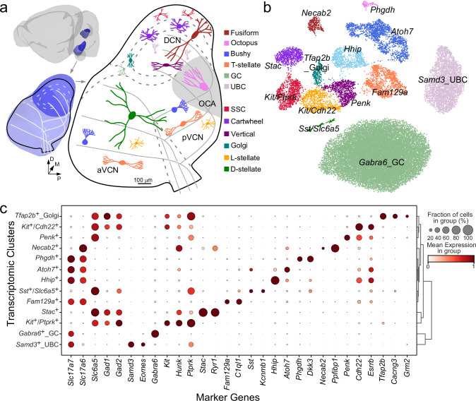
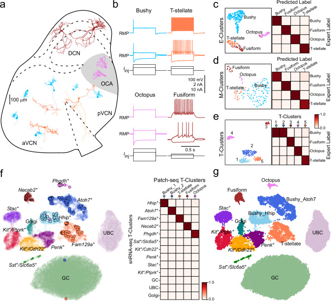
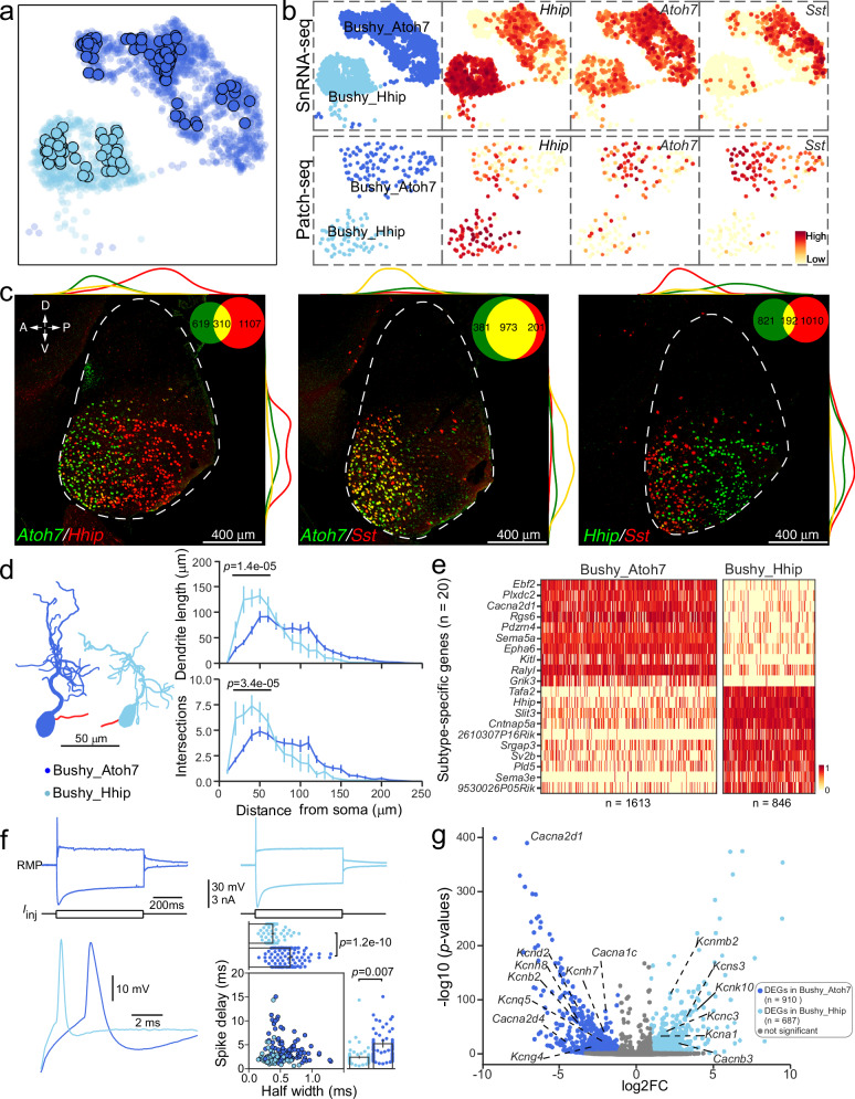
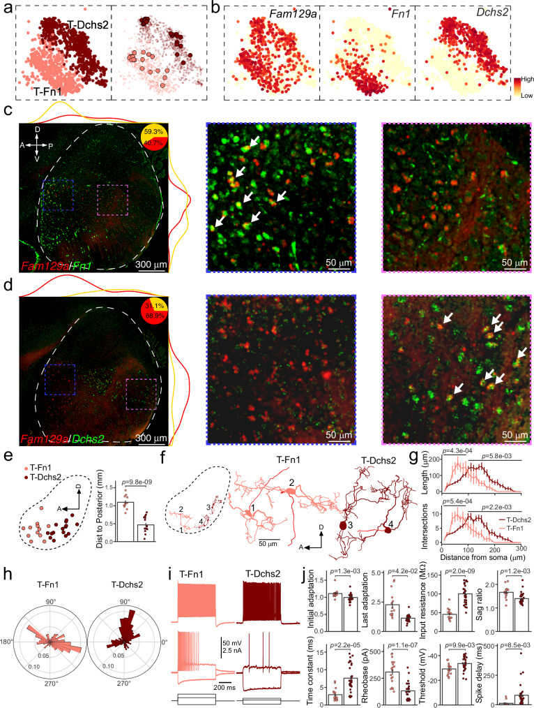
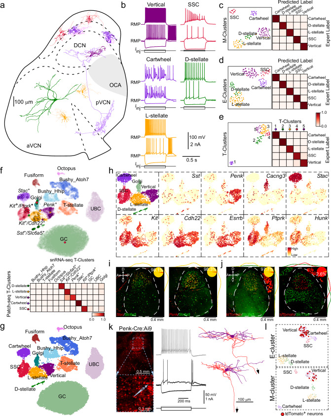
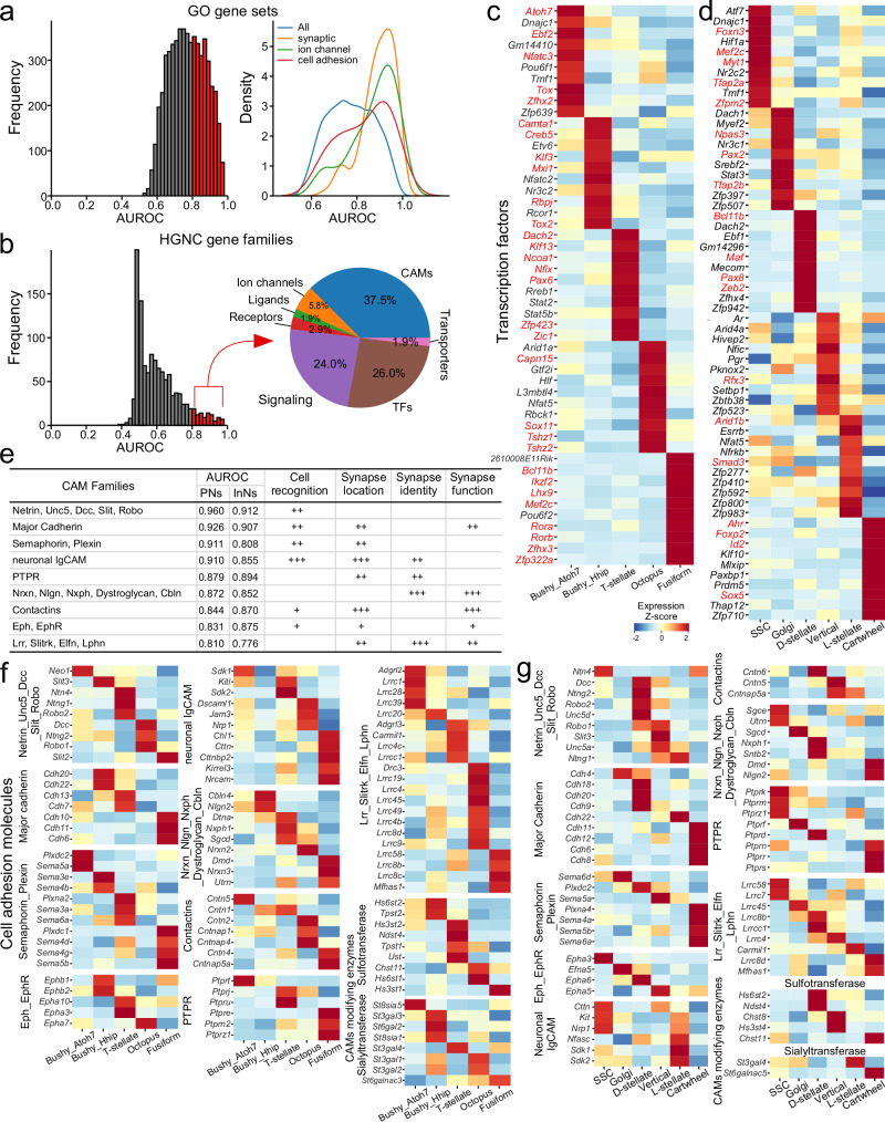
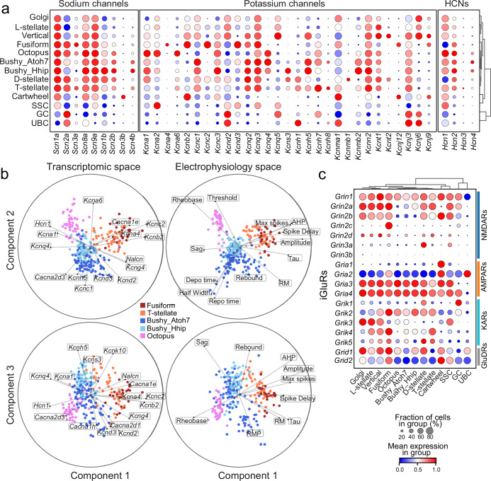
The cochlear nuclear complex (CN), the starting point for all central auditory processing, encompasses a suite of neuronal cell types highly specialized for neural coding of acoustic signals. However, the molecular logic governing these specializations remains unknown. By combining single-nucleus RNA sequencing and Patch-seq analysis, we reveal a set of transcriptionally distinct cell populations encompassing all previously observed types and discover multiple hitherto unknown subtypes with anatomical and physiological identity. The resulting comprehensive cell-type taxonomy reconciles anatomical position, morphological, physiological, and molecular criteria, enabling the determination of the molecular basis of the specialized cellular phenotypes in the CN. In particular, CN cell-type identity is encoded in a transcriptional architecture that orchestrates functionally congruent expression across a small set of gene families to customize projection patterns, input-output synaptic communication, and biophysical features required for encoding distinct aspects of acoustic signals. This high-resolution account of cellular heterogeneity from the molecular to the circuit level reveals the molecular logic driving cellular specializations, thus enabling the genetic dissection of auditory processing and hearing disorders with a high specificity.


Conflict of interest statement

Competing interests: The authors declare no competing interests.

Figures

Fig. 1
Fig. 1. Comprehensive transcriptional profiling of cell types across the mouse CN.
a Anatomy of mouse cochlear nucleus (CN; Light blue: VCN. Dark blue: DCN) and depiction of neuronal cell types (cartoon drawings) across the CN (sagittal view), colored by cell type identity. D dorsal, P posterior, M medial. The 3D brain model was generated with Brainrender (Ver. 2.1.10). b UMAP visualization of 31,639 nuclei from CN neurons clustered by over-dispersed genes, colored by cluster identity. Each cluster labeled by key marker genes. c Right: dendrogram indicating hierarchical relationships between clusters. Left: dot plot of scaled expression of selected marker genes for each cluster shown in (b); see Supplementary Data 1. Circle size depicts percentage of cells in the cluster in which the marker was detected (≥1 UMI), and color depicts the normalized average transcript count in expressing cells. See Figs. S1–3 for more technical details and annotations.
Fig. 2
Fig. 2. Using Patch-seq to identify transcriptomic correspondences for excitatory neurons.
a Examples of reconstructed excitatory neurons positioned at their site of origin in representative mouse CN (sagittal view), colored by cell type identity. b Example responses of CN excitatory cell types to current steps. Bottom traces show the injected currents. Scalebar: 100 mV for potential, 2 nA for injected currents for all cells except octopus cells (10 nA). c Left: UMAP visualization of physiological features of 404 CN excitatory neurons (E-cluster), colored by expert cell type. n = 243 for bushy, n = 67 for T-stellate, n = 58 for octopus, n = 36 for fusiform. Right: Confusion matrix shows performance (an average of 99.2% accuracy) of the cell-type classifier trained with physiological features. See Fig. S4 for more details. d Left: UMAP visualization of morphological features of 157 CN excitatory neurons (M-cluster), colored by expert cell type. n = 105 for bushy, n = 16 for T-stellate, n = 10 for octopus, n = 26 for fusiform. Right: Confusion matrix shows performance (an average of 95.5% accuracy) of the classifier trained with morphological features. See Fig. S4 for more details. e Left: UMAP visualization of 293 Patch-seq excitatory neurons clustered by over-dispersed genes, colored by transcriptomic cluster (T-cluster). Right: The matrix shows proportion of Patch-seq cells assigned to a specific cell type (the expert classification) among each T-cluster. All cells in T-cluster 1 (n = 67/67) and almost all cells in T-cluster 2 (n = 111/113) are bushy cells; all cells in T-cluster 3 are fusiform cells (n = 22/22); all cells in T-cluster 4 are octopus cells (n = 43/43); almost all cells in T-cluster 5 are T-stellate cells (n = 46/48). f Left: Patch-seq neurons (n = 293) mapped to snRNA-seq UMAP space as shown in Fig. 1b. Individual dots represent each cell, colored by T-cluster. Right: Matrix shows proportion of Patch-seq cells assigned to each T-cluster in (e) mapped to one of 13 clusters in UMAP space. g UMAP visualization and annotation of molecular cell types with established morpho-electrophysiological types in CN. Source data are provided in the Source Data file.
Fig. 3
Fig. 3. Two molecular subtypes of bushy cells.
a UMAP visualization of Hhip+ and Atoh7+ clusters and distribution of Patch-seq bushy cells in UMAP space, colored by cluster. Individual dots represent each Patch-seq cell. b UMAP visualization of two bushy cell clusters and normalized expression of Hhip, Atoh7, and Sst. Top: snRNA-seq, Bottom: Patch-seq. c FISH co-staining for Atoh7 and Hhip (left), Atoh7 and Sst (middle), or Hhip and Sst (right) in CN sagittal sections. Lines along the images indicate density of single-labeled or double-labeled cells along two axes of CN. Insets show the total counts of single-labeled and double-labeled cells across eight consecutive sagittal sections for each FISH experiment. d Left: A representative morphology of Hhip+ or Atoh7+ bushy cell. Axon (shown in red) is truncated. Right: Sholl analysis of Hhip+ or Atoh7+ bushy cell dendrites. Data are presented as mean ± SEM. n = 20 for Hhip+ cells from 15 mice, n = 51 for Atoh7+ cells from 20 mice, Two-way mixed model ANOVA. e Heat map displaying normalized expression of the top 20 DEGs for two subtypes. Scale bar: Expression level. Cell number in each subcluster is indicated below the figure. f Top: Example responses of Atoh7+ (left) or Hhip+ (right) bushy cell to a hyperpolarized current step and a near-threshold depolarizing step. Bottom-left: Individual APs from the Hhip+ and Atoh7+ cells shown above, aligned with onset of the depolarizing current. Bottom-right: Two most-discriminating features for Hhip+ and Atoh7+ cells, spike delay and spike duration (half-width). n = 67 for Hhip+ cells from 25 mice, n = 110 for Atoh7+ cells from 29 mice, Two-sided t-test. Data are presented as mean ± SEM. The dots above the bars represent the original values. g Volcano plot showing the log2 fold change (log2FC) and −log10(p-value) of detected genes comparing the two subtypes. The p-values were adjusted by Benjamini–Hochberg correction. Among DEGs are 15 genes encoding voltage-gated ion channels. See Fig. S9 for more analysis. Source data are provided in the Source Data file.
Fig. 4
Fig. 4. Two subtypes of T-stellate cells.
a Left: UMAP visualization of two subclusters of Fam129a+ neurons, colored by subcluster. Right: All Patch-seq cells mapped onto Fam129a+ cluster on the left. Individual dots represent each patch-seq cell. b UMAP visualization of normalized expression of Fam129a, Fn1, and Dchs2 in T-stellate cell cluster. c FISH co-staining for Fam129a and Fn1 in a CN sagittal section. Lines along images depict density of single-labeled or double-labeled neurons along the two axes of CN. Inset pie chart: proportion of double-labeled cells among all Fam129a+ cells counted across eight consecutive sagittal sections. Dashed contours indicate the CN region. Two images on the right are zoomed-in views of boxed regions on the left. Arrows point to double-labeled cells. d FISH co-staining for Fam129a and Dchs2 in a CN sagittal section. Inset pie chart shows the proportion of double-labeled cells among all Fam129a+ cells counted across six consecutive sections. Two images on the right are zoomed-in views. e Left: 2D spatial projection of Patch-seq T-stellate cells onto a sagittal view of CN, colored by subtypes. Right: Comparison of the distance to CN posterior edge between T-Fn1 and T-Dchs2. n = 12 for T-Fn1 cells from 10 mice, and n = 16 for T-Dchs2 cells from 10 mice; Data are presented as mean ± SEM; Two-sided t-test. f Left: Representative morphology of two Fn1+ and two Dchs2+ cells in a CN sagittal view. Right: zoomed-in of T-stellate cells shown on the left. Axons in red. g Sholl analysis of T-stellate cell dendrites. Data are presented as mean ± SEM. n = 6 for T-Fn1 cells from 6 mice, and n = 20 for T-Dchs2 cells from 12 mice; Two-way mixed model ANOVA. h Polar histograms showing the distribution of dendritic branch termination points with respect to soma of T-Fn1 and T-Dchs2. p = 3.0e-19 for the difference between the distributions using the Kolmogorov-Smirnov statistic. i Example responses of two T-stellate subtypes to current steps. j Comparison of electrophysiological features between two T-stellate subtypes. n = 16 for T-Fn1 cells from 13 mice, and n = 29 for T-Dchs2 cells from 16 animals, Mean ± SEM; Two-sided t-test. Source data are provided in the Source Data file.
Fig. 5
Fig. 5. Annotation of molecularly distinct glycinergic cell types in CN.
a Examples of reconstructed inhibitory neurons in a sagittal section, colored by cell type. b Example responses of inhibitory cells to current steps. c Left: UMAP visualization of morphological features of 116 inhibitory neurons (M-cluster), colored by expert cell type. Vertical: n = 38; SSC: n = 13; Cartwheel: n = 15; D-stellate: n = 15, L-stellate: n = 15. Right: Performance (Ave: 96.9%) of the classifier trained with morphological features. d Left: UMAP visualization of electrophysiological features along with anatomic locations (DCN:1; VCN:0) of 172 inhibitory neurons (E-cluster). Vertical: n = 50, SSC: n = 21; cartwheel: n = 21; D-stellate: n = 26, L-stellate: n = 53. Right: Performance (Ave: 97.9%) of the classifier trained with electrophysiological features and anatomic locations. See Fig. S4e, f for more analysis. e Left: UMAP visualization of 94 Patch-seq inhibitory neurons, colored by T-clusters. Right: Proportion of Patch-seq cells in each T-cluster assigned to a specific cell type. T-cluster 1: 12/12 are cartwheel cells; T-cluster 2: 20/21 are D-stellate cells; T-cluster 3: 35/35 are L-stellate cells; T-cluster 4: 10/12 are SSCs; T-cluster 5: 12/14 are vertical cells. f Top: 94 Patch-seq inhibitory neurons mapped onto snRNA-seq UMAP space, colored by T-cluster. Bottom: Proportion of Patch-seq cells assigned to each T-cluster in (e) mapped onto one of 13 distinct clusters in UMAP space. g UMAP visualization of all CN neuronal clusters, along with annotation with morpho-electrophysiological types. h UMAP visualization and annotation of all glycinergic CN cell types, and normalized expression of their respective marker genes. i FISH co-staining for Slc6a5/Stac, or for Slc6a5/Penk in sagittal sections. Lines indicate density of double-labeled neurons along two CN axes. Insets show proportion of double-labeled cells among all Stac+ or Penk+ cells counted across eight or six sections. j FISH co-staining for Slc6a5/tdTomato (left), or for Slc17a6/tdTomato (right) in sagittal sections in Penk-Cre:Ai9 mice. Insets show proportion of double-labeled cells among all tdTomato+ cells counted across six sections. k Micrographs showing labeled cells for recording (left), example responses to current steps (middle), and morphologies of labeled neurons (right) in Penk-Cre:Ai9 mice. l Distribution of labeled cells (red) in electrophysiological or morphological UMAP space of all inhibitory neurons. Source data are provided in the Source Data file.
Fig. 6
Fig. 6. Molecular basis of CN cell type identity.
a Distribution of AUROC values of 5,735 GO terms across all 13 cell types in the snRNA-seq dataset. Red, AUROC ≥ 0.8. Right: GO term probability density by keywords: “synaptic”, “cell adhesion” and “ion channel” are skewed with AUROC ≥ 0.8. b AUROC value distribution of 1424 HGNC gene families across all 13 cell types. 104 families with AUROC ≥ 0.8 (red bars) are classified into seven categories (pie chart). See Supplementary Data 5 for more details. c Heatmap displays the z-scored expression levels of the top 10 specific TFs for each excitatory projection neuron type. TFs highlighted in red show expression in the developing cochlear nucleus, including progenitors and distinct developmental stages of projection neurons (see Supplementary Data 8 for reference support). d Heatmap showing the z-scored expression levels of the top 10 specific TFs for each inhibitory cell type. TFs in red have been shown to be involved in the differentiation and specification of glycinergic/GABAergic interneurons in cochlear nucleus or other brain regions (see Supplementary Data 8 for reference support). e Top-performing cell-adhesion molecule (CAM) families, their CAMAUROC value, and their roles in synaptic connectivity. “+” denotes the degree of involvement in the listed function. f Heatmap showing the differential expression of 9 CAM and 2 carbohydrate-modifying enzyme families across excitatory projection neuron types. g Heatmap showing the differential expression of 9 CAM and 2 carbohydrate-modifying enzyme families across inhibitory cell types.
Fig. 7
Fig. 7. Single-cell transcriptomes provide insights into functional specializations of CN cell types.
a Dot plots showing scaled expression of the genes encoding ion channels across CN cell type (from snRNA-seq dataset). Sodium and potassium channel subtypes or subunits with low expression levels (<20% fraction of the cell in any cell type) were excluded in the figure. Circle size depicts the percentage of cells in the cluster in which the marker was detected (≥1 UMI), and color depicts the normalized average transcript count in expressing cells. b Sparse reduced-rank regression (RRR) model to predict electrophysiological features by the expression patterns of 119 ion channel genes (middle, cross-validated R2 = 0.35). The models selected 25 genes. Cross-validated correlations between the first three pairs of projections were 0.83, 0.65, and 0.61. Both transcriptome (left) and electrophysiology (Right) were embedded in the latent space (bibiplots). In each biplot, lines represent correlations between a feature (gene expression or electrophysiology) and two latent components; the circle corresponds to the maximum attainable correlation (r = 1). Only features with a correlation above 0.4 are shown. Source data are provided in the Source Data file. c Dot plots showing scaled expression of genes encoding ionotropic glutamate receptors in each CN cell type. AMPARs AMPA receptors, NMDARs NMDA receptors, KARs Kainate receptors, GluDRs delta glutamate receptors. See Fig. S12 for additional information.

Update of

Similar articles

Cited by

References

    1. Oertel, D., McGinley, M. & and Cao, X.-J. in Encylopedia of Neuroscience (Springer, 2009).
    1. Grothe, B., Pecka, M. & McAlpine, D. Mechanisms of sound localization in mammals. Physiol. Rev.90, 983–1012 (2010). - PubMed
    1. Chi, T., Ru, P. & Shamma, S. A. Multiresolution spectrotemporal analysis of complex sounds. J. Acoust. Soc. Am.118, 887–906 (2005). - PubMed
    1. Rhode, W. S. & Greenberg, S. in The Mammalian Auditory Pathway: Neurophysiology (eds. Arthur N. Popper & Richard R. Fay) 94–152 (Springer, 1992).
    1. Palmer, A. R. Physiology of the cochlear nerve and cochlear nucleus. Br. Med. Bull.43, 838–855 (1987). - PubMed