. 2025 Jan 9;16(1):540.
doi: 10.1038/s41467-025-55899-7.

The master male sex determinant Gdf6Y of the turquoise killifish arose through allelic neofunctionalization

Affiliations

The master male sex determinant Gdf6Y of the turquoise killifish arose through allelic neofunctionalization

Annekatrin Richter et al. Nat Commun. .

Abstract

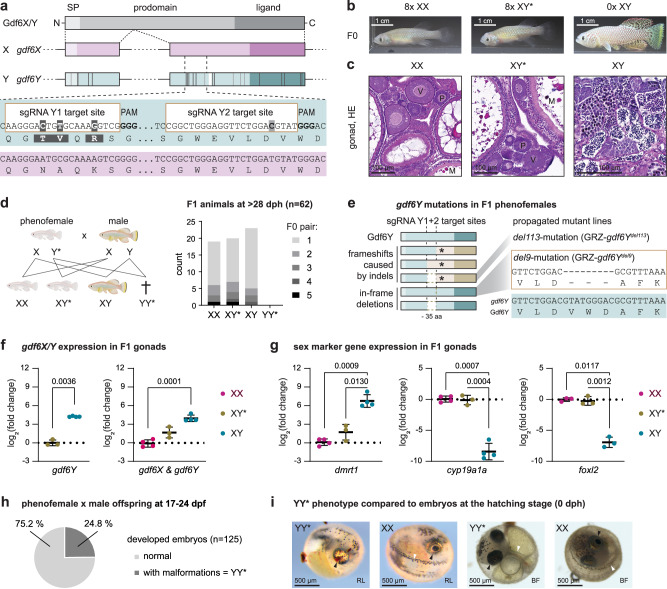
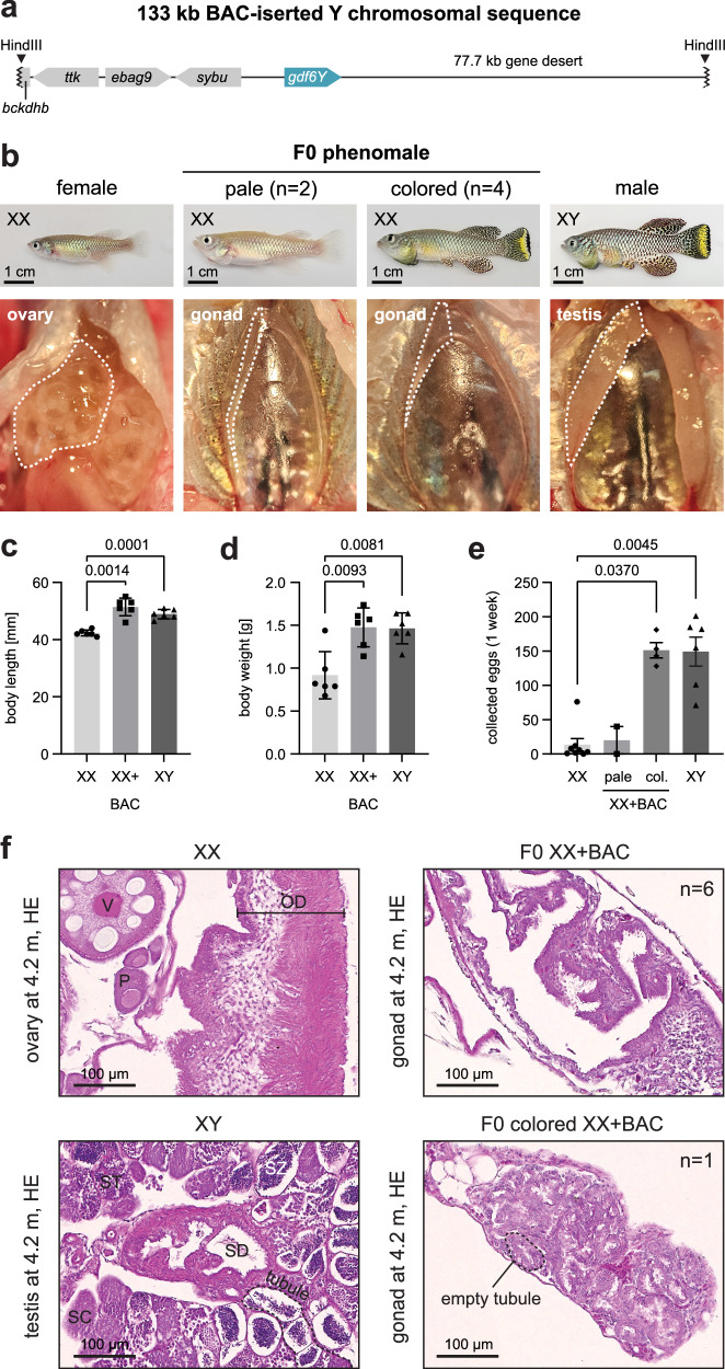
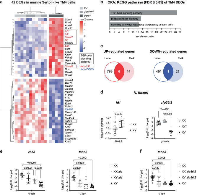
Although sex determination is a fundamental process in vertebrate development, it is very plastic. Diverse genes became major sex determinants in teleost fishes. Deciphering how individual sex-determining genes orchestrate sex determination can reveal new actors in sexual development. Here, we demonstrate that the Y-chromosomal copy of the TGF-β family member gdf6 (gdf6Y) in Nothobranchius furzeri, an emerging model organism in aging research, gained the function of the male sex determinant through allelic diversification while retaining the skeletal developmental function shared with the X-chromosomal gdf6 allele (gdf6X). Concerning sex determination, gdf6Y is expressed by somatic supporting cells of the developing testes. There it induces the male sex in a germ cell-independent manner in contrast to sex determination in zebrafish and the medaka. Looking for downstream effectors of Gdf6Y, we identified besides TGF-β signaling modulators, especially the inhibitor of DNA binding genes id1/2/3, the mRNA decay activator zfp36l2 as a new GDF6 signaling target.


Conflict of interest statement

Competing interests: The authors declare no competing interests.

Figures

Fig. 1
Fig. 1. The inactivation of gdf6Y in Nothobranchius furzeri leads to a full male-to-female sex reversal.
a Schematic of Gdf6X/Y protein domains (SP – signal peptide), exonal distribution, the coding sequence comparison between gdf6X and gdf6Y, and the mutation strategy with two gdf6Y-specific sgRNAs facilitating DNA double-strand breaks at a 109 bp distance. Coding sequence differences between gdf6X and gdf6Y are indicated in gdf6Y’s exons. SNVs: Dark gray – non-synonymous, light gray – synonymous. White – 9 bp deletion. b Phenotype of mosaic F0 GRZ-gdf6Y animals (XY*, phenofemales) compared to unaltered F0 females (XX) and wild-type males (XY) at about 3 months of age. c One exemplarily HE stained F0 XX, F0 XY*, and XY gonad at about 5 months of age. Oocytes: P – previtellogenic, V – vitellogenic, M – mature. SC – spermatocytes, ST – spermatids, SZ – spermatozoa. d Sexually mature offspring (age: 1 month) of phenofemales with males consists of females, phenofemales, and males in equal parts missing YY* animals expected due to Mendelian ratios. Schematic created in BioRender. Richter, A. (2024) https://BioRender.com/n01l529. e Gdf6Y mutant variants causing full male-to-female sex reversal in F1 (frameshifts at either sgRNA position causing early stop codons (asterisks), in-frame deletions of 9 bp at the sgRNA Y2 site or between both sgRNAs). Two mutations were propagated in fish lines. RT-qPCR of (f) gdf6Y separately and together with gdf6X and (g) male and female marker genes in F1 gonads (Supplementary Table 1, XY*: n = 3; XX, XY: n = 4, n = 3 for foxl2) at 2.4 months of age (mean with standard deviation). Statistical testing by a two-tailed Welch’s t-test (gdf6Y) or Welch’s ANOVA and Dunnett’s T3 multiple comparisons test (others). P-values < 0.05 are displayed. h One-quarter of a phenofemale’s (GRZ-gdf6Ydel9) offspring with males develops malformations and carries two Y chromosomes (YY*). i Reflected light (RL) and brightfield (BF) microscopy images of YY* embryos (RL, n = 13; BF, n = 31) within the egg compared to normally developed animals (RL, n = 45; BF, n = 94; here: XX at 0 dph) from independent samplings from two phenofemales (RL, GRZ-gdf6Ydel6, del8; BF, GRZ-gdf6Ydel9 from h). Arrows: Black – eye, white – tail. d, f–h Source data are provided as a Source Data file.
Fig. 2
Fig. 2. Gdf6Y can functionally cover for Gdf6X.
Schematic of (a) the gdf6X inactivation and (b) the obtained open reading frame disruption of the propagated mutant line GRZ-gdf6Xdel113. c Phenotype of heterozygous GRZ-gdf6Xdel113 animals, of which both sexes, females (XX*) and males (X*Y), were 1st observed in F2 due to the X-linked heredity of the mutation that was transferred from an F0 father. d Homozygous GRZ-gdf6Xdel113 F3 animals (X*X*) were generated by an incross of heterozygous F2 animals and have a curled phenotype after hatching (brightfield microscopy), while siblings (X*X, X*Y, XY) are normal. X*Y is shown for comparison. Schematic created in BioRender. Richter, A. (2024) https://BioRender.com/x35g473. e Phenotype of a 2-month-old homozygous GRZ-gdf6Xdel113 F3 female (X*X*). f Dissected X*X* compared to XX* F3 ovaries at 2 months of age. g Pearson correlation (two-tailed p-value) of body and ovarian weight of homozygous (X*X*) and heterozygous (XX*) GRZ-gdf6Xdel113 F3 females (n = 8 each) at 2 months of age. h HE stained X*X* and XX* F3 ovary at 2 months of age. Oocytes: P – previtellogenic, V – vitellogenic, M – mature. i Luciferase reporter assay to test the differential activation of Smad1, 2, 3, 5, and 9 upon expression of gdf6X or gdf6Y compared to gdf6Y mutant variants (gdf6Ydel9 and gdf6Yins25; n = 5 each). Gdf6Ydel9 has an in-frame 9 bp deletion in the prodomain’s CDS, while gdf6Yins25 harbors a 25 bp insertion at the same position leading to a frameshift and early stop codon. Stratified statistical testing per Smad by Kruskal–Wallis tests and Dunn’s multiple comparisons tests with the grouped gdf6Y mutant variants. P-values < 0.05 are displayed. g, i Source data are provided as a Source Data file.
Fig. 3
Fig. 3. Localization of the gonadal gdf6Y expression in XY animals.
Transcripts of gdf6Y (magenta) and the GC marker ddx4 (green) as well as the (a) Sertoli or (b) somatic supporting cell marker amh (orange) were detected in N. furzeri testes at (a) sexually mature (n = 9; here: 3 months old) and (b) 0 dph stage (n = 7). a, b White arrows – cells co-expressing gdf6Y and amh. c Minimal distances of gdf6Y signals to ddx4 and amh signals from 4 XY gonads at 0 dph including (b). Left: Violin plots of minimal distances <8 µm (n = 212 per gene, equals lowest 95%) with median, quartiles, and respective modes (bin width: 0.1 µm) from the 4 individual gonads (circle, square, triangle, and diamond). Statistical testing by a two-tailed Mann–Whitney test. Right: Frequency distribution of all measured minimal distances (bin width: 0.1 µm, n = 224). Source data are provided as a Source Data file.
Fig. 4
Fig. 4. Oogenesis starts before 0 dph, depends on foxl2l, and can be measured and intercepted by genetic modification in N. furzeri.
a Volcano plots of DEGs (FDR < 0.05) between XX and XY animals in the whole embryo at 10 dpf and in trunk parts at 0 and 3 dph as derived from a respective previously published data set (XX at 0 dph n = 2, others n = 3; Supplementary Data 1). b Overlap of female DEGs between 0 and 3 dph. c Overrepresentation analysis (ORA) of KEGG pathways within the overlap of female DEGs between 0 and 3 dph. d Schematic of CRISPR/Cas9-based chimeric mutant (CRISPant) assay to address the influence of a goi (gene of interest) on oogenesis onset as a proxy for sexual differentiation. T7EI – T7 endonuclease I. Created in BioRender. Richter, A. (2024) https://BioRender.com/t02m033. e Expression of female marker genes on mRNA-level in trunks of females (XX; n = 8), phenofemales (GRZ-gdf6Ydel9 XY*; n = 4), and males (XY; tacc3, n = 5; zp4, n = 6) and their mosaic foxl2l counterparts (XX, n = 6; XY*, n = 5; XY, n = 3) at 0 dph. Welch’s ANOVA and Dunnett’s T3 multiple comparisons test with XX animals. P-values < 0.05 are displayed. c, e Source data are provided as a Source Data file.
Fig. 5
Fig. 5. A gdf6Y-containing Y-chromosomal fragment leads to a female-to-male sex reversal.
a Schematic of the Y-chromosomal sequence inserted via HindIII into the bacterial artificial chromosome (BAC) GRZ-B-a-208Dg03. b Phenotypes and streak gonads in situ of partially sex-reversed mosaic XX animals (phenomales) created by zygotic BAC injection compared to an XX female and an XY male at 4.2 months of age. Body length (c) and body weight (d) of phenomales (XX + BAC) compared to females and males at 4.2 months of age (n = 6 each). Statistical testing by Welch’s ANOVA and Dunnett’s T3 multiple comparisons test. e Numbers of all eggs (dead or alive) collected after 1 week of constant breeding of a single female (2.5 months) with another female (XX, 2.5-5 months, n = 8), a pale (4 months, n = 2) or a colored phenomale (XX + BAC, 4 months, n = 4), or a male (XY, 4.2 months, n = 6). Statistical testing by a Kruskal–Wallis test and Dunn’s multiple comparisons test with XX. ce Mean with standard deviation. P-values < 0.05 are displayed. Source data are provided as a Source Data file. f HE stained gonadal sections of two BAC-positive individuals with the observation frequency in all phenomales. Ovary and testis connecting to the respective gonadal duct are given for comparison. Oocytes: P – previtellogenic, V – vitellogenic; OD - oviduct. SC – spermatocytes, ST – spermatids, SZ – spermatozoa; SD – sperm duct.
Fig. 6
Fig. 6. Gdf6Y leads to the specific regulation of certain genes independent of the targeted cell’s identity.
a Heatmap of DEGs (FDR < 0.05) between murine Sertoli-like TM4 cells transfected with a gdf6Y expression plasmid (n = 6) and TM4 cells transfected with either a gdf6Y mutant variant (gdf6Ydel9, n = 5) expression plasmid or an empty vector (EV, n = 6). Genes of the TGF-beta signaling pathway are boxed. b Overrepresentation analysis (ORA) of KEGG pathways within the 42 DEGs identified in TM4 cells. FDR – false discovery rate. c Overlap of the up- or downregulated DEGs identified in TM4 cells with those identified in human HeLa cells. The common genes are highlighted in red (upregulated genes) or blue (the downregulated gene) in (a). d RT-qPCR analyses of the expression of id1 and zfp36l2 in female (XX, n = 8) and male (XY, n = 4) embryos at 10 dpf (stage 4) or gonads of 3 months old females (XX, n = 7), males (XY, n = 5), and phenofemales (GRZ-gdf6Ydel9 XY*, n = 7), respectively. Two-tailed Welch’s t-test (id1) or Welch’s ANOVA and Dunnett’s T3 multiple comparisons test (zfp36l2). e RT-qPCR analyses of female marker genes in trunks of uninjected (n = 6 each) and indel-negative (XX, n = 3; XY, n = 4) females and males and id1 CRISPants of both sexes (XX, n = 5; XY, n = 7) at 0 dph. Welch’s ANOVA and Dunnett’s T3 multiple comparisons test. f RT-qPCR analyses of female marker genes in trunks of uninjected (n = 9 each) and indel-negative (n = 3 each) females and males and zfp36l2 CRISPants of both sexes (XX, n = 8; XY, n = 11) at 0 dph. Kruskal–Wallis test and Dunn’s multiple comparisons test. d–f Mean with standard deviation. P-values < 0.05 are displayed. a, b, d–f Source data are provided as a Source Data file.

Similar articles

Cited by

  • Multiple Origins of Sex Chromosomes in Nothobranchius Killifishes.
    Hospodářská M, Mora P, Voleníková AC, Al-Rikabi A, Altmanová M, Simanovsky SA, Tolar N, Pavlica T, Janečková K, Štundlová J, Bobryshava K, Jankásek M, Hiřman M, Liehr T, Reichard M, Krysanov EY, Ráb P, Englert C, Nguyen P, Sember A. Hospodářská M, et al. Mol Ecol. 2025 Aug;34(16):e70029. doi: 10.1111/mec.70029. Epub 2025 Jul 24. Mol Ecol. 2025. PMID: 40702874 Free PMC article.

References

    1. Reuter, H., Krug, J., Singer, P. & Englert, C. The African turquoise killifish Nothobranchius furzeri as a model for aging research. Drug Discov. Today. Dis. Models27, 15–22 (2018).
    1. Jubb, R. A new Nothobranchius (Pisces, Cyprinodontidae) from Southeastern Rhodesia. J. Am. Killifish Assoc.8, 12–19 (1971).
    1. Terzibasi, E. et al. Large differences in aging phenotype between strains of the short-lived annual fish Nothobranchius furzeri. PLoS ONE3, e3866 (2008). - PMC - PubMed
    1. Tozzini, E. T. et al. Parallel evolution of senescence in annual fishes in response to extrinsic mortality. BMC Evol. Biol.13, 77 (2013). - PMC - PubMed
    1. Api, M. et al. Effects of parental aging during embryo development and adult life: the case of Nothobranchius furzeri. Zebrafish15, 112–123 (2018). - PubMed

Substances

Supplementary concepts