. 2025 Jan 9;15(1):1423.
doi: 10.1038/s41598-025-85895-2.

Inhibition of skin fibrosis via regulation of Th17/Treg imbalance in systemic sclerosis

Affiliations

Inhibition of skin fibrosis via regulation of Th17/Treg imbalance in systemic sclerosis

Akiko Sekiguchi et al. Sci Rep. .

Abstract

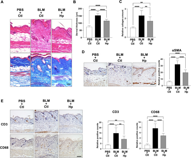
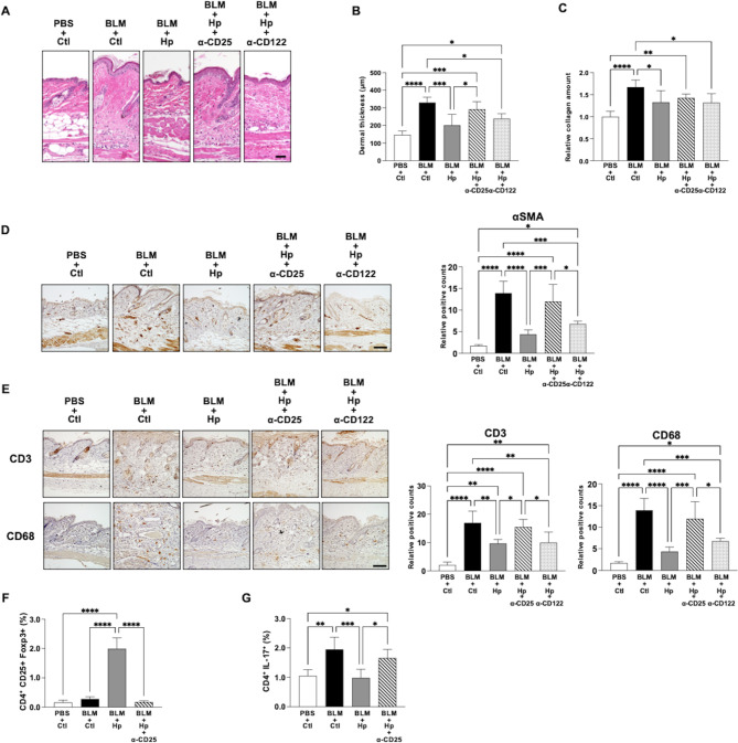
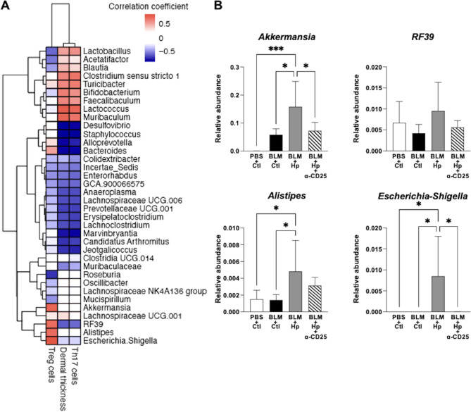
Systemic sclerosis (SSc) is an idiopathic systemic connective tissue disorder characterized by fibrosis of the skin and internal organs, with growing interest in the imbalance between Th17 cells and regulatory T cells (Tregs) in the disease's pathogenesis. Heligmosomoides polygyrus (Hp), a natural intestinal parasite of mice, is known to induce Tregs in the host. We aimed to investigate the effects of Hp-induced Tregs on bleomycin-induced dermal fibrosis and clarify the role of the Th17/Treg balance in SSc fibrosis. Infection with Hp suppressed the development of bleomycin-induced dermal fibrosis and the infiltration of CD3+ T cells and CD68+ macrophages. Flow cytometric analysis revealed that Hp infection increased Tregs and inhibited the induction of bleomycin-induced Th17 cells. Treg depletion nullified these effects, suggesting that Hp-induced Tregs may prevent bleomycin-induced dermal fibrosis and inflammation. Analysis of the intestinal microbiota showed that bacteria positively correlated with Tregs exhibited a negative correlation with Th17 cells and dermal fibrosis in mice. SSc patients with severe fibrosis displayed a distinct microbiota profile. These results suggest that alterations in the intestinal microbiota may contribute to the Th17/Treg imbalance in SSc and its progression. Enhancing Tregs to regulate the Th17/Treg imbalance may present a promising strategy for suppressing fibrosis in SSc.

Keywords: Heligmosomoides polygilus; Bleomycin; Fibrosis; Microbiota; Systemic sclerosis; Th17/Treg balance.


Conflict of interest statement

Declarations. Competing interests: The authors declare no competing interests.

Figures

Fig. 1
Fig. 1
Helminth infection suppressed the bleomycin-induced dermal fibrosis and inflammation in mice. (A) Representative images of H&E staining (top) or Masson trichrome staining (bottom) of skin sections taken 14 days after initiation of subcutaneous injection of bleomycin or control PBS into Hp-infected or uninfected mice. Scale bar = 50 μm. (B) Quantification of dermal thickness of the lesional skin in mice. Values were determined in 6–8 random microscopic fields in n = 9 mice per groups. (C) Relative amounts of soluble collagen in 5 mm diameter skin sections. n = 10 mice per groups. (D) Representative immunochemical staining images of skin sections to show the numbers of αSMA+ myofibroblasts. Right panel shows quantification of positive cells in 6 random microscopic fields in the dermis. n = 7–10 mice per groups. Scale bar = 50 μm. (E) Representative immunochemical staining images of skin sections taken 5 days after the initiation of subcutaneous injection of bleomycin or control PBS into Hp-infected or uninfected mice to show the numbers of CD3+ T cells (top) and CD68+ macrophages (bottom). Right panels show quantification of each positive cells in 6 random microscopic fields in the dermis. n = 8–10 mice per groups. Scale bar = 50 μm. Values represent mean ± SEM. **P < 0.01 and ****P < 0.0001. One-way ANOVA followed by Tukey-Kramer test was used for (BE). Hp, Heligmosomoides polygilus; BLM, bleomycin; Ctl, control.
Fig. 2
Fig. 2
CD4+ Treg cells are required for the suppressive effect of helminth infection on bleomycin-induced dermal fibrosis and for regulating the Th17/Treg imbalance. (A) Representative images of H&E staining of skin sections taken 14 days after the initiation of subcutaneous injection of bleomycin or control PBS into Hp-infected or uninfected mice injected intraperitoneally with anti-CD25 antibody, anti-CD122 antibody or control PBS. Scale bar = 50 μm. (B) Quantification of dermal thickness of the lesional skin in mice. Values were determined in 6 random microscopic fields in n = 5 mice per groups. (C) Relative amounts of soluble collagen in 5 mm diameter skin sections. n = 10 mice per groups. (D) Representative immunochemical staining images of skin sections to show the numbers of αSMA+ myofibroblasts. Right panel shows quantification of positive cells in 6 random microscopic fields in the dermis. n = 4–5 mice per groups. Scale bar = 50 μm. (E) Representative immunochemical staining images of skin sections taken 5 days after the initiation of subcutaneous injection of bleomycin or control PBS into Hp-infected or uninfected mice injected intraperitoneally with anti-CD25 antibody, anti-CD122 antibody or control PBS to show the numbers of CD3+ T cells (top) and CD68+ macrophages (bottom). Right panels show quantification of each positive cells in 6 random microscopic fields in the dermis. n = 5 mice per groups. Scale bar = 50 μm. (F,G) Quantification by flow cytometry of Treg cells defined as CD25+Foxp3+ cells (F) and Th17 cells defined as Th17+ cells (G) among gated CD4+ T cells in the inguinal lymph node taken 14 days after the initiation of subcutaneous injection of bleomycin or control PBS into Hp-infected or uninfected mice injected intraperitoneally with anti-CD25 antibody, anti-CD122 antibody or control PBS. Values represent mean ± SEM. *P < 0.05, **P < 0.01, ***P < 0.001 and ****P < 0.0001. One-way ANOVA followed by Tukey-Kramer test was used for (BG). Hp, Heligmosomoides polygilus; BLM, bleomycin; Ctl, control.
Fig. 3
Fig. 3
Association of intestinal microbiota in bleomycin-induced dermal fibrosis and Th17/Treg imbalance in SSc mouse model. (A,B) Investigations of bacterial genera in faeces taken 14 days after the initiation of subcutaneous injection of bleomycin or control PBS into Hp-infected or uninfected mice injected intraperitoneally with anti-CD25 antibody or control PBS. (A) Heatmap showing the strength of correlation based on Pearson correlation coefficient on a color scale between abundance of faecal bacterial genera and the number of Treg cells, dermal thickness, and the number of Th17 cells, respectively. (B) Relative abundance of bacteria in mouse faeces that were positively correlated with the number of Treg cells. n = 5–6 mice per group. Values represent mean ± SEM. *P < 0.05, ***P < 0.001. One-way ANOVA followed by Tukey-Kramer test was used for (B). Hp, Heligmosomoides polygilus; BLM, bleomycin; Ctl, control; Treg, regulatory T.
Fig. 4
Fig. 4
Characteristics of the microbiota in patients with SSc. (A,B) The faecal bacterial composition of 36 SSc patients and 20 healthy individuals is shown in hierarchical clustering trees (A) and Principal Coordinate Analysis (PCoA) plots (B), based on Bray-Curtis distances calculated from the OTU distribution across samples. (C) Relative abundance of Bifidobacterium characterized by a decrease in the intestinal microbiota of SSc patients in group A. (D) Model of inhibition of bleomycin-induced dermal fibrosis by helminth infection-mediated regulation of Th17/Treg balance. Values represent mean ± SEM. One-way ANOVA followed by Tukey-Kramer test was used for (C). *P < 0.05 and ****P < 0.0001. SSc, systemic sclerosis; PCoA, principal coordinates analysis; OTU, operational taxonomic unit; Treg, regulatory T; HV, healthy volunteer.

Similar articles

References

    1. Denton, C. P. & Khanna, D. Systemic sclerosis. Lancet390, 1685–1699 (2017). - PubMed
    1. Okamoto, Y. et al. Potential roles of interleukin-17A in the development of skin fibrosis in mice. Arthritis Rheum.64, 3726–3735 (2012). - PubMed
    1. Jin, W., Zheng, Y. & Zhu, P. T cell abnormalities in systemic sclerosis. Autoimmun. Rev.21, 103185 (2022). - PubMed
    1. Hemdan, N. Y. et al. Interleukin-17-producing T helper cells in autoimmunity. Autoimmun. Rev.9, 785–792 (2010). - PubMed
    1. Zambrano-Zaragoza, J. F., Romo-Martínez, E. J., Durán-Avelar, Mde, J. & García-Magallanes, N. & Vibanco-Pérez, N. Th17 cells in autoimmune and infectious diseases. Int. J. Inflamm.2014, 651503 (2014). - PMC - PubMed

Publication types