. 2025 Jan 9;27(1):4.
doi: 10.1186/s13058-024-01958-8.

Secreted LGALS3BP facilitates distant metastasis of breast cancer

Affiliations

Secreted LGALS3BP facilitates distant metastasis of breast cancer

Seung-Su Kim et al. Breast Cancer Res. .

Abstract

Background: Patients with estrogen receptor (ER)-positive breast cancer (BC) can be treated with endocrine therapy targeting ER, however, metastatic recurrence occurs in 25% of the patients who have initially been treated. Secreted proteins from tumors play important roles in cancer metastasis but previous methods for isolating secretory proteins had limitations in identifying novel targets.

Methods: We applied an in situ secretory protein labeling technique using TurboID to analyze secretome from tamoxifen-resistant (TAMR) BC. The increased expression of LGALS3BP was validated using western blotting, qPCR, ELISA, and IF. Chromatin immunoprecipitation was applied to analyze estrogen-dependent regulation of LGALS3BP transcription. The adhesive and angiogenic functions of LGALS3BP were evaluated by abrogating LGALS3BP expression using either shRNA-mediated knockdown or a neutralizing antibody. Xenograft mouse experiments were employed to assess the in vivo metastatic potential of TAMR cells and the LGALS3BP protein. Clinical evaluation of LGALS3BP risk was carried out with refractory clinical specimens from tamoxifen-treated ER-positive BC patients and publicly available databases.

Results: TAMR secretome analysis revealed that 176 proteins were secreted at least 2-fold more from MCF7/TAMR cells than from sensitive cells, and biological processes such as cell adhesion and angiogenesis were associated with the TAMR secretome. Galectin-3 binding protein (LGALS3BP) was one of the top 10 most highly secreted proteins in the TAMR secretome. The expression level of LGALS3BP was suppressed by estrogen signaling, which involves direct ERα binding to its promoter region. Secreted LGALS3BP in the TAMR secretome helped BC cells adhere to the extracellular matrix and promoted the tube formation of human umbilical vein endothelial cells. Compared with sensitive cells, xenograft animal experiments with MCF7/TAMR cells showed increased pulmonary metastasis, which completely disappeared in LGALS3BP-knockdown TAMR cells. Finally, higher levels of LGALS3BP were associated with poor prognosis in ER-positive BC patients treated with adjuvant tamoxifen in the clinic.

Conclusion: TAMR secretome analysis identified secretory proteins, such as LGALS3BP, that are involved in biological processes closely related to metastasis. Secreted LGALS3BP from the TAMR cells promoted adhesion of the cells to the extracellular matrix and vasculature formation, which may support metastasis of TAMR cells.

Keywords: LGALS3BP; Metastasis; Secretome; Tamoxifen-resistant breast cancer.


Conflict of interest statement

Declarations. Ethics approval: The animal experiments were performed in accordance with the guidelines of the Seoul National University Animal Care and Use Committee (SNU-210906-2-3 and SNU-240124-3). Competing interests: The authors declare no competing interests.

Figures
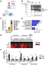
Fig. 1
Fig. 1
Isolation and characterization of the secretome from the MCF7/TAMR cells. (A) Schematic representation of the TAMR cell-specific secretory protein labeling using the Sec61b-TurboID system. ER: Endoplasmic reticulum, AdV: Adeno-virus (B) The culture supernatants of the AdV-Sec61b-TurboID infected MCF7/TAMR cells and those of the parental cells were collected, concentrated, and subjected to western blotting (WB) using streptavidin-HRP. (C) The relative abundance of the top 10 secretory proteins upregulated in terms of biotinylated spectral counts in the MCF7/TAMR cells. (D) GO Biological process analysis for the 176 upregulated genes in the MCF7/TAMR (fold change ≥ 2). Bar graph showing the most enriched biological process based on p-value. The number of annotated genes in each ontology term is shown in parenthesis. (E) Heat map representation of differentially regulated genes associated with “cell adhesion”, “angiogenesis” and “wound healing” biological processes. The full names of the genes are annotated in Table S3. (F) Total RNA extract obtained from the MCF7/TAMR cells and their parental cells were subjected to qPCR analysis. Data presented as mean ± SD (n = 3). *, P < 0.05; **, P < 0.01, and ***, P < 0.001
Fig. 2
Fig. 2
Secretion of LGAL3BP increased from the MCF7/TAMR and T47D/TR cells. (A) Brief experimental scheme for Fig. 2. (B) Whole cell lysates obtained from the MCF7/TAMR and T47D/TR cells and their parental cells were subjected to western blotting (upper). The proteins in the culture supernatants were TCA precipitated and immunoblotted with anti-LGALS3BP antibodies (lower). Band intensity was quantified and normalized to the intensity of the corresponding β-actin band. (C) The amounts of LGALS3BP in the culture supernatants were measured by ELISA. (D) Expression of LGALS3BP was visualized by green immunofluorescence. The nuclei were stained with DAPI. Scale bars: 20 μm. Data presented as mean ± SD (n = 3). *, P < 0.05; **, P < 0.01, and ***, P < 0.001
Fig. 3
Fig. 3
Expression of LGAL3BP is regulated by E2 in the MCF7/TAMR cells. (A and B) The MCF7/TAMR cells and their parental cells were treated with 1 and 10 nM of E2 for 24 h. Whole cell lysates or total RNA were obtained and subjected to western blotting (A) or qPCR analysis (B), respectively. *, P < 0.05 and **, P < 0.01 (n = 4 for WB and n = 3 for qPCR). (C and D) The MCF7/TAMR cells and their parental cells were treated with 0.5 and 5 µM tamoxifen for 4 h prior to treatment with 10 nM E2. The cells were further incubated for an additional 24 h. At the end of incubation, whole cell lysates or total RNA were obtained and subjected to western blotting (C) or qPCR analysis (n = 3) (D), respectively. *, P < 0.05, **, P < 0.01 and ***, P < 0.001. (E) MCF7/S0.5 and MCF7/TAMR-8 cells were treated with 10 nM E2 or vehicle (100% EtOH). After 24 h, the cells were subjected to ChIP-qPCR analysis. Data were normalized as percentages relative to 1 µg input. Data presented as mean ± SD (n = 3). *, P < 0.05, **, P < 0.01 and ***, P < 0.001
Fig. 4
Fig. 4
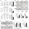
LGALS3BP enhances cell adhesion of MCF7/TAMR cells and tube formation of HUVEC. (A) Attachment of the MCF7/TAMR, shLG, and their matched control cells was evaluated by staining with crystal violet 2 h after seeding (upper). Or MCF7/TAMR-8 cells were pre-treated with 1 µg/mL of anti-LGALS3BP antibodies or IgG control for 10 min before the adhesion assay was performed (lower). Adherent cells were quantified by dissolving them in MeOH before the absorbance at 570 nm was detected. Scale bars = 500 μm. (B) HUVECs were cultured in conditioned media obtained from MCF7/TAMR, shLG, and their matched control cells (upper). Or HUVECs were cultured in MCF7/TAMR-8 conditioned media treated with 1 µg/mL of anti-LGALS3BP antibodies or IgG control (lower). Tube formation was quantified by counting the number of meshes formed. Scale bars = 100 μm. (C) HUVECs were cultured in serum-free or conditioned media obtained from shLG #1 cells and treated with recombinant LGALS3BP protein as indicated. Tube formation was quantified by counting the number of meshes formed. Scale bars = 100 μm. rLG, recombinant LGALS3BP (D) HUVECs were treated with recombinant LGALS3BP protein as indicated. The cell growth was determined using MTT assay. Data presented as mean ± SD. *, P < 0.05, **, P < 0.01, and ***, P < 0.001 (n = 3 for A and D and n = 4 for B and C)
Fig. 5
Fig. 5
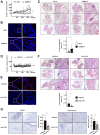
Depletion of LGALS3BP suppresses pulmonary metastasis of the MCF7/TAMR-8 in xenograft experiments. (A) MCF7/S0.5 or TAMR-8 cells were inoculated into the fourth mammary fat pads of female athymic nude mice. When the tumor volume reached approximately 50 mm3, the tumor diameter was measured with a caliper (n = 9–10). (B) Representative images of LGALS3BP immunofluorescent staining in the primary tumor sections. Scale bars = 20 μm. (C) The spontaneous lung metastasis developed from the MCF7/TAMR cells and their parental cell xenografts. Hematoxylin and eosin staining of lung sections are shown. Arrowheads indicate metastatic lesions. Scale bars = 800 μm for full images and 200 μm for inserts. The area of metastatic lesions in the lungs were quantified using ImageJ. Data presented as mean ± SD (n = 9–10). *, P < 0.05. (D) MCF7/TAMR-8-shScramble or MCF7/TAMR-8-shLG cells were inoculated into the fourth mammary fat pads of female athymic nude mice. When the tumor volume reached approximately 50 mm3, the tumor diameter was measured with a caliper. (n = 9) (E) Representative images of LGALS3BP immunofluorescent staining in the primary tumor sections. Scale bars = 20 μm. (F) The spontaneous lung metastasis developed from the shLG and its control cell xenograft. Hematoxylin and eosin staining of lung sections are shown. Arrowheads indicate metastatic lesions. Scale bars = 800 μm for full images and 200 μm for inserts. The area of the metastatic lesions in the lung was quantified using ImageJ. Data presented as mean ± SD (n = 9). *, P < 0.05. (G) Immunohistochemistry of CD31 or fibronectin of shScrb and shLG primary tumors are shown. Scale bars = 100 μm. At least two images of 2 mm3 in size from each primary tumor sections were quantified using ImageJ. Data presented as mean ± SD (n = 7–8). *, P < 0.05
Fig. 6
Fig. 6
Expression level of LGALS3BP is enhanced in the relapsed BC in clinic. (A) The expression level of LGALS3BP was evaluated by IHC in a total of 16 sets of primary and relapsed BC samples that obtained from patients with ER-positive BC. All patients received tamoxifen as adjuvant treatment [22]. Representative images for immunohistochemical staining of LGALS3BP in the human breast cancer specimens. LGALS3BP staining intensity was scored by a board-certified pathologist. The comparison was made between primary tumor and matching relapsed tumor. Scale bars = 100 μm. *P < 0.05. (B) Kaplan-Meier analyses (log-rank tests) for relapse-free and distant-metastasis free survival of patients with ER-positive BC treated with tamoxifen were performed using KM plotter (GSE17705, GSE12093, GSE9195, GSE2990, and GSE45255) and GSE9893 dataset. High and low groups were defined based on maximal cut-off values

References

    1. Siegel RL, Giaquinto AN, Jemal A. Cancer statistics, 2024. CA Cancer J Clin. 2024;74(1):12–49. - PubMed
    1. Waks AG, Winer EP. Breast Cancer Treatment: a review. JAMA. 2019;321(3):288–300. - PubMed
    1. Davies C, Godwin J, Gray R, Clarke M, Cutter D, Darby S, et al. Relevance of breast cancer hormone receptors and other factors to the efficacy of adjuvant tamoxifen: patient-level meta-analysis of randomised trials. Lancet. 2011;378(9793):771–84. - PMC - PubMed
    1. Richman J, Dowsett M. Beyond 5 years: enduring risk of recurrence in oestrogen receptor-positive breast cancer. Nat Rev Clin Oncol. 2019;16(5):296–311. - PubMed
    1. Pan H, Gray R, Braybrooke J, Davies C, Taylor C, McGale P, et al. 20-Year risks of breast-Cancer recurrence after stopping endocrine therapy at 5 years. N Engl J Med. 2017;377(19):1836–46. - PMC - PubMed

MeSH terms

LinkOut - more resources