. 2025 Jan 10:31:e944521.
doi: 10.12659/MSM.944521.

Impact of rTMS and iTBS on Cerebral Hemodynamics and Swallowing in Unilateral Stroke: Insights from fNIRS

Affiliations

Impact of rTMS and iTBS on Cerebral Hemodynamics and Swallowing in Unilateral Stroke: Insights from fNIRS

Qian Zhang et al. Med Sci Monit. .

Abstract

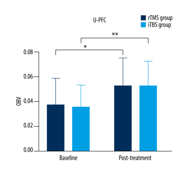
BACKGROUND Swallowing is a complex behavior involving the musculoskeletal system and higher-order brain functions. We investigated the effects of different modalities of repetitive transcranial magnetic stimulation (rTMS) on the unaffected hemisphere and observed correlation between suprahyoid muscle activity and cortical activation in unilateral stroke patients when swallowing saliva, based on functional near-infrared spectroscopy (fNIRS). MATERIAL AND METHODS From November 2022 to March 2023, twenty-five patients with unilateral stroke were screened using computed tomography or magnetic resonance imaging and identified via a video fluoroscopic swallow study. Finally, patients were divided into rTMS (n=10) and iTBS (n=10) groups. Both groups received 2 weeks of stimulation on unaffected suprahyoid motor cortex. Surface electromyographic measured peak amplitude and swallowing time of bilateral suprahyoid muscles, and penetration-aspiration scale was assessed at baseline and after treatment. fNIRS monitored oxyhemoglobin beta values (OBV) in the primary motor, sensory, and bilateral prefrontal cortex (PFC). RESULTS Both groups showed significant improvements in penetration-aspiration scale, peak amplitude, and swallowing time, compared with baseline (P<0.001), and increased OBV in unaffected regions (P<0.05), especially PFC (P<0.001). No significant OBV increases were seen in affected regions (P>0.05). After treatment, OBV in the unaffected PFC was significantly higher than in the unaffected primary sensory and motor cortex regions for both groups (P<0.05). No significant differences were observed between groups in outcome measures (P>0.05). CONCLUSIONS rTMS and iTBS significantly improved swallowing function in unilateral stroke, relying on compensation by the unaffected cortex, particularly the PFC. iTBS may outperform rTMS by shortening treatment sessions and improving efficiency.


Conflict of interest statement

Conflict of interest: None declared

Figures

Figure 1
Figure 1
Participant flow diagram of repetitive transcranial magnetic stimulation (rTMS) and intermittent theta burst stimulation (iTBS) groups. Created with www.home-for-researchers.com/index.html#/.
Figure 2
Figure 2
Configuration of fNIRS channels. ROI – region of interest; LPFC – left prefrontal cortex; RPFC – right prefrontal cortex; LM1 – left primary motor cortex; RM1 – right primary motor cortex; LS1 – left primary sensory cortex; RS1 – right primary sensory cortex. Created with www.home-for-researchers.com/index.html#/.
Figure 3
Figure 3
Experimental procedure of swallowing blocking with fNIRS. Created with www.home-for-researchers.com/index.html#/.
Figure 4
Figure 4
Comparison of oxyhemoglobin beta values (OBV) of affected prefrontal cortex (A-PFC) region in the 2 groups before and after treatment. rTMS – repetitive transcranial magnetic stimulation; iTBS – intermittent theta burst stimulation. The figure was created using GraphPad Prism version 9 for Windows (www.graphpad.com; San Diego, CA, USA).
Figure 5
Figure 5
Compared with oxyhemoglobin beta values (OBV) of affected primary motor cortex (A-M1) region in the 2 groups before and after treatment. rTMS – repetitive transcranial magnetic stimulation; iTBS – intermittent theta burst stimulation. The figure was created using GraphPad Prism version 9 for Windows (www.graphpad.com; San Diego, CA, USA).
Figure 6
Figure 6
Compared with oxyhemoglobin beta values (OBV) of affected primary sensory cortex (A-S1) region in the 2 groups before and after treatment. rTMS – repetitive transcranial magnetic stimulation; iTBS – intermittent theta burst stimulation. The figure was created using GraphPad Prism version 9 for Windows (www.graphpad.com; San Diego, CA, USA).
Figure 7
Figure 7
Comparison of oxyhemoglobin beta values (OBV) of the unaffected prefrontal cortex (U-PFC) region in the 2 groups before and after treatment. * P<0.05, ** P<0.001. rTMS – repetitive transcranial magnetic stimulation; iTBS – intermittent theta burst stimulation. The figure was created using GraphPad Prism version 9 for Windows (www.graphpad.com; San Diego, CA, USA).
Figure 8
Figure 8
Comparison of oxyhemoglobin beta values (OBV) of the unaffected primary motor cortex (U-M1) region in the 2 groups before and after treatment. * P<0.05. rTMS – repetitive transcranial magnetic stimulation; iTBS – intermittent theta burst stimulation. The figure was created using GraphPad Prism version 9 for Windows (www.graphpad.com; San Diego, CA, USA).
Figure 9
Figure 9
Comparison of oxyhemoglobin beta values (OBV) of the unaffected primary sensory cortex (U-S1) region in the 2 groups before and after treatment. * P<0.05. rTMS – repetitive transcranial magnetic stimulation; iTBS – intermittent theta burst stimulation. The figure was created using GraphPad Prism version 9 for Windows (www.graphpad.com; San Diego, CA, USA).
Figure 10
Figure 10
Comparison of oxyhemoglobin beta values (OBV) of the affected (A) and unaffected (U) prefrontal cortex (PFC), primary motor cortex (M1), and primary sensory cortex (S1) in the 2 groups after treatment. * P<0.05. rTMS – repetitive transcranial magnetic stimulation; iTBS – intermittent theta burst stimulation. The figure was created using NirSpark (www.hcmedx.cn; NirScan Danyang Huichuang Medical Equipment Co Ltd, Danyang, China) software.

Similar articles

References

    1. Qin Y, Tang Y, Liu X, Qiu S. Neural basis of dysphagia in stroke: A systematic review and meta-analysis. Front Hum Neurosci. 2023;17:1077234. - PMC - PubMed
    1. Kim DY, Park HS, Park SW, Kim JH. The impact of dysphagia on quality of life in stroke patients. Medicine (Baltimore) 2020;99(34):e21795. - PMC - PubMed
    1. Omari TI, Jones CA, Hammer MJ, et al. Predicting the activation states of the muscles governing upper esophageal sphincter relaxation and opening. Am J Physiol Gastrointest Liver Physiol. 2016;310(6):G359–66. - PMC - PubMed
    1. Al Rjoob M, Hassan NFHN, Aziz MAA, et al. Quality of life in stroke patients with dysphagia: A systematic review. Tunis Med. 2022;100(10):664–69. - PMC - PubMed
    1. Arnold M, Liesirova K, Broeg-Morvay A, et al. Dysphagia in acute stroke: Incidence, burden and impact on clinical outcome. PLoS One. 2016;11(2):e0148424. - PMC - PubMed