. 2025 Feb;21(7):e2407742.
doi: 10.1002/smll.202407742. Epub 2025 Jan 10.

Photochemical Stabilization of Self-Assembled Spherical Nucleic Acids

Affiliations

Photochemical Stabilization of Self-Assembled Spherical Nucleic Acids

Sepideh Kaviani et al. Small. 2025 Feb.

Abstract

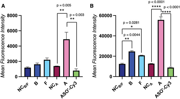
Oligonucleotide therapeutics, including antisense oligonucleotides and small interfering RNA, offer promising avenues for modulating the expression of disease-associated proteins. However, challenges such as nuclease degradation, poor cellular uptake, and unspecific targeting hinder their application. To overcome these obstacles, spherical nucleic acids have emerged as versatile tools for nucleic acid delivery in biomedical applications. Our laboratory has introduced sequence-defined DNA amphiphiles which self-assemble in aqueous solutions. Despite their advantages, self-assembled SNAs can be inherently fragile due to their reliance on non-covalent interactions and fall apart in biologically relevant conditions, specifically by interaction with serum proteins. Herein, this challenge is addressed by introducing two methods of covalent crosslinking of SNAs via UV irradiation. Thymine photodimerization or disulfide crosslinking at the micellar interface enhance SNA stability against human serum albumin binding. This enhanced stability, particularly for disulfide crosslinked SNAs, leads to increased cellular uptake. Furthermore, this crosslinking results in sustained activity and accessibility for release of the therapeutic nucleic acid, along with improvement in unaided gene silencing. The findings demonstrate the efficient stabilization of SNAs through UV crosslinking, influencing their cellular uptake, therapeutic release, and ultimately, gene silencing activity. These studies offer promising avenues for further optimization and exploration of pre-clinical, in vivo studies.

Keywords: crosslinking; delivery; disulfide; gene silencing; oligonucleotides; self‐assembly; spherical nucleic acids.


Conflict of interest statement

The authors declare no conflict of interest.

Figures

Figure 1
Figure 1
A) Structure of the DNA‐polymer conjugate non‐crosslinked control NCB/F used in this study with a thymidine repeat (light blue) for photocyclization, an endonuclease‐cleavable ATAT spacer* (red) and a FASO strand against firefly luciferase (dark blue). Capital letters = PO DNA, lower case letters = PS DNA, underlined = 2′‐fluoroarabinose (FANA). B) Structure of a DNA‐polymer conjugate containing 4 primary amines for conjugation with activated esters in solution or by resonant acoustic mixing (RAM) for subsequent ring‐opening disulfide polymerization. Because partial disulfide polymerization was observed with these methods, another non‐crosslinked control (NCA) was synthesized as a control non‐crosslinked compound.*When crosslinked with UVB/F, the thymines are covalently bound together and ATAT is used as the cleavable spacer.
Figure 2
Figure 2
A) Schematic representation of UV crosslinking of self‐assembled SNA via UVB or UVF irradiation (thymidine dimerization). B) Denaturing PAGE (16%) tracking UVB and UVF crosslinking over time and C) quantification of crosslinking yield over time by calculating the ratio of the intensity of all higher order bands on the gel to the intensity of all the bands in a lane. D) Schematic representation of UV crosslinking of self‐assembled SNA via UVA irradiation (1,2‐dithiolane ring‐opening polymerization). E) Denaturing PAGE (16%) tracking UVA crosslinking over time. F,G) Representative AFM images of non‐crosslinked control NCB/F (F) and UVB crosslinked SNAs (B SNA).
Figure 3
Figure 3
A,B) Normalized size exclusion chromatograms of non‐crosslinked control NCB/F and UVB crosslinked SNA (B SNA) SNA after incubation with HSA at 37 °C for 0 to 24 h. C) Possible outcomes from SNA binding to human serum albumin (HSA), either maintaining its integrity and binding to albumin or disassembling to DNA conjugates bound to albumin. D) Ratio of the protein‐coated SNA peak on SEC compared to the albumin‐bound portions for non‐crosslinked and crosslinked SNAs.
Figure 4
Figure 4
Mean fluorescence intensity measured by flow cytometry in HeLa CRM cells after incubation with 1 µm of SNA hybridized with 10% complementary ASO’‐Cy3 strands for A) 4 h and B) 24 h to demonstrate cellular uptake of crosslinked and non‐crosslinked SNAs with time. Error bars represent SD of duplicate experiments for each sample. ****: p < 0.0001; ***: p < 0.0002; **: p < 0.0021; *: p < 0.033; ns: p > 0.123.
Figure 5
Figure 5
Normalized fluorescence intensity measured by flow cytometry in HeLa CRM cells after co‐incubation of 1 µm of 10 % ASO’‐Cy3 hybridized SNA without (control) or with inhibitor polyinosinic acid (Poly I, 500 µg mL−1). The Cy3 unit was placed at the interface between the DNA corona and the core to avoid nuclease‐induced dissociation. The data were normalized for each sample type using its own control. For example, the uptake of inhibited SNA was normalized to the uptake of the same SNA without inhibition. Error bars represent SD of duplicate experiments for each sample. ****: p < 0.0001.
Figure 6
Figure 6
A) Schematic representation of the enzymatic cleavage of polythymidine spacer in the SNA and release of the gapmer FASO. B) Denaturing PAGE (16%) for assessment of FASO cleavage from tetrathymidine spacer after 0, 5, 15, 30, 45, or 60 min of incubation with DNAse I (4 U nmol−1 of SNA). C) Comparison of FASO release rate by measuring the band intensity of FASO as compared to all the other bands in each lane.
Figure 7
Figure 7
A) Firefly luciferase knockdown activity of crosslinked and non‐crosslinked SNAs and FASO (1 µm) after 72 h of incubation in HeLa X1/5 cells, without transfection agent. Each sample is normalized to the cell viability and the buffer control. Error bars represent SD of triplicate experiments for each sample. B) Firefly luciferase knockdown activity after incubation for 24 h in HeLa X1/5 cells with varying concentrations of non‐crosslinked and crosslinked samples (0–300 nm) with transfection using LipofectamineTM 3000. This data was used to extrapolate the IC50 of the different constructs. Error bars represent SD of duplicate experiments for each sample.

References

    1. Anwar S., Mir F., Yokota T., Pharmaceutics 2023, 15, 1130. - PMC - PubMed
    1. Bost J. P., Barriga H., Holme M. N., Gallud A., Maugeri M., Gupta D., Lehto T., Valadi H., Esbjorner E. K., Stevens M. M., ACS Nano 2021, 15, 13993. - PMC - PubMed
    1. Hammond S. M., Aartsma‐Rus A., Alves S., Borgos S. E., Buijsen R. A., Collin R. W., Covello G., Denti M. A., Desviat L. R., Echevarría L., EMBO Mol. Med. 2021, 13, 13243. - PMC - PubMed
    1. Huang S., Hao X.‐Y., Li Y.‐J., Wu J. Y., Xiang D.‐X., Luo S., Biomater. Res. 2022, 26, 49. - PMC - PubMed
    1. Samaridou E., Heyes J., Lutwyche P., Adv. Drug Delivery Rev. 2020, 154, 37. - PubMed

LinkOut - more resources