. 2023 Feb 27;1(2):226-230.
doi: 10.1016/j.atssr.2023.02.009. eCollection 2023 Jun.

Improved Right Ventricular Diastolic Function Assessed by Hepatic Vein Flow After Pectus Excavatum Repair

Affiliations

Improved Right Ventricular Diastolic Function Assessed by Hepatic Vein Flow After Pectus Excavatum Repair

Juan M Farina et al. Ann Thorac Surg Short Rep. .

Abstract

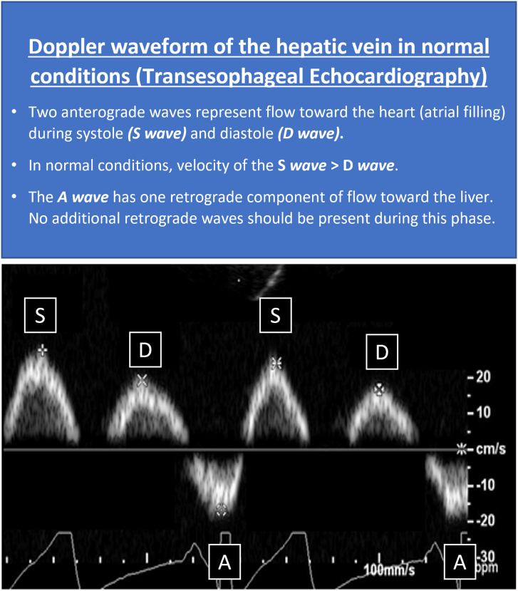
Background: The cardiovascular benefits of surgical repair in pectus excavatum (PEx) continue to be debated, with limited data supporting repair in adult patients. Hepatic vein flow is used to identify right-sided diastolic dysfunction in cardiovascular disorders, including tricuspid stenosis, cardiac tamponade, and constrictive pericarditis. This study evaluates the effects of cardiac compression on diastolic function (as assessed by hepatic vein flow patterns and velocities) before and after repair of PEx.

Methods: A retrospective study was performed of intraoperative transesophageal echocardiograms including hepatic vein assessments of adult patients who underwent preoperative and postoperative evaluations during repair of PEx from 2018 to 2021.

Results: In total, 127 patients were included (median age, 29.0 [15.4] years; median Haller index, 4.2 [1.7]; 60.6% male). Statistically significant improvements were seen after pectus repair of right ventricular stroke volume and diastolic function as measured by increased postoperative velocities for hepatic vein waves (P < .001 for all comparisons). Preoperatively, 5.5% of patients had constrictive-like physiology with end-diastolic retrograde flow that normalized postoperatively (P = .016). Approximately 10% of patients changed their pattern of hepatic vein flow after surgical procedure. Patients with more proximal cardiac compression had greater improvements in hepatic vein velocities after repair.

Conclusions: Surgical relief of cardiac compression resulted in an immediate improvement in hepatic vein flow and right ventricle stroke volume in patients with PEx. These results support diastolic dysfunction in a large number of patients, with improvement in function and compliance after the surgical relief of cardiac compression.


Figures

None
Graphical abstract
Figure 1
Figure 1
Classification of the patients according to the site of maximal cardiac compression. (A) Transesophageal echocardiography (TEE) and computed tomography (CT) showing no significant compression of right-sided heart chambers. (B) TEE and chest CT with contrast enhancement showing that the site of maximal compression is located at the level of the tricuspid annulus (proximal compression). (C) TEE and CT demonstrating compression of the right ventricle free wall and apex by left costochondral junction (distal compression).
Figure 2
Figure 2
Abnormalities in hepatic vein flow in patients with pectus. (A) A 23-year-old woman with pectus excavatum. Hepatic vein flow analysis at preoperative transesophageal echocardiography showed diastolic predominance (D wave > S wave), with low anterograde systolic velocities. This abnormal pattern in hepatic vein flow has been described in patients with obstruction to the right ventricle inflow tract, such as cases of tricuspid stenosis. Postoperatively, hepatic vein flow changed to a normal pattern (S wave > D wave) with increased systolic forward velocities. (B) A 44-year-old woman with pectus excavatum. Preoperative evaluation of the hepatic vein flow revealed the presence of an end-diastolic retrograde wave before atrial contraction (pre-A wave, arrow) in a manner similar to that seen in constrictive pericarditis. After repair, the end-diastolic retrograde wave was no longer present.
Supplemental Figure 1

References

    1. Jaroszewski D.E., Farina J.M., Gotway M.B., et al. Cardiopulmonary outcomes after the Nuss procedure in pectus excavatum. J Am Heart Assoc. 2022;11 doi: 10.1161/JAHA.121.022149. - DOI - PMC - PubMed
    1. Jaroszewski D.E., Velazco C.S., Pulivarthi V.S., Arsanjani R., Obermeyer R.J. Cardiopulmonary function in thoracic wall deformities: what do we really know? Eur J Pediatr Surg. 2018;28:327–346. doi: 10.1055/s-0038-1668130. - DOI - PubMed
    1. Raggio I.M., Martínez-Ferro M., Bellía-Munzón G., et al. Diastolic and systolic cardiac dysfunction in pectus excavatum: relationship to exercise and malformation severity. Radiol Cardiothorac Imaging. 2020;2 doi: 10.1148/ryct.2020200011. - DOI - PMC - PubMed
    1. Rodriguez-Granillo G.A., Raggio I.M., Deviggiano A., et al. Impact of pectus excavatum on cardiac morphology and function according to the site of maximum compression: effect of physical exertion and respiratory cycle. Eur Heart J Cardiovasc Imaging. 2020;21:77–84. doi: 10.1093/ehjci/jez061. - DOI - PubMed
    1. Appleton C.P., Hatle L.K., Popp R.L. Superior vena cava and hepatic vein Doppler echocardiography in healthy adults. J Am Coll Cardiol. 1987;10:1032–1039. doi: 10.1016/s0735-1097(87)80343-1. - DOI - PubMed

LinkOut - more resources