. 2025 Feb;103(4):1112-1125.
doi: 10.1177/13872877241309120. Epub 2025 Jan 10.

Impaired cerebral microvascular reactivity and endothelial SK channel activity in a streptozotocin-treated mouse model of Alzheimer's disease

Affiliations

Impaired cerebral microvascular reactivity and endothelial SK channel activity in a streptozotocin-treated mouse model of Alzheimer's disease

Hang Xing et al. J Alzheimers Dis. 2025 Feb.

Abstract

Background: Alzheimer's disease (AD) is a complex neurodegenerative disease marked by increased amyloid-β (Aβ) deposition, tau hyperphosphorylation, impaired energy metabolism, and chronic ischemia-type injury. Cerebral microvascular dysfunction likely contributes to AD pathology, but its precise pathogenic role has been poorly defined.

Objective: To examine microvascular reactivity to endothelium-dependent vasodilators and small conductance calcium-activated potassium (SK) channel activity in an intracerebral streptozotocin (STZ)-induced AD mouse model.

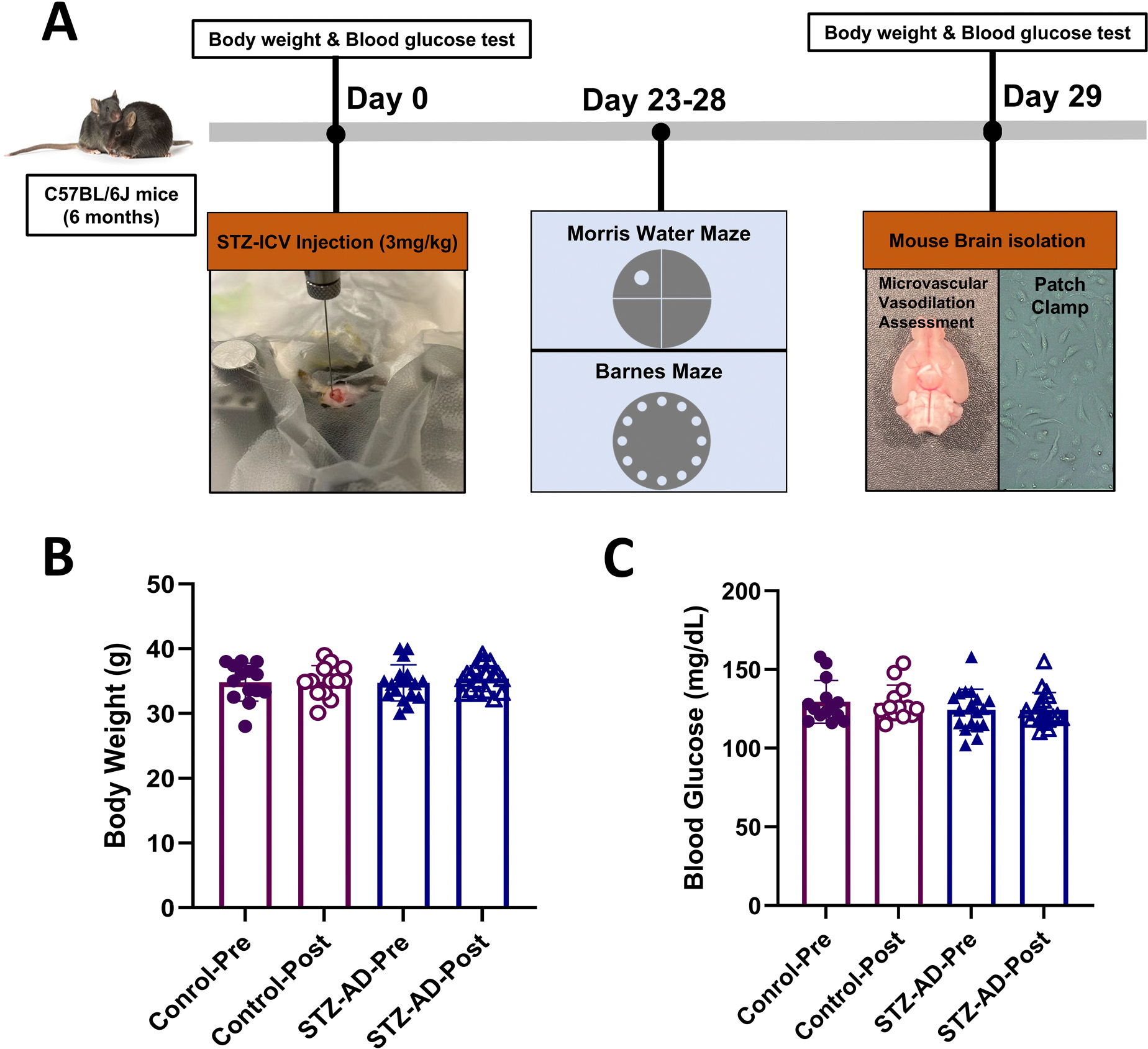
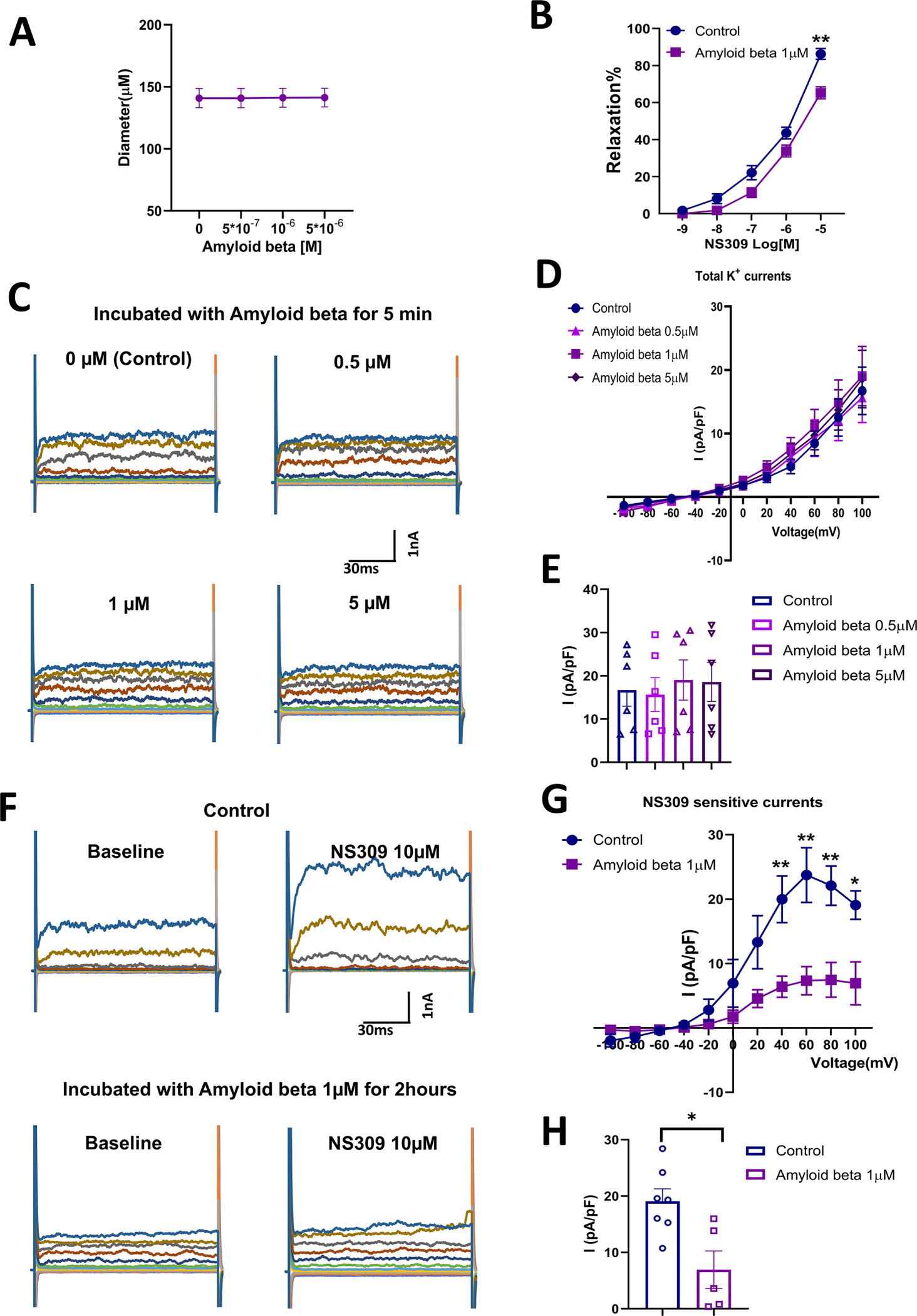
Methods: Control and STZ-AD mice underwent Morris Water Maze and Barnes testing, after which cerebral microvascular and brain microvascular endothelial cells (MBMECs) were dissected to assess microvascular reactivity, responses to SK channel activator NS309, and ion-channel current recordings using whole-cell patch clamp methodology. Control mouse cerebral microvascular and human brain microvascular endothelial cells (HBMECs) were treated with soluble Aβ1-42 peptide to characterize microvascular reactivity and endothelial potassium currents.

Results: STZ-AD mice exhibited impaired performance vs control mice in behavioral testing. STZ-AD mice also exhibited diminished cerebral microvascular responsiveness and MBMECs potassium current augmentation in response to NS309 compared with control mice. Incubation of control mouse cerebral micro-vessels and HBMECs with soluble Aβ (1 µM) for 2 h attenuated relaxation responses to NS309 and diminished NS309-sensitive endothelial potassium currents.

Conclusions: STZ-AD mice exhibited impaired microvascular relaxation responses to endothelium-dependent vasodilators; SK/IK channel dysfunction may be involved in the mechanism of this impairment. Acute treatment with Aβ produced dysregulated cerebrovascular endothelial SK/IK channels. Further elucidation of the role of microvascular dysfunction in AD is needed to prevent the chronic ischemia-type injury that contributes to cognitive decline.

Keywords: Alzheimer's disease; Morris water maze; NS309; SK channels; amyloid-β; cerebral endothelial dysfunction; cerebral microvascular dysfunction; streptozotocin.


Conflict of interest statement

Declaration of conflicting interestsThe authors declared no potential conflicts of interest with respect to the research, authorship, and/or publication of this article.

Figures

Figure 1.
Figure 1.
(A) Illustration of overall experimental design. STZ-AD mice received a single intracerebroventricular (ICV) injection of 3.0 mg/kg STZ in 3.0 μl cold citrate buffer (pH 4.5) into the right ventricle of the brain. Control mice also received ICV injection of 3.0 μl cold citrate buffer (pH 4.5). Twenty-three days after ICV injection, the mice were subjected to Morris water maze testing over the course of 5 days. Immediately after this, on day 29, all mice were anesthetized by inhaled isoflurane, after which a decapitation was performed, and brains were removed from the cranium. Brains were either placed in cold artificial cerebrospinal fluid (ACSF) buffer for in vitro microvascular experiments, or preserved in cell culture medium in preparation for endothelial cell isolation. (B) The mean body weights of control and STZ-AD mice were not significantly different before and after ICV-injection. (C) Blood glucose levels among control and STZ-AD mice were not significantly different before and after ICV-injection. Data are shown as mean ± SD.
Figure 2.
Figure 2.
The Morris water maze (A–I) test: (A) Representative paths taken by mice on the first trial on days 1–5. (B) Escape latencies during days 1–5. (C) Mean speed during testing on days 1–5. (D) Path efficiency during days 1–5. (E) Ratio of time spent in the target quadrant to total duration in the maze during days 1–5. (F) Rotation times during days 1–5. (G–I) Time in target quadrant (%), mean speed (m/s), and times of crossing platform zone during day 6 of probe trial without platform. Visible platform, day 1; hidden platform, days 2–5. Barnes maze (J-M): (J) Escape latency across three consecutive days of training (Day 1, Day 2, and Day 3) measured in seconds, comparing Control and STZ-AD groups. (K) Representative probe trail images showing the path taken by Control and STZ-AD mice during the Barnes maze test. (L) Percentage of time spent in the target quadrant on Day 5 during the probe trials, with a significant reduction observed in STZ-AD mice compared to Control (*p < 0.05). (M) Distance traveled (in meters) during the probe trials on Day 5, showing no significant difference between Control and STZ-AD groups. Data are shown as the mean ± SEM, with a sample size of n = 14 for Male control and n = 18 for Male STZ-AD in the water maze test. For the Barnes maze test, there were n = 4 in the Male control and n = 5 in the Male STZ-AD group. p values were obtained using two-way ANOVA followed post hoc by Tukey’s test. **p < 0.01.
Figure 3.
Figure 3.
Expression of p-tau and soluble amyloid in the cortex and hippocampus of mice across groups. (A) Western blot analysis of p-tau. (B) Quantification of p-tau Ser 396, p-tau Ser 202/Thr 205, and p-tau Thr 181 band density. (C, D) Demonstration of amyloid deposits by Congo red staining of mouse hippocampal sections. A representative sample is shown for normal mouse brains and for STZ-AD brains. Arrows indicate amyloid plaques. The bar graph represents the significantly higher amyloid plaque count in the STZ-AD group when compared with control animals. (E, F) Quantitative assessment of soluble Aβ levels in the lysates of the hippocampus and cortex using the Elisa. The levels of Aβ in STZ-AD were significantly higher than that in control (p < 0.01). Data are shown as the mean ± SEM, n = 4 in Control, n = 3 in STZ-AD; **p < 0.01. p values were obtained using two-tailed Student’s t tests.
Figure 4.
Figure 4.
(A) In vitro cerebral microvascular reactivity studies determined that the vascular relaxation response to increasing concentrations of the SK/IK channel activator NS309 was significantly impaired in STZ-AD male mice (n = 6) compared with control counterparts (n = 6). (B) In contrast, there was no substantial change in the vascular relaxation response to incubation with increasing concentrations of the endothelium-independent vasodilator SNP between STZ-AD (n = 6) and control mice (n = 6). (C–E) The selective small-/intermediate-conductance calcium-activated potassium (SK/IK) channel activator NS309 increased whole-cell K + currents in MBMECs from STZ-AD and control mice. (C) Representative current traces were obtained under control conditions in the presence of NS309 in STZ-AD (n = 8) and control (n = 6) animals. (D, E) Current-voltage plot of currents shown in C demonstrated reduced current in STZ-AD mice. Data are shown as mean ± SEM. *p < 0.05. p values were obtained using two-way ANOVA with post hoc Tukey’s test.
Figure 5.
Figure 5.
(A) Incubation of cerebral micro-vessels isolated from control male mice (n = 6) with soluble Aβ for 5 min did not result in a significant change in vessel diameter. (B) Soluble Aβ (1 μM) pre-treatment of cerebral micro-vessels isolated from mice resulted in an attenuated relaxation in response to the SK/IK channel activator NS309 (n = 6 in control group and n = 6 in Aβ treatment group). (C–E) Total K+ currents were not significantly altered following a five-minute incubation with soluble Aβ at various concentrations (0 μM, 0.5 μM, 1 μM, 5 μM) in HBMECs (n = 6). (F–H) Incubation of HBMECs in soluble Aβ (1 μM) for 2 h resulted in impaired SK currents (n = 7) when compared to controls that were not pre-treated with soluble Aβ (n = 5). Data are shown as mean ± SEM. *p < 0.05, **p < p-values were obtained using two-way ANOVA with post hoc Tukey’s test.
Figure 6.
Figure 6.
Expression of SK3 and SK4 across different groups. (A) Western blot analysis of SK3 and SK4 in mouse cortical and hippocampal tissue. (B, C) Quantification of SK3 and SK4 band density derived from this tissue. (D) Western blot analysis of SK3 and SK4 in control and Aβ-treated HBMECs. (E, F) Quantification of SK3 and SK4 band density in control and Aβ treated HBMECs. Data are shown as mean ± SEM. N = 6 in each group.

Similar articles

Cited by

References

    1. Alzheimer’s disease facts and figures. Alzheimer’s Disease and Dementia. https://www.alz.org/alzheimers-dementia/facts-figures#prevalence (accessed 18 March 2023)
    1. Wong W Economic burden of Alzheimer disease and managed care considerations. Am J Manag Care 2020; 26: S177–S183. - PubMed
    1. Digma LA, Madsen JR, Reas ET, et al. Tau and atrophy: domain-specific relationships with cognition. Alzheimer Res Ther 2019; 11: 65. - PMC - PubMed
    1. Tiwari S, Atluri V, Kaushik A, et al. Alzheimer’s disease: pathogenesis, diagnostics, and therapeutics. Int J Nanomed 2019; 14: 5541–5554. - PMC - PubMed
    1. Fan L, Mao C, Hu X, et al. New insights into the pathogenesis of Alzheimer’s disease. Front Neurol 2020; 10: 1312. - PMC - PubMed

MeSH terms

Substances

LinkOut - more resources