. 2024 Dec 27;14(1):17.
doi: 10.3390/cells14010017.

Unravelling Secondary Brain Injury: Insights from a Human-Sized Porcine Model of Acute Subdural Haematoma

Affiliations

Unravelling Secondary Brain Injury: Insights from a Human-Sized Porcine Model of Acute Subdural Haematoma

Thomas Kapapa et al. Cells. .

Abstract

Traumatic brain injury (TBI) remains one of the leading causes of death. Because of the individual nature of the trauma (brain, circumstances and forces), humans experience individual TBIs. This makes it difficult to generalise therapies. Clinical management issues such as whether intracranial pressure (ICP), cerebral perfusion pressure (CPP) or decompressive craniectomy improve patient outcome remain partly unanswered. Experimental drug approaches for the treatment of secondary brain injury (SBI) have not found clinical application. The complex, cellular and molecular pathways of SBI remain incompletely understood, and there are insufficient experimental (animal) models that reflect the pathophysiology of human TBI to develop translational therapeutic approaches. Therefore, we investigated different injury patterns after acute subdural hematoma (ASDH) as TBI in a post-hoc approach to assess the impact on SBI in a long-term, human-sized porcine TBI animal model. Post-mortem brain tissue analysis, after ASDH, bilateral ICP, CPP, cerebral oxygenation and temperature monitoring, and biomarker analysis were performed. Extracerebral, intraparenchymal-extraventricular and intraventricular blood, combined with brainstem and basal ganglia injury, influenced the experiment and its outcome. Basal ganglia injury affects the duration of the experiment. Recognition of these different injury patterns is important for translational interpretation of results in this animal model of SBI after TBI.

Keywords: acute subdural haematoma; animal model; guidelines; intracranial pressure; management; multimodal brain monitoring; outcome; secondary brain injury; traumatic brain injury.


Conflict of interest statement

Author Claire Hartmann conducted all experiments as an employed physician of the University Hospital Ulm. After finishing the series of experiment, she was employed by the company Boehringer Ingelheim Pharma. The remaining authors declare that the research was conducted in the absence of any commercial or financial relationships that could be construed as a potential conflict of interest. The Boehringer Ingelheim Pharma had no role in the design of the study; in the collection, analyses, or interpretation of data; in the writing of the manuscript, or in the decision to publish the results.

Figures

Figure 1
Figure 1
Macroscopic findings of a pig brain removed after acute subdural hematoma (ASDH) with the view (A) from above, (B) from the right, (C) from the left side and (D) in coronal sections from frontal to sub-occipital. In A–C fronto-parietal cortex after implantation of the neuro-monitoring probes and the right-sided ASDH. In (D), blood deposits on the right side of the cortex as evidence of intraparenchymal bleeding. (E,F): Exemplary courses of intracranial pressure (ICP) and partial oxygen pressure (PtO2, PO2) in mmHg over time for the right side (with ASDH) and the left side (control) of the animals analysed. There was an increase in ICP values and a decrease in the PtO2 values for the right ASDH hemisphere and subsequently for the control side after ASDH is applied (E). In the further course (F), ICP was significantly higher on the right than on the left side and the continuous drop of PtO2 values was more pronounced on the right than on the left.
Figure 2
Figure 2
Injury pattern and distribution of haemorrhage after the experiment: (A) only extracerebral, (B) intraparenchymal–extraventricular, (C) intraventricular, (D) brain stem involvement (arrow) and (E) basal ganglia involvement (arrow).
Figure 3
Figure 3
Median and interquartile representation of the duration of the experiment (survival of the animal) according to the division into (A) haemorrhage distributions, (B) occurrence of brainstem injuries and (C) occurrence of basal ganglia injuries. The significance of the difference between the injury patterns with and without basal ganglia injury in (C) is p = 0.0001 = *** (Mann–Whitney U test).
Figure 4
Figure 4
The exclusion of the animals from the experiment and the length of the experiment after application of the acute subdural hematoma. (A) There was a difference of animals with basal ganglia lesions and without basal ganglia injuries (p = 0.004, Log rank (Mantel–Cox)). (B) Animals with intraventricular blood distribution dropped out earlier than other animals, followed by animals with intraparenchymal blood distribution (p = 0.228, Log rank (Mantel–Cox). (C) Animals with brainstem lesions dropped out earlier than those without (p = 0.072, Log rank (Mantel–Cox). The dotted lines show the corresponding 95% confidence interval.
Figure 5
Figure 5
Total modified Glasgow Coma Scale scores (MGCS) over time (3 = minimum, 18 = maximum), showing scores at 4, 30 and 54 h of the current study (AC). In general, the animals show a deterioration in scores after the application of trauma (acute subdural haematoma), in this case at hour 30, and then a recovery. Both non-extracerebral damage and damage to the brainstem and basal ganglia resulted in lower scores than without such damage. This suggests a clinical equivalent of brain damage.
Figure 6
Figure 6
Body temperature of animals differentiated by (A) occurrence of basal ganglia injury, (B) occurrence of brainstem injuries and (C) different haemorrhage distribution. Results of the mixed-model approach (restricted maximum likelihood, REML): animals without basal ganglia injury showed higher body temperature than animals with basal ganglia injuries (p = 0.007) (A). The distribution for brainstem injuries and haemorrhage type revealed no significant results. The conditional R2 values are 0.489 for basal ganglia injury, 0.612 for brainstem injury and 0.609 for haemorrhage distributions.
Figure 7
Figure 7
Time course of the neuromonitoring values over the experiment (0 to 54 h) separated by intraparenchymal and intraventricular haemorrhage. Extracerebral (N = 3) cases were excluded due to early dropout and the low number of values. (A) Intracranial pressure = ICP, (B) cerebral perfusion pressure = CPP, (C) partial tissue (brain) oxygen saturation = PtO2, (D) brain temperature in grade Celsius. ICP 8 h: haemorrhage side and control side and ICP 24 h: haemorrhage side, p ≤ 0.029 (**); Mann–Whitney-U Test (A). CPP 24 h: haemorrhage side, p = 0.05 (**) and CPP 48 h: control side, p = 0.046 (**); Mann–Whitney-U Test (B).
Figure 8
Figure 8
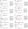
Time course of the neuromonitoring values over the experiment (0 to 54 h) separated by the occurrence of brainstem (left) and basal ganglia (right) injury. (A,B) Intracranial pressure = ICP, (C,D) cerebral perfusion pressure = CPP, (E,F) partial tissue (brain) oxygen saturation = PtO2, and (G,H) brain temperature in grade Celsius. Significant differences in animals with and without basal ganglia injury: ICP, p = 0.044 (12 h, control side) (B), CPP, p = 0.027 (24 h, control side) (D), PtO2, p = 0.044 (12 h, control side) and 0.017 (36 h, haemorrhage side) (F), temperature, p = 0.012 (24 h, haemorrhage side) and p = 0.002 (24 h, control side) (H). Significant differences in animals with and without brainstem injury: temperature, p = 0.036 (36 h, haemorrhage side) (G).
Figure 9
Figure 9
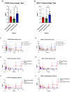
Time course of the biomarkers (1): S110ß and, (2): MAP2 separated based on the injury patterns. (A,D) Haemorrhage type, (B,E) occurrence of brainstem injuries and (C,F) occurrence of basalganglia injuries. The biomarkers S100ß (p = 0.0153 = *, Kruskal–Wallis-H) and MAP2 (p = 0.0126 = *, Kruskal–Wallis-H) showed significantly higher cumulative concentrations in animals with intraventricular injuries.
Figure 10
Figure 10
Time course of the biomarkers (1): NSE and (2): GFAP separated based on the injury patterns. (A,D) Haemorrhage type, (B,E) occurrence of brainstem injuries and (C,F) occurrence of basalganglia injuries. The biomarker GFAP (p = 0.0003 = ***, Kruskal–Wallis-H) showed significantly higher cumulative concentrations in animals with intraventricular injuries.

References

    1. Guan B., Anderson D.B., Chen L., Feng S., Zhou H. Global, regional and national burden of traumatic brain injury and spinal cord injury, 1990–2019: A systematic analysis for the Global Burden of Disease Study 2019. BMJ Open. 2023;13:e075049. doi: 10.1136/bmjopen-2023-075049. - DOI - PMC - PubMed
    1. Dewan M.C., Rattani A., Gupta S., Baticulon R.E., Hung Y.C., Punchak M., Agrawal A., Adeleye A.O., Shrime M.G., Rubiano A.M., et al. Estimating the global incidence of traumatic brain injury. J. Neurosurg. 2019;130:1080–1097. doi: 10.3171/2017.10.JNS17352. - DOI - PubMed
    1. GBD 2021 Nervous System Disorders Collaborators Global, regional, and national burden of disorders affecting the nervous system, 1990–2021: A systematic analysis for the Global Burden of Disease Study 2021. Lancet Neurol. 2024;23:344–381. doi: 10.1016/S1474-4422(24)00038-3. - DOI - PMC - PubMed
    1. Maas A.I.R., Menon D.K., Manley G.T., Abrams M., Akerlund C., Andelic N., Aries M., Bashford T., Bell M.J., Bodien Y.G., et al. Traumatic brain injury: Progress and challenges in prevention, clinical care, and research. Lancet Neurol. 2022;21:1004–1060. doi: 10.1016/S1474-4422(22)00309-X. - DOI - PMC - PubMed
    1. Daugherty J., Waltzman D., Sarmiento K., Xu L. Traumatic Brain Injury-Related Deaths by Race/Ethnicity, Sex, Intent, and Mechanism of Injury—United States, 2000–2017. MMWR Morb Mortal Wkly Rep. 2019;68:1050–1056. doi: 10.15585/mmwr.mm6846a2. - DOI - PMC - PubMed

Publication types

LinkOut - more resources