Review
. 2025 Jan 4;14(1):49.
doi: 10.3390/cells14010049.

Mechanisms of Rhodopsin-Related Inherited Retinal Degeneration and Pharmacological Treatment Strategies

Affiliations
Review

Mechanisms of Rhodopsin-Related Inherited Retinal Degeneration and Pharmacological Treatment Strategies

Maria Azam et al. Cells. .

Abstract

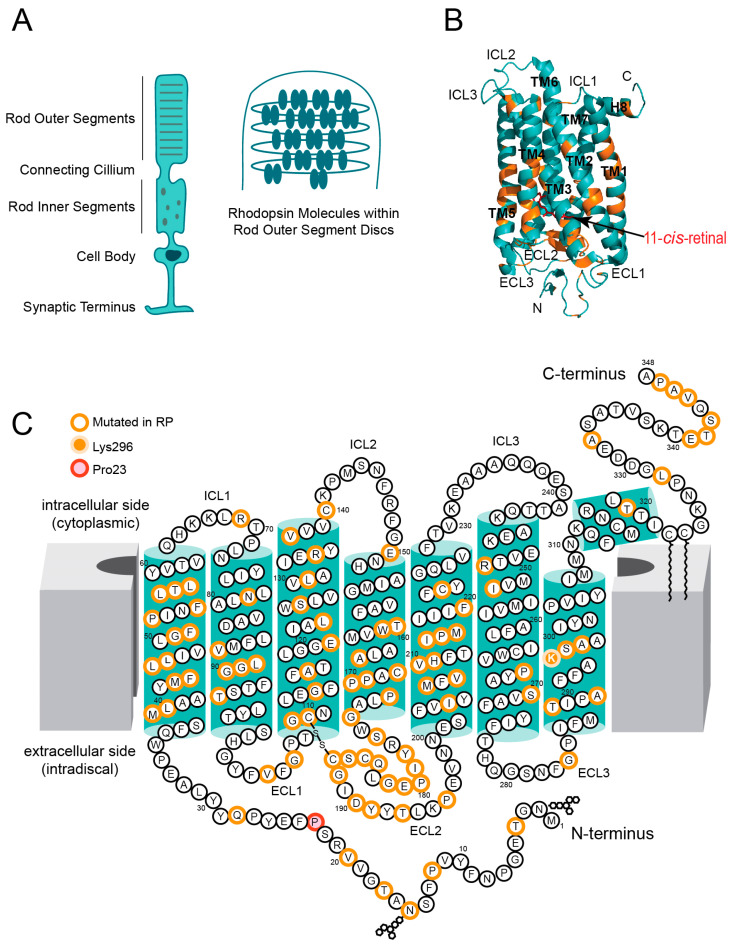
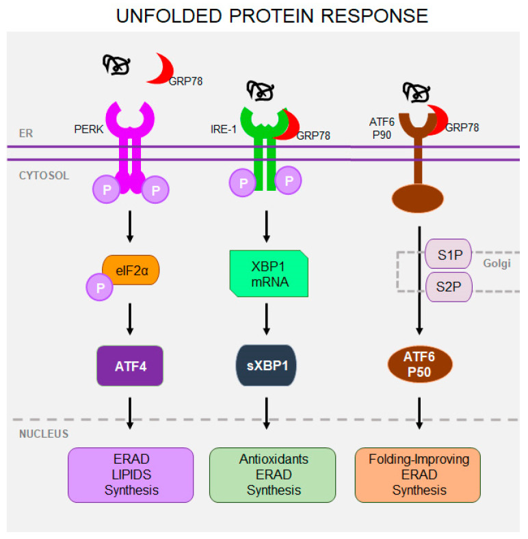
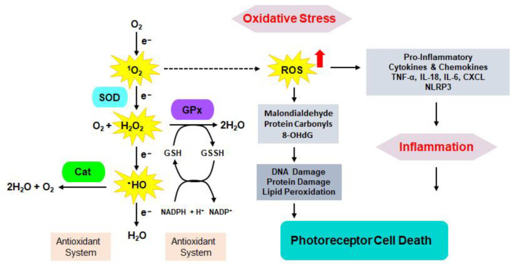
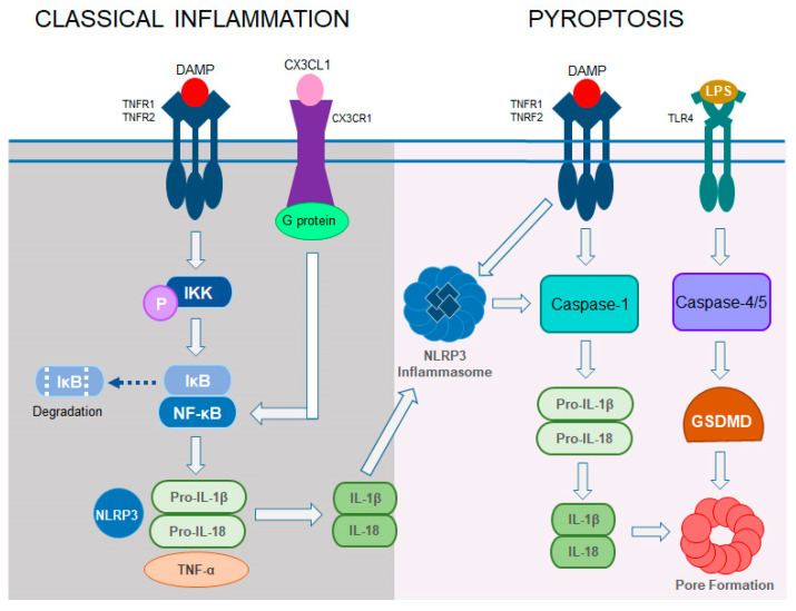
Retinitis pigmentosa (RP) is a hereditary disease characterized by progressive vision loss ultimately leading to blindness. This condition is initiated by mutations in genes expressed in retinal cells, resulting in the degeneration of rod photoreceptors, which is subsequently followed by the loss of cone photoreceptors. Mutations in various genes expressed in the retina are associated with RP. Among them, mutations in the rhodopsin gene (RHO) are the most common cause of this condition. Due to the involvement of numerous genes and multiple mutations in a single gene, RP is a highly heterogeneous disease making the development of effective treatments particularly challenging. The progression of this disease involves complex cellular responses to restore cellular homeostasis, including the unfolded protein response (UPR) signaling, autophagy, and various cell death pathways. These mechanisms, however, often fail to prevent photoreceptor cell degradation and instead contribute to cell death under certain conditions. Current research focuses on the pharmacological modulation of the components of these pathways and the direct stabilization of mutated receptors as potential treatment strategies. Despite these efforts, the intricate interplay between these mechanisms and the diverse causative mutations involved has hindered the development of effective treatments. Advancing our understanding of the interactions between photoreceptor cell death mechanisms and the specific genetic mutations driving RP is critical to accelerate the discovery and development of therapeutic strategies for this currently incurable disease.

Keywords: misfolding; neuroinflammation; oxidative stress; photoreceptor; retinal degeneration; rhodopsin.


Conflict of interest statement

The authors declare no conflicts of interest.

Figures

Figure 1
Figure 1
Schematic rod photoreceptor and rhodopsin structure. (A) The schematic representation of the rod photoreceptor cell (left panel) and a close-up of rod outer segment disc membranes with rhodopsin (Rho) molecules. (B) The structure of bovine Rho. The PDB ID:1GZM was used to show the side view of bovine Rho in the dark state. Transmembrane helices are labeled TM1–7. Cytoplasmic helix 8 is labeled H8. Extracellular (intradiscal) loops connecting TM helices on the ligand-binding site of the receptor are labeled ECL1, ECL2, and ECL3. Intracellular (cytoplasmic) loops, connecting TM helices on the effector binding site of the receptor are labeled ICL1, ICL2, and ICL3. 11-cis-retinal is shown as red sticks. The location of residues mutated in retinitis pigmentosa (RP) is shown in orange. (C) Two-dimensional representation of human Rho structure. Residues mutated in RP are indicated with orange circles. The Lys296, which covalently binds the 11-cis-retinal, is shown with a yellow circle filled with orange. The P23H mutation is shown with a red circle filled with orange.
Figure 2
Figure 2
Unfolded protein response. The unfolded protein response (UPR) involves three primary sensor receptors within the ER membranes: protein kinase RNA-like ER kinase (PERK), inositol-requiring enzyme 1 (IRE1), and activating transcription factor 6 (ATF6). PERK phosphorylates eIF2α, which reduces protein translation and upregulates ATF4 transcription factor, which activates the expression of antioxidants and components of the ER-associated degradation ERAD signaling. Activated by unfolded proteins, IRE1 activates transcription factor sXBP1 which stimulates the synthesis of protein folding regulators, ERAD, and lipid biosynthesis. ATF6 (P90), upon activation, translocates to the Golgi apparatus, where it is cleaved to P50 form by proteases S1P and S2P. Cleaved ATF6 stimulates the expression of ERAD and folding regulators.
Figure 3
Figure 3
Schematic interplay between oxidative stress, inflammation, and photoreceptor cell death. Oxidative radicals are generated during respiration in mitochondria. Under normal physiological conditions, superoxide dismutase (SOD) catalyzes superoxide radicals (1O2) into hydrogen peroxide (H2O2) and oxygen (O2), while catalase breaks down hydroxyl radicals ·OH to water (H2O) and O2. H2O2 is converted by glutathione peroxidase to H2O. During this reaction, GSH is converted to its reduced form GSSH. The back conversion of GSSH → GSH involves NADPH → NAD+ change. Excess of reactive oxygen species (ROS) accumulated under chronic conditions of genetic mutation leads to damage of cellular content and release of pro-inflammatory markers that aggravate inflammation, ultimately leading to cell death.
Figure 4
Figure 4
Classical inflammation and pyroptosis signaling. In classical inflammation, damage-associated molecular patterns (DAMPs) activate phosphorylation of IκB kinase (IKK), which degrades IκB from the IκB/NFκB complex leading to the activation of NFκB. Activated NFκB stimulates the expression of proinflammatory cytokines, including IL-1β, IL-18, and TNF-α, as well as the expression of NLRP3, which leads to the formation of inflammasome. In addition, chemokine receptor CX3CR1 activated by CX3CL1 stimulates NFκB through G protein signaling. Pyroptosis is activated by DAMPs through death receptors; for example, tumor necrosis factor receptors (TNFR1 and TNRF2), which stimulate the expression of NOD-like receptor protein 3 (NLRP3) and inflammasome formation that activates caspase-1, which activates IL-1β and IL-18. Alternatively, pyroptosis is activated through Toll-like receptor 4 (TLR4) stimulated by bacterial lipopolysaccharides (LPS). Caspase-4 and -5 are activated in this pathway leading to the activation of gasdermin (GSDMD), which inserts into the membrane forming a pore that allows for the release of pro-inflammatory cytokines activated by caspase-1.
Figure 5
Figure 5
Apoptosis pathway. Extrinsic apoptosis is activated by extrinsic signals through death receptors (TNFRs), which recruit adaptor proteins like the Fas-associated death domain (FADD), followed by pro-caspase-8 activation. Active caspase-8 directly stimulates executioner caspase-3 and -7, leading to apoptosis. Caspase-8 can also stimulate BID, which activates BAX and BAK to permeabilize the mitochondrial membrane, linking the extrinsic and intrinsic pathways. The intrinsic pathway is activated by cellular stressors like damaged DNA or oxidative stress, which activates BAX and BAK. Permeabilized mitochondria release cytochrome c, which binds to apoptotic protease activating factor APAF1 and triggers activation of caspase-9 followed by activation of executioner caspase-3 and -7.
Figure 6
Figure 6
Necroptosis signaling. Necroptosis is triggered by the activation of death receptors, particularly tumor necrosis factor receptor 1 (TNFR1) upon binding of TNF-α. It could also be activated by Toll-receptor 4 (TLR4). TNFR1 recruits adaptor proteins TRADD, TRAF2, and RIPK1. In apoptosis, receptor-interacting protein kinase-1 (RIPK1) is polyubiquitinated and promotes cell survival. When caspase-8 is blocked, RIPK1 interacts with RIPK3, forming a necrosome complex. RIPK3 autophosphorylates and then phosphorylates mixed-lineage kinase domain-like protein (MLKL), a necroptosis key effector, which isomerizes and translocates to the membrane where it forms a pore enabling the release of cellular content. This can further lead to the activation of inflammatory response through released DAMPs.
Figure 7
Figure 7
Ferroptosis signaling. Cellular iron is imported via the transferrin receptor (TFR1), which binds Fe3+ (ferric iron)-loaded transferrin. Inside the cell, Fe3+ became reduced to Fe2+ (ferrous iron). Free Fe2+ can catalyze the Fenton reaction leading to the generation of reactive oxygen species (ROS) production, which oxidizes unsaturated membrane phospholipids. Under normal physiological conditions, an antioxidant system involving glutathione peroxidase (GPx) prevents lipid peroxidation using its cofactor GSH, which is generated in exchange for glutamate transported out of the cell through the antiporter SLC7A11. Under chronic stress of pathogenic mutations, unchecked lipid peroxidation disrupts membrane integrity and leads to photoreceptor cell death.

Similar articles

Cited by

References

    1. Kolb H. Simple Anatomy of the Retina. In: Kolb H., Fernandez E., Jones B., Nelson R., editors. Webvision: The Organization of the Retina and Visual System. University of Utah Health Sciences Center; Salt Lake City, UT, USA: 1995.
    1. Wong-Riley M.T. Energy metabolism of the visual system. Eye Brain. 2010;2:99–116. doi: 10.2147/EB.S9078. - DOI - PMC - PubMed
    1. Sakami S., Imanishi Y., Palczewski K. Muller glia phagocytose dead photoreceptor cells in a mouse model of retinal degenerative disease. FASEB J. 2019;33:3680–3692. doi: 10.1096/fj.201801662R. - DOI - PMC - PubMed
    1. Ramirez A.I., de Hoz R., Salobrar-Garcia E., Salazar J.J., Rojas B., Ajoy D., Lopez-Cuenca I., Rojas P., Trivino A., Ramirez J.M. The Role of Microglia in Retinal Neurodegeneration: Alzheimer’s Disease, Parkinson, and Glaucoma. Front. Aging Neurosci. 2017;9:214. doi: 10.3389/fnagi.2017.00214. - DOI - PMC - PubMed
    1. Jastrzebska B., Debinski A., Filipek S., Palczewski K. Role of membrane integrity on G protein-coupled receptors: Rhodopsin stability and function. Prog. Lipid Res. 2011;50:267–277. doi: 10.1016/j.plipres.2011.03.002. - DOI - PMC - PubMed

Publication types

MeSH terms

LinkOut - more resources