. 2025 Jan 6;14(1):56.
doi: 10.3390/cells14010056.

Small Extracellular Vesicles Promote Axon Outgrowth by Engaging the Wnt-Planar Cell Polarity Pathway

Affiliations

Small Extracellular Vesicles Promote Axon Outgrowth by Engaging the Wnt-Planar Cell Polarity Pathway

Samar Ahmad et al. Cells. .

Abstract

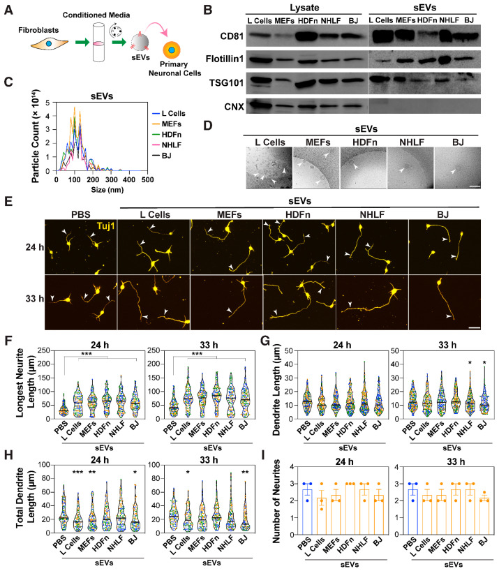
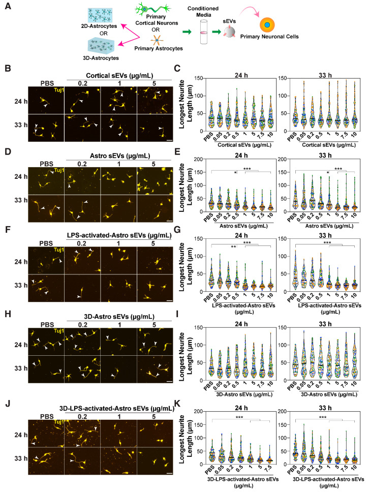
In neurons, the acquisition of a polarized morphology is achieved upon the outgrowth of a single axon from one of several neurites. Small extracellular vesicles (sEVs), such as exosomes, from diverse sources are known to promote neurite outgrowth and thus may have therapeutic potential. However, the effect of fibroblast-derived exosomes on axon elongation in neurons of the central nervous system under growth-permissive conditions remains unclear. Here, we show that fibroblast-derived sEVs promote axon outgrowth and a polarized neuronal morphology in mouse primary embryonic cortical neurons. Mechanistically, we demonstrate that the sEV-induced increase in axon outgrowth requires endogenous Wnts and core PCP components including Prickle, Vangl, Frizzled, and Dishevelled. We demonstrate that sEVs are internalized by neurons, colocalize with Wnt7b, and induce relocalization of Vangl2 to the distal axon during axon outgrowth. In contrast, sEVs derived from neurons or astrocytes do not promote axon outgrowth, while sEVs from activated astrocytes inhibit elongation. Thus, our data reveal that fibroblast-derived sEVs promote axon elongation through the Wnt-PCP pathway in a manner that is dependent on endogenous Wnts.

Keywords: axon outgrowth; cortical neurons; extracellular vesicles; neurite outgrowth; planar cell polarity.


Conflict of interest statement

The authors declare no conflicts of interest.

Figures

Figure 1
Figure 1
sEVs promote the growth of the prospective axon. (A) A schematic of the experimental set up. Mouse cortical neurons (E15.5-16.5) are treated with sEVs isolated from fibroblast-conditioned media (CM) using differential centrifugation. (B) Representative immunoblotting of cell lysates and sEV pellets (100,000× g) from the indicated fibroblast cell lines for EV markers CD81, Flotillin1, and TSG101, and the ER protein, calnexin (CNX). (C) Nanoparticle tracking analysis (NTA) of differential centrifugation pellets. A representative plot indicating the particle size distribution from three independent purifications is shown. (D) Representative transmission electron microscopy (TEM) images of sEV-containing pellets. Arrowheads indicate round vesicles. Scale bar, 200 nm. (EI) Cortical neurons were treated with sEVs (5 μg/mL) purified from the indicated fibroblast cell lines, 4 h after plating. Neurons were fixed at 24 and 33 h, and neuronal morphology was examined after staining for Tuj1. (E) Representative images are shown. Arrowheads mark the longest neurite. Scale bar, 40 μm. (FI) The length of the longest neurite (prospective axon; F), individual neurite/dendrite lengths (G), total dendrite length (longest neurite excluded; H), and total number of neurites (I) were quantified from a minimum of 90 neurons per condition from three independent experiments. Neurite lengths are plotted as a violin plot with values from each experiment distinctly colored and the median marked by a black line (F,G,H). The number of neurites is plotted as the average of the median ± SEM (I), where each dot represents the median from 30 neurons from one of the three independent experiments. Statistical significance: * p < 0.05, ** p < 0.01, *** p < 0.001 using one-way ANOVA with Dunnett’s post-test.
Figure 2
Figure 2
sEVs promote the growth of the prospective axon. The PCP components, Pk1/2 and Vangl2, are required for sEV-induced growth of the longest neurite. (AC,EG) Pk1/2 and Vangl2 promote sEV-induced neurite outgrowth. Dissociated E15.5-16.5 mouse cortical neurons were electroporated with siRNA against Pk1 (siPk1) and Pk2 (siPk2) (AC), or Vangl1 (siVangl1) and Vangl2 (siVangl2) (EG) individually or in combination or with siControl (siCtl) along with a GFP-expressing plasmid and were treated with sEVs (5 μg/mL) from L cells, 4 h after plating. Neurons were fixed at 24 and 33 h, and neuronal morphology was examined in GFP-positive neurons. (A,E) Representative images are shown. Arrowheads mark the longest neurite. Scale bar, 40 μm. (B,F) The length of the longest neurites was quantified. (C,G) Knockdown efficiency for Pk1/Pk2 (C) and Vangl1/2 (G) was determined in GFP-positive neurons isolated by FACS. Relative mRNA expression was determined by qPCR. (D,I) Pk1/2 and Vangl2 promote sEV-induced neurite outgrowth in mutant mouse models. Cortical neurons (E15.5-16.5) were isolated from Pk1 and Pk2 conditional knockout mice obtained by crossing Pk floxed mice with a Nestin-Cre line (D) or Vangl2 mutant littermates obtained by crossing heterozygous loop-tail mutants (Vangl2+/Lp) (I). Neurons were treated with sEVs from L cells, 4 h after plating, fixed at 24 and 33 h, and morphology examined in Tuj1 stained neurons. The length of the longest neurite was quantified from 40 neurons per embryo, and the total number of embryos analyzed is indicated below the genotypes. Violin plots with the median marked by a black line are shown. (H) Loop-tail embryos (E15.5-E16.5) exhibit an open neural tube. Representative images are shown from a minimum of four independent experiments. Arrowheads mark the open neural tube. In siRNA experiments, neurite lengths are quantified from a minimum of 90 neurons per condition from three independent experiments and plotted as a violin plot with values from each experiment distinctly colored and the median marked by a black line (B,F). For qPCR plots, data is presented as the mean ± SEM from 3 independent experiments (C,G). Statistical significance: * p < 0.05, ** p < 0.01, *** p < 0.001 using one-way ANOVA with Dunnett’s post-test (C,G), or two-way ANOVA with Tukey’s post-test (B,D,F,I).
Figure 3
Figure 3
sEVs promote the growth of the prospective axon. (AD) Dissociated E15.5-16.5 mouse cortical neurons were treated with sEVs (5 μg/mL) from L cells, 4 h after plating. (A,B) Neurons were co-treated with IgG or an Fzd blocking antibody, F2.A (at 50 nM and 100 nM), along with sEVs. (C,D) Neurons were electroporated with siRNA against Fzds (siFzds) or siControl in combination with a GFP-expressing plasmid prior to the addition of sEVs. Neurons were fixed at 24 and 33 h, and neuronal morphology was examined in Tuj1 stained (A,B) or GFP-positive neurons (C,D), with representative images shown. Arrowheads mark the longest neurite. Scale bar, 40 μm. (BD) The length of the longest neurite was quantified. Neurite lengths are quantified from a minimum of 90 neurons per condition from three independent experiments and plotted as a violin plot with values from each experiment distinctly colored, and the median marked by a black line (B,D). Statistical significance: *** p < 0.001 using one-way ANOVA with Dunnett’s post-test (B), or two-way ANOVA with Tukey’s post-test (D).
Figure 4
Figure 4
sEVs promote the growth of the prospective axon. (A,B) Dissociated E15.5-16.5 mouse cortical neurons were treated with sEVs (5 μg/mL) from L cells, 4 h after plating. Neurons were electroporated with siRNA against Dvls (siDvl) or siControl in combination with a GFP-expressing plasmid prior to the addition of sEVs. Neurons were fixed at 24 and 33 h, and neuronal morphology was examined in GFP-positive neurons, with representative images shown (A). Arrowheads mark the longest neurite. Scale bar, 40 μm. (B) The length of the longest neurite was quantified. (C,D) sEVs promote localization of Vangl2 to the distal axon. Cortical neurons were treated with sEVs from L cells, 4 h after plating and fixed after 24 h. Representative confocal images of neurons stained with DAPI (blue), Vangl2 (green), and Tuj1 (red) are shown. Arrowheads mark the Vangl2 localization. Scale bar, 50 μm. (D) The ratio of distal/proximal Vangl2 intensity and the relative intensity of Vangl2 in the soma is quantified from 30 neurons from three independent experiments. Neurite lengths are quantified from a minimum of 90 neurons per condition from three independent experiments and plotted as a violin plot with values from each experiment distinctly colored, and the median marked by a black line (B). For Vangl2 plots, data are presented as the mean ± SEM from 3 independent experiments (D). Statistical significance: ** p < 0.01, *** p < 0.001 using unpaired t-test (D), or two-way ANOVA with Tukey’s post-test (B).
Figure 5
Figure 5
Neuronal Wnts mediate sEV-induced growth of the longest neurite. (A,E) Schematics illustrating the experimental setup. (AG) Dissociated E15.5-16.5 mouse cortical neurons were treated with sEVs isolated from L cells transfected with siCtl or siPorcupine (AD) or with sEVs isolated from regular L cells (EG). Neurons were treated with PBS as a control and with Porcupine (Porcn) inhibitors, IWP2 (10 μM) or LGK974 (1 and 5 μM) (EG), 4 h after plating and co-incubated with sEVs. (HL) Cortical neurons were electroporated with siRNAs against Wls (siWls) (H,I) or Wnts (siWnts) (K,L) or siControl (siCtl) along with a GFP-expressing plasmid and then treated with sEVs (5 μg/mL) from L cells, 4 h after plating. Neurons were fixed at 24 and 33 h, and neuronal morphology was examined in Tuj1 stained neurons (B,C,F,G) or GFP-positive neurons for siRNA experiments (H,I,K,L). Representative images (B,F,H,K) and quantifications of the longest neurite (C,G,I,L) are shown. Arrowheads mark the longest neurite. Scale bar, 40 μm. (D,J) Knockdown efficiency for Porcupine (D) and Wls (J) was determined in L cells and GFP-positive neurons isolated by FACS, respectively. Relative mRNA expression was determined by qPCR. Neurite lengths are quantified from a minimum of 90 neurons per condition from three independent experiments and plotted as a violin plot with values from each experiment distinctly colored and the median marked by a black line (C,G,I,L). For all other plots, data is presented as the mean ± SEM from three independent experiments (D,J). Statistical significance: *** p < 0.001 using unpaired t-test (D,J), one-way ANOVA with Dunnett’s post-test (C,G), or two-way ANOVA with Tukey’s post-test (I,L).
Figure 6
Figure 6
sEVs promote neurite elongation and can colocalize with Wnt7b. (AC) sEVs promote neurite elongation. (A) A schematic illustration of the two-compartment Xona microfluidic device. The somal compartment is connected to the axonal compartment through a 150 μm microgroove. (B) Cortical neurons (E15.5-16.5) were seeded in the somal compartment and cultured for 5 days prior to the addition of L cell-derived sEVs in either the somal or axonal compartment. Neurons were fixed 24 h later, and neuronal morphology was examined in Tuj1 stained neurons. Representative images are shown. Scale bar, 200 μm. (C) The length of the neurites growing in the microgroove and emerging in the axonal compartment was quantified for a minimum of 90 neurites. A dotted line marks both ends of the microgroove (150 μm). (DI) sEVs can be internalized by neurons and colocalize with Wnts. Cortical neurons were treated with 10X concentrated conditioned media (CM) from L cells stably expressing CD81-EYFP, 4 h after plating for 29 h (F) or 24 h after plating for 30 min (GI). In panel (G), after 30 min of treatment, neurons were washed and subsequently treated with regular complete media for 0, 2, 4, and 24 h. Representative images of neurons immunostained with GFP and Tuj1 (F,G) or GFP, Tuj1, and Wnt7b (H) are shown. Dashed boxes (H) indicate higher magnification of neurons. Arrowheads mark GFP puncta of internalized sEVs. Scale bar, 20 μm (F) or 40 μm (G,H). (E) Characterization of sEVs. The concentrated CM (10X) and sEV pellet from L cells were immunoblotted with anti-GFP antibody. (I) Pearson’s colocalization coefficient for (H). Neurons were identified using Tuj1 as a reference channel, and the colocalization coefficient was quantified using Nikon NIS-Elements software. Images (F,G,H) and the quantification (I) are representative of 30 neurons from 3 independent experiments. In all the violin plots, values are distinctly colored for each experiment, and the median is marked by a black line. Statistical significance: *** p < 0.001 using unpaired t-test (I) or one-way ANOVA with Dunnett’s post-test (C).
Figure 7
Figure 7
sEVs from neurons and astrocytes do not promote the growth of the longest neurite. (A) A schematic illustration of the experimental set up. Dissociated E15.5-16.5 cortical neurons were treated with sEVs purified from primary cortical neurons or primary astrocytes. (BK) Cortical neurons were treated with various concentrations (0.05–10 μg/mL) of sEVs purified from cortical neurons (B,C), astrocytes (D,E), LPS-activated astrocytes (F,G), 3D-astrocytes grown in collagen gel (H,I), and LPS-activated 3D-astrocytes (J,K) 4 h after plating. Neurons were fixed at 24 and 33 h, and neuronal morphology was examined in Tuj1 stained neurons. Representative images (B,D,F,H,J) and quantifications (C,E,G,I,K) are shown. Arrowheads mark the longest neurite. Scale bar, 40 μm. Neurite lengths are quantified from a minimum of 90 neurons per condition from 3 independent experiments and plotted as a violin plot, with values from each experiment distinctly colored and the median marked by a black line. Statistical significance: * p < 0.05, ** p < 0.01, *** p < 0.001 using one-way ANOVA with Dunnett’s post-test.
Figure 8
Figure 8
A model depicting the effect of sEVs in promoting axon outgrowth and polarized neuronal morphology through Wnt-PCP signaling. sEVs secreted by L cells engage Wnt-PCP signaling in neurons to promote axon outgrowth that results in the acquisition of a polarized neuronal morphology. sEVs induce a shift in Vangl2 localization towards the distal axon. sEVs can be internalized by neurons and can colocalize with Wnt7b to promote the growth of the prospective axon. In contrast to fibroblast-derived sEVs, those isolated from activated astrocytes inhibit neurite outgrowth.

References

    1. Takano T., Funahashi Y., Kaibuchi K. Neuronal Polarity: Positive and Negative Feedback Signals. Front. Cell Dev. Biol. 2019;7:69. doi: 10.3389/fcell.2019.00069. - DOI - PMC - PubMed
    1. Arimura N., Kaibuchi K. Neuronal polarity: From extracellular signals to intracellular mechanisms. Nat. Rev. Neurosci. 2007;8:194–205. doi: 10.1038/nrn2056. - DOI - PubMed
    1. Namba T., Funahashi Y., Nakamuta S., Xu C., Takano T., Kaibuchi K. Extracellular and Intracellular Signaling for Neuronal Polarity. Physiol. Rev. 2015;95:995–1024. doi: 10.1152/physrev.00025.2014. - DOI - PubMed
    1. Schelski M., Bradke F. Neuronal polarization: From spatiotemporal signaling to cytoskeletal dynamics. Mol. Cell Neurosci. 2017;84:11–28. doi: 10.1016/j.mcn.2017.03.008. - DOI - PubMed
    1. Bradke F. Mechanisms of Axon Growth and Regeneration: Moving between Development and Disease. J. Neurosci. 2022;42:8393–8405. doi: 10.1523/JNEUROSCI.1131-22.2022. - DOI - PMC - PubMed

Publication types

Grants and funding

LinkOut - more resources