. 2025 Jan 1;15(1):56.
doi: 10.3390/nano15010056.

Detonation Nanodiamond Soot-A Structurally Tailorable Hybrid Graphite/Nanodiamond Carbon-Based Material

Affiliations

Detonation Nanodiamond Soot-A Structurally Tailorable Hybrid Graphite/Nanodiamond Carbon-Based Material

Tikhon S Kurkin et al. Nanomaterials (Basel). .

Abstract

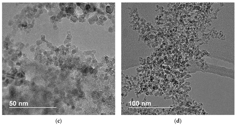
The results of a comprehensive investigation into the structure and properties of nanodiamond soot (NDS), obtained from the detonation of various explosive precursors (trinitrotoluene, a trinitrotoluene/hexogen mixture, and tetryl), are presented. The colloidal behavior of the NDS particles in different liquid media was studied. The results of the scanning electron microscopy, dynamic light scattering, zeta potential measurements, and laser diffraction analysis suggested a similarity in the morphology of the NDS particle aggregates and agglomerates. The phase composition of the NDS nanoparticles was studied using X-ray diffraction, Raman spectroscopy, electron diffraction, transmission electron microscopy, atomic force microscopy, and scanning tunneling microscopy. The NDS particles were found to comprise both diamond and graphite phases. The ratio of diamond to graphite phase content varied depending on the NDS explosive precursor, while the graphite phase content had a significant impact on the electrical conductivity of NDS. The study of the mechanical and tribological characteristics of polymer nanocomposites, modified with the selected NDS particles, indicated that NDS of various types can serve as a viable set of model nanofillers.

Keywords: graphite nanoribbons; nanodiamond; nanodiamond soot; nanoparticles; polymer nanocomposites.


Conflict of interest statement

The authors declare no conflicts of interest.

Figures

Figure 1
Figure 1
SEM images of the NDS powders: (a,b)—NDS-1; (c,d)—NDS-2; (e,f)—NDS-3.
Figure 1
Figure 1
SEM images of the NDS powders: (a,b)—NDS-1; (c,d)—NDS-2; (e,f)—NDS-3.
Figure 2
Figure 2
Distributions of hydrodynamic diameter of the (a,d,g,j) NDS-1, (b,e,h) NDS-2, and (c,f,i,k) NDS-3 nanoparticles in (ac) ethanol, (df) isopropanol, (gi) DMSO, and (j,k) acetone, obtained using the DLS technique.
Figure 3
Figure 3
Results of zeta potential measurements for (a) NDS-1, (b) NDS-2, and (c) NDS-3 nanoparticles dispersed in different media versus the time between the ultrasonication procedure and the measurement of zeta potential of the NDS aggregates/agglomerates in the dispersions.
Figure 4
Figure 4
Distributions of diameter of the NDS particles in a suspension of NDS powders in water, obtained with the LD method for all the investigated NDS powders.
Figure 5
Figure 5
TEM images of the NDS powders: (a)—NDS-1; (b)—NDS-2; (c,d)—NDS-3.
Figure 5
Figure 5
TEM images of the NDS powders: (a)—NDS-1; (b)—NDS-2; (c,d)—NDS-3.
Figure 6
Figure 6
XRD patterns (CuKα-radiation) for various NDS types in (a) I − 2θ and in (b) I(s)s2dss coordinates. The peaks that are colored blue correspond to nanographite, while the peaks that are colored purple correspond to detonation nanodiamonds. Dashed vertical lines in (a) indicate the centers of the peaks.
Figure 7
Figure 7
ND weight fraction values in the NDS powders of various types calculated using integration of the corresponding peaks in I − 2θ (“XRD”) and I(s)s2dss (“XRD s2”) coordinates. Literature data were taken from the publication of Dolmatov et al. [22].
Figure 8
Figure 8
Raman spectroscopy results for NDSs of different types. Dotted lines represent the obtained experimental data, and solid lines are peaks from decomposition of the experimental data. Dashed vertical lines indicate the centers of the peaks.
Figure 9
Figure 9
Electron diffraction results for the (a) NDS-1, (b) NDS-2, and (c) NDS-3. The insets in the diffractograms show distributions of normalized intensity along the dashed yellow lines. The yellow arrows point at the scattering peaks, corresponding to the diamond 111 and 022, and graphite 002 planes of carbon atoms.
Figure 10
Figure 10
(ac) Topography maps, (df) tunnel current distributions, (gi) overlays of the corresponding tunnel current distributions with the topography maps, and (jl) voltage-current curves obtained using the STM method for the (a,d,g,j) NDS-1, (b,e,h,k) NDS-2, and (c,f,i,l) NDS-3. The voltage-current curves correspond to the points indicated with the same number and color in the overlayed tunnel current maps (gi).
Figure 11
Figure 11
(a) Schematic image of the apparatus used for measuring specific resistivity of the NDS powders. (b) Conductivity of the NDS powders of various types versus volume fraction of the NDS nanoparticles in the measurement cell.
Figure 12
Figure 12
The assumed packaging of the ND (a hexagonal with a triangle inside) and nanographite (a hexagonal with three lines inside) subunits for the (a) NDS-1, (b) NDS-2, and (c) NDS-3 nanoparticles.
Figure 13
Figure 13
Dependencies of the friction coefficient on the ND content in the NDS powder (Table 2) in nanocomposites based on PP modified with (a) 2.5 and (b) 10 wt.% of NDS of various types. The curves were obtained for different durations (1 and 120 min) of annealing at 205 °C.
Figure 14
Figure 14
Dependence of the tensile strength on the homogeneous shear deformation ratio for the composites based on disentangled UHMWPE reactor powder and obtained using the solid-state processing approach without (“UHMWPE”) and with the addition of 10 wt.% NDS of various types.

References

    1. Laurila T., Sainio S., Caro M.A. Hybrid carbon based nanomaterials for electrochemical detection of biomolecules. Prog. Mater. Sci. 2017;88:499–594. doi: 10.1016/j.pmatsci.2017.04.012. - DOI
    1. Navrotskaya A.G., Aleksandrova D.D., Krivoshapkina E.F., Sillanpää M., Krivoshapkin P. V Hybrid Materials Based on Carbon Nanotubes and Nanofibers for Environmental Applications. Front. Chem. 2020;8:546. doi: 10.3389/fchem.2020.00546. - DOI - PMC - PubMed
    1. He Z., Zhao Z., Xiao S., Yang J., Zhong M. Preparation of carbon-based hybrid particles and their application in microcellular foaming and flame-retardant materials. RSC Adv. 2018;8:26563–26570. doi: 10.1039/C8RA03007C. - DOI - PMC - PubMed
    1. Ferrero G.A., Sevilla M., Fuertes A.B. Free-standing hybrid films based on graphene and porous carbon particles for flexible supercapacitors. Sustain. Energy Fuels. 2017;1:127–137. doi: 10.1039/C6SE00047A. - DOI
    1. Kiany P., Goharpey F., Karimi M., Foudazi R. Hybrids of Bowl-like and Crumpled Hollow Carbon Particles Synthesized through Encapsulation Templating. Langmuir. 2020;36:130–140. doi: 10.1021/acs.langmuir.9b01835. - DOI - PubMed

LinkOut - more resources