

Infectious keratitis following photorefractive keratectomy: a 13-year study at a tertiary center
- PMID: 39792283
- PMCID: PMC11723873
- DOI: 10.1186/s12348-025-00452-2
Infectious keratitis following photorefractive keratectomy: a 13-year study at a tertiary center
Abstract
Introduction: Infectious keratitis is a rare but devastating complication following photorefractive keratectomy (PRK) that may lead to visual impairment. This study assessed the clinical features, treatment strategies, and outcomes of post-PRK infectious keratitis.
Methods: This retrospective study was conducted on patients with post-PRK infectious keratitis presenting to Khalili Hospital, Shiraz, Iran, from June 2011 to March 2024. The study was conducted in two stages: the first stage assessed the incidence of post-PRK infectious keratitis among patients who underwent PRK at our center, while the second stage included all patients with post-PRK infectious keratitis, regardless of where their PRK was performed. The following data were collected: demographics, post-surgery presentation time, risk factors, culture results, treatments, follow-up duration, complications, and corrected distance visual acuity (CDVA) at admission and the last follow-up.
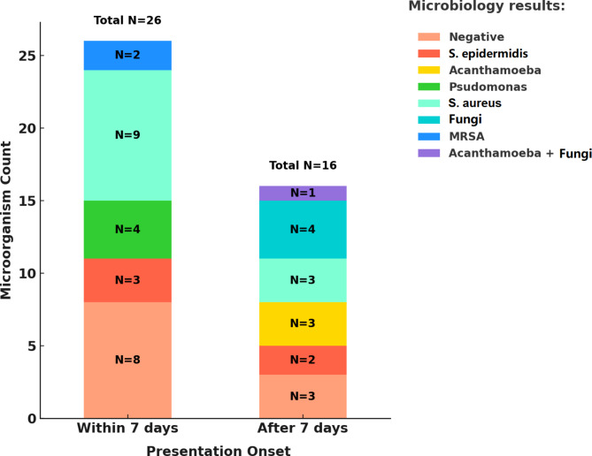
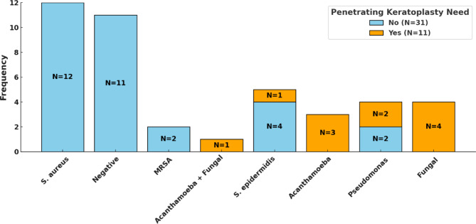
Results: Forty-two patients (42 eyes) with a mean age of 28.74 years (male-to-female ratio of 1.2:1) were included. Among 38,938 PRK procedures performed at our center, the incidence of keratitis was estimated to be 0.018% (7/38,938). The odds of keratitis during the COVID-19 pandemic were 7.05 times higher (95% CI: 1.58 to 31.52, p-value = 0.015) than outside this timeframe (February 2020 to August 2023). Gram-positive bacteria were the most commonly isolated pathogens in microbiological studies, accounting for 45.2% (19/42) of cases. Early-onset infections were primarily caused by Staphylococcus aureus (9/26, 34.6%), Staphylococcus epidermidis (4/26, 15.4%), and Pseudomonas aeruginosa (4/26, 15.4%), whereas all of the cases with fungi (4/4, 100% (and Acanthamoeba (3/3, 100%) infections caused late-onset infections. All patients received broad-spectrum antibiotic therapy, followed by adjusted treatment based on microbial results. Cases developing endophthalmitis and those not responding to treatment or having non-resolving corneal scars required further interventions, such as penetrating keratoplasty and deep vitrectomy. The mean follow-up duration was 40.81 months, and 97.6% (41/42) of cases experienced CDVA improvement at follow-up.
Conclusion: This long-term study found a post-PRK keratitis rate of 0.018%, with gram-positive bacteria as the most common pathogens. Prompt management and regular follow-up assessments are essential for achieving satisfactory outcomes.
Keywords: COVID-19; Infectious keratitis; Management; PRK; Photorefractive Keratectomy; Refractive surgery; Risk factors; Visual acuity.
© 2025. The Author(s).
Conflict of interest statement
Declarations. Ethics approval and consent to participate: The current study was conducted in accordance with the principles of the Declaration of Helsinki, and the study protocol received approval from the Ethics Committee of Shiraz University of Medical Sciences, Shiraz, Iran (ethics code: IR.SUMS.MED.REC.1402.381). Written informed consent was obtained from all participants included in the study. Consent for publication: Not applicable. Conflict of interest: All authors declared that there are no conflicts of interest.
Figures
References
-
- Flitcroft D (2012) The complex interactions of retinal, optical and environmental factors in myopia aetiology. Prog Retin Eye Res 31(6):622–660 - PubMed
-
- Holden BA, Fricke TR, Wilson DA, Jong M, Naidoo KS, Sankaridurg P et al (2016) Global prevalence of myopia and high myopia and temporal trends from 2000 through 2050. Ophthalmology 123(5):1036–1042 - PubMed
-
- Vitale S, Sperduto RD, Ferris FL (2009) Increased prevalence of myopia in the United States between 1971–1972 and 1999–2004. Arch Ophthalmol 127(12):1632–1639 - PubMed
-
- Kim T-i, del Barrio Alió, Wilkins JL, Cochener M, Ang B (2019) Refractive surgery. Lancet 393(10185):2085–2098 - PubMed
-
- Alexander JK, Davidson RS (2016) Managing expectations in refractive surgery. Int Ophthalmol Clin 56(2):1–17 - PubMed
LinkOut - more resources
Full Text Sources
