

Familial Mediterranean fever with sigmoid colon stricture
- PMID: 39792323
- PMCID: PMC11923005
- DOI: 10.1007/s12328-025-02095-1
Familial Mediterranean fever with sigmoid colon stricture
Abstract
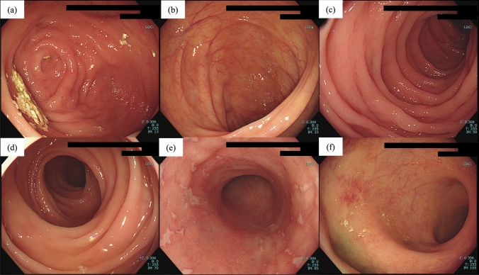
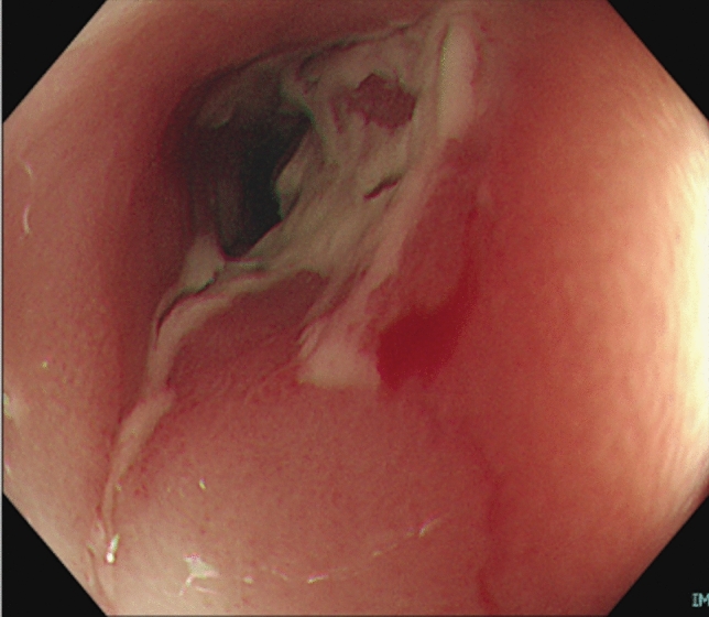
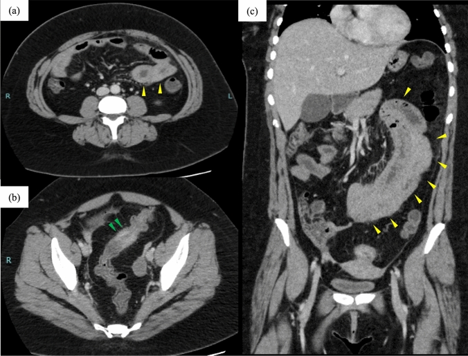
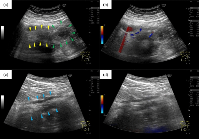
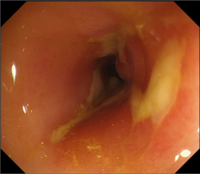
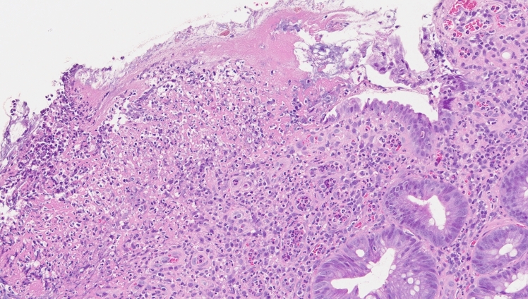
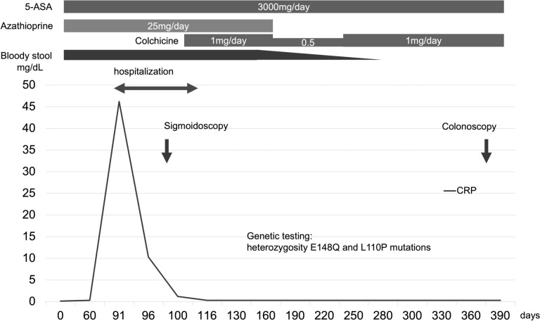
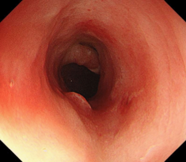
We describe a case of familial Mediterranean fever (FMF) with sigmoid colon stricture. The patient, a woman in her 30 s, had a 12-year history of ileocolitis-type Crohn's disease. The colonoscope could not pass because of the sigmoid colon stricture, and the patient was referred to our hospital with complaints of abdominal pain and fever. At 2-month postreferral, the patient presented with severe abdominal pain and fever. Computed tomography and intestinal ultrasonography revealed no bowel obstruction, whereas wall thickening was observed in the sigmoid colon and small bowel. Our medical interview revealed a cyclical nature to the symptoms. We diagnosed FMF and initiated colchicine. Subsequently, for more than 2 years, the patient remained asymptomatic, and the sigmoid colon stricture improved. FMF should be considered in patients with inflammatory bowel disease with periodic abdominal pain and fever.
Keywords: Colchicine; Crohn’s disease; Familial Mediterranean fever; Inflammatory bowel disease; Intestinal stricture.
© 2025. The Author(s).
Conflict of interest statement
Declarations. Conflict of interest: The authors declare that they have no conflicts of interest. Human/animal rights: All procedures followed were in accordance with the ethical standards of the responsible committee on human experimentation (institutional and national) and with the Helsinki Declaration of 1964, as revised in 2013. Informed consent: This study does not contain identifying information of the patient.
Figures
Similar articles
-
Familial Mediterranean Fever with Small Bowel Stenosis.Intern Med. 2019;58(14):2025-2028. doi: 10.2169/internalmedicine.2293-18. Epub 2019 Jul 15. Intern Med. 2019. PMID: 31308342 Free PMC article.
-
Adhesive small-bowel obstruction as a challenging complication of familial Mediterranean fever: A case-based review.Int J Rheum Dis. 2024 Jan;27(1):e14867. doi: 10.1111/1756-185X.14867. Epub 2023 Aug 14. Int J Rheum Dis. 2024. PMID: 37575017 Review.
-
Familial Mediterranean Fever without Fever.Intern Med. 2020 May 15;59(10):1267-1270. doi: 10.2169/internalmedicine.3175-19. Epub 2020 Feb 12. Intern Med. 2020. PMID: 32051376 Free PMC article.
-
Regular abdominal pain and fever in each menstruation onset: an unusual menses-associated familial Mediterrenean fever attacks and a favor result on colchicine treatment.Rheumatol Int. 2006 Jun;26(8):760-1. doi: 10.1007/s00296-005-0055-6. Epub 2005 Sep 23. Rheumatol Int. 2006. PMID: 16179998
-
Familial Mediterranean fever--a not so unusual cause of abdominal pain.Best Pract Res Clin Gastroenterol. 2005 Apr;19(2):199-213. doi: 10.1016/j.bpg.2004.11.009. Best Pract Res Clin Gastroenterol. 2005. PMID: 15833688 Review.
References
-
- Alghamdi M. Familial Mediterranean fever, review of the literature. Clin Rheumatol. 2017;36:1707–13. - PubMed
-
- Migita K, Uehara R, Nakamura Y, et al. Familial Mediterranean fever in Japan. Medicine. 2012;91:337–43. - PubMed
-
- Fidder HH, Chowers Y, Lidar M, et al. Crohn disease in patients with familial Mediterranean fever. Medicine. 2002;81:411–6. - PubMed
-
- Cattan D, Notarnicola C, Molinari N, et al. Inflammatory bowel disease in non-Ashkenazi Jews with familial Mediterranean fever. Lancet. 2000;355:378–9. - PubMed
Publication types
MeSH terms
Substances
LinkOut - more resources
Full Text Sources
Medical
