. 2025 Apr;197(4):2713-2733.
doi: 10.1007/s12010-024-05141-9. Epub 2025 Jan 10.

Metformin Alleviates Doxorubicin-Induced Cardiotoxicity via Preserving Mitochondrial Dynamics Balance and Calcium Homeostasis

Affiliations

Metformin Alleviates Doxorubicin-Induced Cardiotoxicity via Preserving Mitochondrial Dynamics Balance and Calcium Homeostasis

Nashwa Maghraby et al. Appl Biochem Biotechnol. 2025 Apr.

Abstract

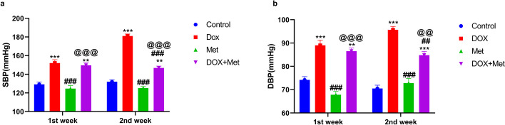
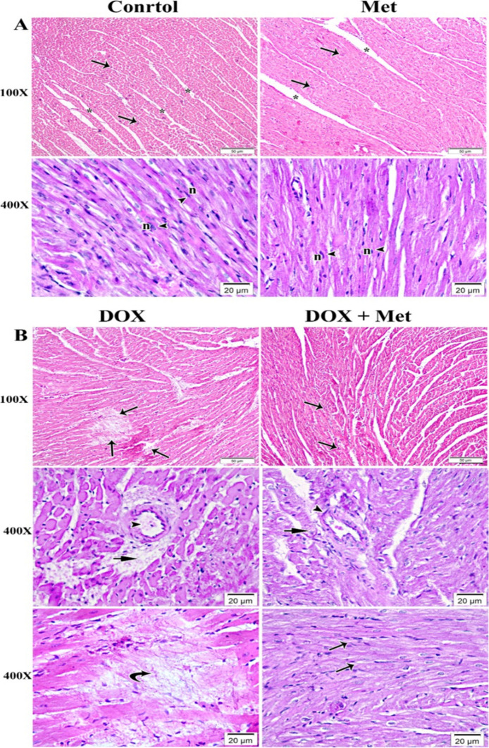
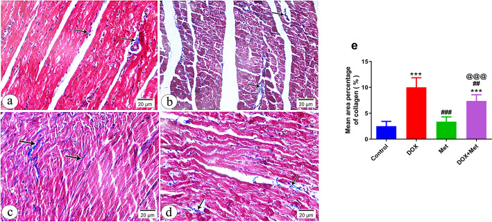
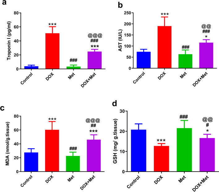
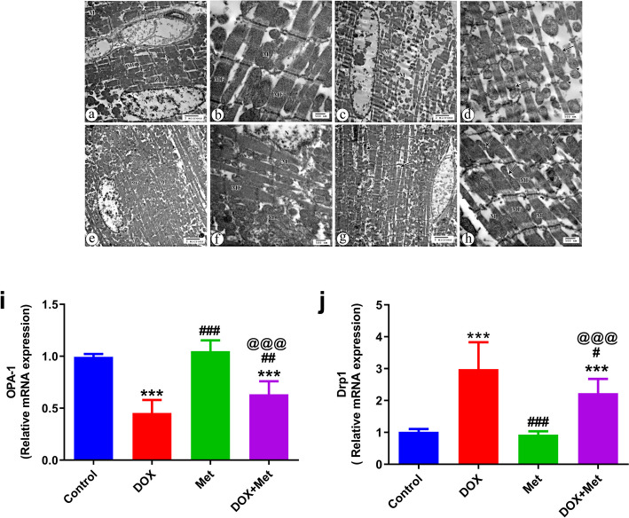
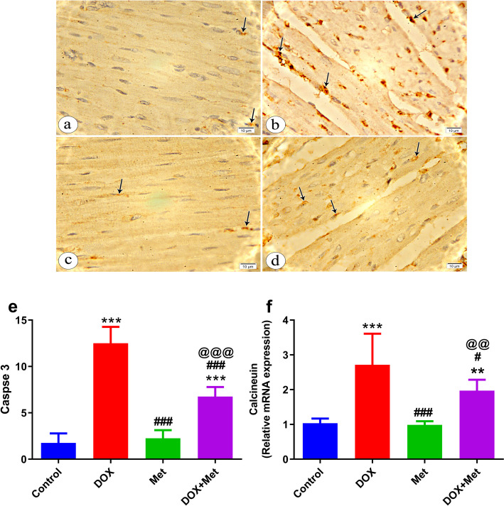
Doxorubicin (DOX) is a commonly used chemotherapeutic medication for treating malignancies, although its cardiotoxicity limits its use. There is growing evidence that alteration of the mitochondrial fission/fusion dynamic processes accompanied by excessive reactive oxygen species (ROS) production and alteration of calcium Ca2+ homeostasis are potential underlying mechanisms of DOX-induced cardiotoxicity (DIC). Metformin (Met) is an AMP-activated protein kinase (AMPK) activator that has antioxidant properties and cardioprotective effects. The purpose of the study is to assess Met's possible cardioprotective benefits against DOX-induced cardiotoxicity. The study included 32 adult male rats. They were randomly divided into four groups: administered saline, DOX, Met, or DOX combined with Met respectively. Heart tissues were used for biochemical assays that measured oxidative stress markers, malondialdehyde (MDA), reduced glutathione (GSH), mitochondrial dynamics markers, optic atrophy-1(OPA-1) and dynamin-1-like protein (Drp1), calcineurin and caspase-3. Serum levels of myocardial injury markers, cardiac troponin I (cTn-I), and aspartate aminotransferase (AST), were also measured. The results revealed that DOX intoxication was associated with a significant increase in the levels of serum cTn-I and AST, increased cardiac MDA level, increased cardiac Drp1, calcineurin, and caspase-3 expressions, as well as reduced cardiac GSH level and cardiac OPA-1 expression. On the other hand, Met treatment significantly reduced DIC by decreasing oxidative stress, apoptosis, and improving mitochondrial and calcium balance. Finally, this study shows that Met may be able to protect the heart from damage caused by DOX by working as an antioxidant and anti-apoptotic agent and keeping the balance of calcium and mitochondria.

Keywords: Calcineurin; Cardiotoxicity; Doxorubicin; Metformin; Mitochondrial dynamics.


Conflict of interest statement

Declarations. Ethics Approval: This study was performed according to the principles and guidelines given by the Animal Research Ethics Committee, Faculty of Medicine, Assiut University (26/6/2023, IRB local approval number 04–2023-300201). Consent to Participate: Not applicable. Consent for Publication: Not applicable. Competing Interests: The authors declare no competing interests.

Figures

Fig. 1
Fig. 1
The impact of metformin (Met) on the systolic (a) and diastolic (b) blood pressure in the DOX-intoxicated rats. n = 8 per group. The data represents the means ± SD. **p ≤ 0.01, ***p ≤ 0.001 vs. control group; ##p ≤ 0.01, ##p ≤ 0.001 vs. DOX-treated group; @@p ≤ 0.01, @@@p ≤ 0.001 vs. DOX: doxorubicin group; Met: metformin group; DOX + Met: doxorubicin + metformin group
Fig. 2
Fig. 2
Photomicrograph of myocardium of left ventricle stained with hematoxylin and eosin (H&E). n = 8 per group. A The myocardium of the control and Met groups, showed normal architectural features of cylindrical irregular branched muscle fibers (arrows) separated by slit-like narrow spaces of endomysium (*). The branched myofibers contained central oval vesicular nuclei (n) and acidophilic sarcoplasm (arrow heads). B The myocardium of DOX group showed marked deterioration within the myocardial fibers (arrows). With high magnification, endothelial cell injury of the blood vessels (arrow heads) and perivascular hemorrhage (short arrows) were associated with deteriorated myofibers. Replacement of necrotic myocardial muscles with activation of a fibrocystic cell reaction was also revealed (curved arrows). In the group that received DOX and Met, the myocardium showed a small area of cellular deterioration (arrow heads) and mild vascular wall degeneration (short arrow) that was associated with many normally arranged myocardial fibers (arrows). DOX: doxorubicin group; Met: metformin group; DOX + Met: doxorubicin + metformin group
Fig. 3
Fig. 3
A photomicrograph of myocardium of left ventricle of different groups stained with Masson Trichrome stain at 400 × magnification (a-d). n = 8 per group. a Control group showing fine collagen fibers (arrows) between the cardiac muscle fibers and around blood vessels (arrowhead). b Group received DOX only showing increased amount of collagen fibers (arrows) between the muscle fibers compared to the control. c Group received Met only exhibiting minimal amount of collagen fibers (arrows) between the cardiac muscle fibers. d Group received DOX and Met showing moderate amount of collagen fibers (arrows) between the muscle fibers. e Histogram showing the quantitative estimation of the mean area percentage of collagen fibers in Masson Trichrome-stained sections. n = 8 per group. Data are expressed as mean ± SD. ***p ≤ 0.001 vs. control; ##p ≤ 0.01, ###p ≤ 0.001 vs. the DOX-treated group; @@@p ≤ 0.001 vs. Met-treated group. DOX: doxorubicin group; Met: metformin group; DOX + Met: doxorubicin + metformin group
Fig. 4
Fig. 4
The effects of metformin treatment on cardiac injury and oxidative stress in the DOX-intoxicated rats. n = 8 per group. a Serum cTn-I levels, b Serum AST levels, c Tissue MDA levels, d Tissue GSH levels. n = 8 per group. Data are expressed as mean ± SD. *p ≤ 0.05, ***p ≤ 0.001 vs. control group; #p ≤ 0.05, ##p ≤ 0.01, ###p ≤ 0.001 vs. DOX-treated group; @@p ≤ 0.01, @@@p ≤ 0.001 vs. Met-treated group. AST: Aspartate aminotransferase; cTn-I: cardiac troponin-I; DOX: doxorubicin; DOX + Met: doxorubicin + metformin group, GSH: glutathione; MDA: malondialdehyde; Met: metformin
Fig. 5
Fig. 5
The effects of metformin treatment on cardiac mitochondrial dynamics in the DOX-treated rats. a-h Analysis of mitochondrial dynamics using transmission electron microscopy (TEM). The control group shows large euchromatic nucleus (N), parallel arrangement of myofibers (MF). In addition, the mitochondria (M) are observed arranged perinuclear and in rows between the myofibers with regularly arranged sarcomeres between Z lines (Z), regular arrangement of the myofibrils (F) within the sarcomeres between Z lines (Z), and endomysium blood capillaries (arrow) (X 4800) (a). Distribution of mitochondria (M) in between well-organized myofibers (F) (X 14000) is also noticed (b). The group that received Dox only shows a prominent myofibrillar loss and disarray (MF) with distribution of spaced mitochondria (M) in between disorganized and destructed myofibers (X 4800) (c). Marked abnormalities in mitochondrial morphology that include contour irregularities with fission (arrows) and fusion (arrow heads) in shape are also noticed (X 14000) (d). Group received Met only, shows equal or more apparent pathological changes in myofibers arrangement (MF) with mitochondria (M) distributed perinuclear and in rows between the myofibers (X 4800) (e). The group received DOX and Met, shows the distribution of mitochondria (M) between myofibers (MF) with relatively normal configuration and part of euchromatic nucleus (N) (X 14000) (f). Some pathological changes with disorganization and destruction of few myofibers (arrows) and thinning of others (arrowhead) are present. Notice the distribution of the mitochondria (M) in between the myofibers (X 4800) (g). More or less parallel arrangement of myofibers (MF) and distribution of mitochondria (M) in rows in between the myofibers, and some mitochondria with fission morphology are observed (arrows) (X 14000) (h). i and j Relative gene expression of mitochondrial dynamics markers, OPA-1 and Drp1. β actin was utilized to normalize expression data. n = 8 per group. Data are expressed as mean ± SD. ***p ≤ 0.001 vs. control group; #p ≤ 0.05, ##p ≤ 0.01, ###p ≤ 0.001 vs. DOX-treated group; @@@p ≤ 0.001 vs. Met-treated group. DOX: doxorubicin; DOX + Met: doxorubicin + metformin group; Drp1: dynamin-related protein 1; Met: metformin; OPA-1: optic atrophy type 1
Fig. 6
Fig. 6
The effects of metformin treatment on cardiac calcium homeostasis and apoptosis in DOX-intoxicated rats. a-e A photomicrograph of myocardium of left ventricle immunohistochemically stained with caspase 3 (× 1000). Control group shows faint positive immunostaining reaction within the sarcoplasm of minimal myocardial cells (arrows) (a). Group received DOX only shows marked positive immunostaining reaction within the sarcoplasm of many myocardial cells (arrows) (b). Group received Met only treated group shows mild positive immunostaining reaction within the sarcoplasm of few myocardial cells (arrows) (c). Group received DOX and Met showing moderate positive immunostaining reaction in the sarcoplasm of some myocardial cells (arrows) (d). Histogram showing the quantitative estimation of the mean count of caspase-3 positive cells (e). f Relative gene expression of calcineurin. β actin was utilized to normalize expression data. n = 8 per group. Data are expressed as mean ± SD. **p ≤ 0.01, ***p ≤ 0.001 vs. control group; #p ≤ 0.05, ###p ≤ 0.001 vs. DOX-treated group; @@p ≤ 0.01, @@@p ≤ 0.001 vs. Met-treated group. DOX: doxorubicin; Met: metformin; DOX + Met: doxorubicin + metformin group

Similar articles

Cited by

References

    1. Xia, P., Chen, J., Liu, Y., Fletcher, M., Jensen, B. C., & Cheng, Z. (2020). Doxorubicin induces cardiomyocyte apoptosis and atrophy through cyclin-dependent kinase 2–mediated activation of forkhead box O1. Journal of Biological Chemistry,295, 4265–4276. - PMC - PubMed
    1. Christidi, E., & Brunham, L. R. (2021). Regulated cell death pathways in doxorubicin-induced cardiotoxicity. Cell Death & Disease,12, 339. - PMC - PubMed
    1. Harahap, Y., Ardiningsih, P., Corintias Winarti, A., & Purwanto, D. J. (2020). Analysis of the doxorubicin and doxorubicinol in the plasma of breast cancer patients for monitoring the toxicity of doxorubicin. Drug Design, Development and Therapy, 3469–3475.‏ 10.2147/DDDT.S251144 - PMC - PubMed
    1. Cardinale, D., Colombo, A., Bacchiani, G., Tedeschi, I., Meroni, C. A., Veglia, F., Civelli, M., Lamantia, G., Colombo, N., & Curigliano, G. (2015). Early detection of anthracycline cardiotoxicity and improvement with heart failure therapy. Circulation,131, 1981–1988. - PubMed
    1. Jordan, J. H., Castellino, S. M., Meléndez, G. C., Klepin, H. D., Ellis, L. R., Lamar, Z., Vasu, S., Kitzman, D. W., Ntim, W. O., & Brubaker, P. H. (2018). Left ventricular mass change after anthracycline chemotherapy. Circulation: Heart Failure,11, e004560. - PMC - PubMed

LinkOut - more resources