

Nasopharyngeal tuberculosis suspected of malignancy: A case report
- PMID: 39792743
- PMCID: PMC11730819
- DOI: 10.1097/MD.0000000000040920
Nasopharyngeal tuberculosis suspected of malignancy: A case report
Abstract
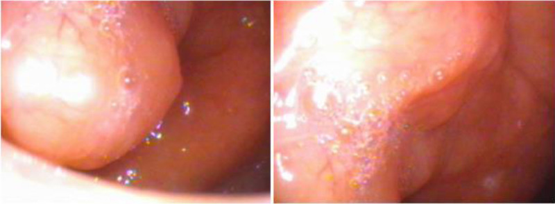
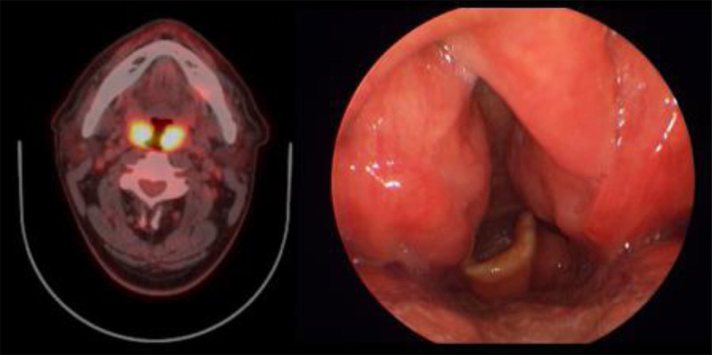
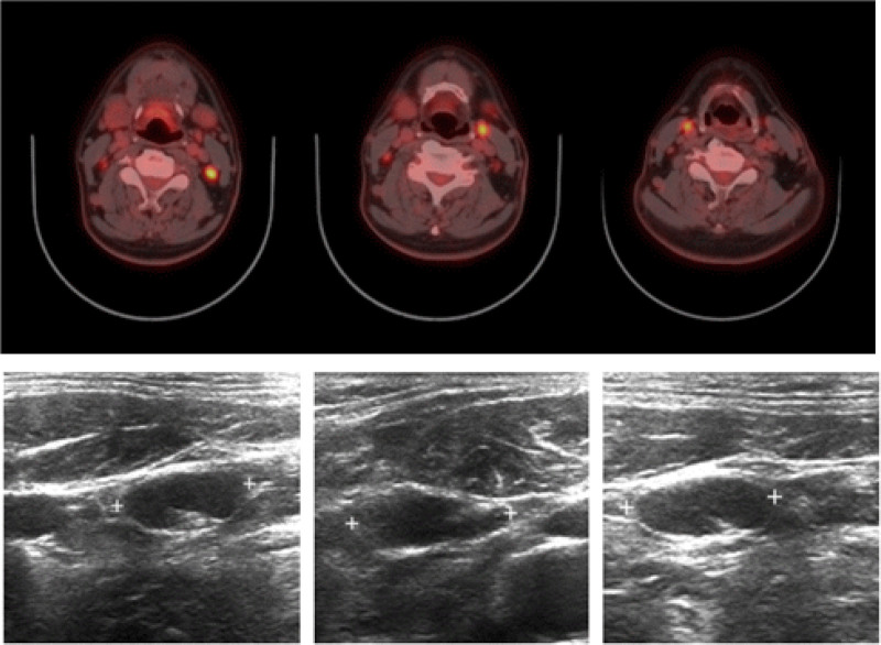
Rationale: Nasopharyngeal tuberculosis (TB), a rare form of tuberculosis outside the lungs, can affect any organ or tissue in the body. It is difficult to diagnose because of nonspecific symptoms, often leading to delayed confirmation after the initial patient visit. Clinical manifestations such as cervical lymphadenopathy and irregular mucosal surfaces can be challenging to distinguish from nasopharyngeal cancer or malignant lymphoma.
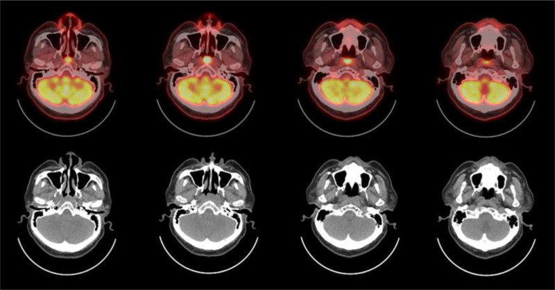
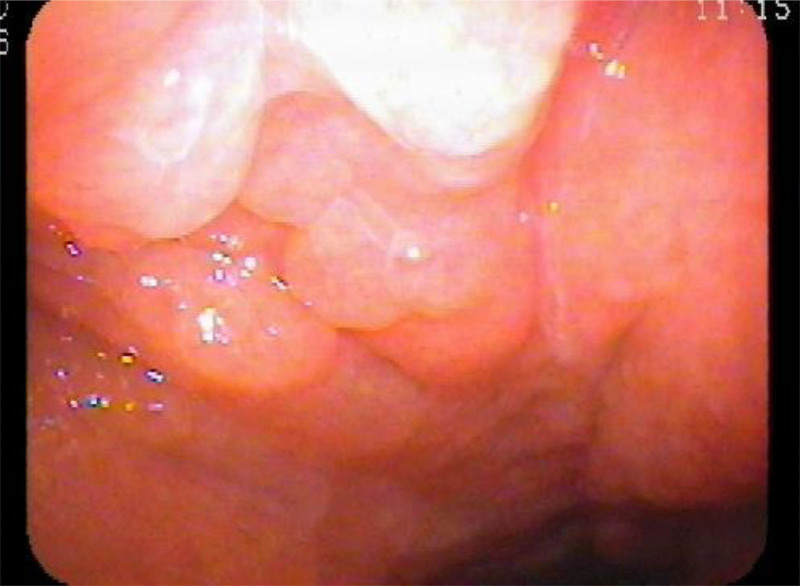
Patient concerns: In this case report, we present a patient initially suspected of having a malignant disease based on abnormal nasopharyngeal imaging findings.
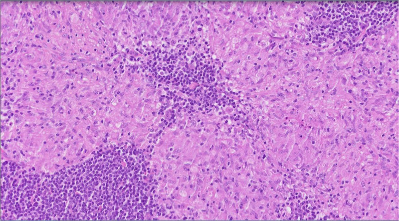
Diagnoses: Further examination revealed chronic granulomatous inflammation, and subsequent tuberculosis polymerase chain reaction (TB-PCR) confirmed the diagnosis of tuberculosis.
Interventions: The patient is currently receiving anti-TB treatment with a 4-drug regimen, which has shown a good response with continuous reduction in lesion size.
Outcomes: After anti-TB treatment, the lesion size gradually decreased and continued to decrease, showing a significant response.
Lessons: Awareness and precise evaluation are key to avoiding misdiagnosis, particularly when confronted with diverse clinical presentations. Extrapulmonary tuberculosis, although relatively rare, presents unique diagnostic challenges. Nasopharyngeal tuberculosis, in particular, lacks a definitive diagnostic method, often necessitating a combination of clinical suspicion, imaging studies, microbiological tests, and histopathological examination for confirmation. The absence of specific symptoms and the variability in presentation further compound the diagnostic dilemma. Given the potential consequences of misdiagnosis, further exploration and discussion on this issue are warranted. Enhanced awareness among healthcare providers, coupled with advancements in diagnostic modalities, are essential in ensuring timely and accurate differentiation between nasopharyngeal malignancies and tuberculosis, thereby facilitating appropriate management and improving patient outcomes.
Copyright © 2025 the Author(s). Published by Wolters Kluwer Health, Inc.
Conflict of interest statement
The authors have no funding and conflicts of interest to disclose.
Figures
References
-
- Min HJ, Kim KS. Primary nasopharyngeal tuberculosis: a case report focused on nasopharyngoscopic features and CT findings. Ear Nose Throat J. 2021;100(10_suppl):949S–52S. - PubMed
Publication types
MeSH terms
Substances
LinkOut - more resources
Full Text Sources
