. 2025 Feb;12(8):e2405988.
doi: 10.1002/advs.202405988. Epub 2025 Jan 10.

Highly Self-Adhesive and Biodegradable Silk Bioelectronics for All-In-One Imperceptible Long-Term Electrophysiological Biosignals Monitoring

Affiliations

Highly Self-Adhesive and Biodegradable Silk Bioelectronics for All-In-One Imperceptible Long-Term Electrophysiological Biosignals Monitoring

Seyed Sajjad Mirbakht et al. Adv Sci (Weinh). 2025 Feb.

Abstract

Skin-like bioelectronics offer a transformative technological frontier, catering to continuous and real-time yet highly imperceptible and socially discreet digital healthcare. The key technological breakthrough enabling these innovations stems from advancements in novel material synthesis, with unparalleled possibilities such as conformability, miniature footprint, and elasticity. However, existing solutions still lack desirable properties like self-adhesivity, breathability, biodegradability, transparency, and fail to offer a streamlined and scalable fabrication process. By addressing these challenges, inkjet-patterned protein-based skin-like silk bioelectronics (Silk-BioE) are presented, that integrate all the desirable material features that have been individually present in existing devices but never combined into a single embodiment. The all-in-one solution possesses excellent self-adhesiveness (300 N m-1) without synthetic adhesives, high breathability (1263 g h-1 m-2) as well as swift biodegradability in soil within a mere 2 days. In addition, with an elastic modulus of ≈5 kPa and a stretchability surpassing 600%, the soft electronics seamlessly replicate the mechanics of epidermis and form a conformal skin/electrode interface even on hairy regions of the body under severe perspiration. Therefore, coupled with a flexible readout circuitry, Silk-BioE can non-invasively monitor biosignals (i.e., ECG, EEG, EOG) in real-time for up to 12 h with benchmarking results against Ag/AgCl electrodes.

Keywords: biopotential monitoring; flexible electronics; functional biomaterials; stretchable electronics; wearable silk.


Conflict of interest statement

The authors declare no conflict of interest.

Figures

Figure 1
Figure 1
Schematic illustration of the synthesis, fabrication, and application of the introduced skin‐like silk bioelectronics (Silk‐BioE). a) Graphical representation of the effect of Ca2+ on the protein structure transition from crystalline to amorphous. b) Silk‐BioE patterning: (1) inkjet‐printing of AgNP on donor PET substrate, (2) drop‐casting silk solution on the printed AgNP mesh, (3) degassing and drying of the silk solution, and (4) transferring the AgNP pattern to silk by peeling the silk from PET donor substrate. c) Photographic depiction of the Silk‐BioE positioned on the original Bombyx mori silk cocoons. d) Comparative analysis of Silk‐BioE and existing silk‐based skin electronics for physiological monitoring. Details are given in Table 1. e) Schematic representation of key features of Silk‐BioE. f) Potential applications of the unit from healthcare to human‐computer interaction (HCI) to fitness and wellness to emerging novel applications in virtual reality. g) A prototype of the Silk‐BioE connected with a flexible wireless electronic circuitry.
Figure 2
Figure 2
Characterization of silk as a soft, self‐adhesive, biodegradable, transparent, and stretchable substrate for skin‐like bioelectronics. a) The Young's modulus of the silk substrate corresponds to different CaCl2 values at a relative humidity of ≈45% (n = 5), where the inset shows the tensile stress‐strain curve of 45 wt.% CaCl2/silk weight ratio. b) The strength of losing and capturing the ambient water molecules of 5 and 45 wt.% silk substrates (n = 3). c) The interface of red‐dyed 100 µm silk substrate with PDMS skin replica. d) FTIR spectra of different CaCl2/silk ratios illustrate the effect of Ca2+ ions on forming beta‐sheets and random coils. e) Raman spectra of the 45 wt.% CaCl2/silk substrate. f) Transmittance analysis of the silk substrates with different CaCl2/silk weight ratios. g) Adhesion force of silk substrates and commercial Kapton tape with respect to glass.
Figure 3
Figure 3
Surface topography and water‐resistant adhesion of the skin‐like silk bioelectronics (Silk‐BioE). SEM images of a) the pristine silk substrate section of the Silk‐BioE, b) Silk‐BioE with inkjet‐printed AgNP patterns, and c) zoom‐in view of the AgNP section of the Silk‐BioE. AFM images showing: d) the surface topology, e) 3D topography of the silk substrate, f) Evaluation of the adhesion quality of Silk‐BioE on skin under water prior to submersion in water, during full submersion of the hand in water, with the hand partially submerged and partially exposed to air, and after removal from the water, and g) Silk‐BioE on skin under running water.
Figure 4
Figure 4
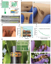
Mechanical properties of the skin‐like silk bioelectronics (Silk‐BioE) and their flexible readout circuitry. a) Geometry of the serpentine design. b) AgNP jetting profile of the inkjet‐printer head. c) Silk‐BioE on the skin under (i) stretching, (ii) compressing, and (iii) twisting motions. d) Feasibility of attaching Silk‐BioE to hairy skin areas. e) Resistance variation of the patch under 20% stretching and releasing tests. f) Resistance variation profile of the patch in 100 times cyclic stretching‐releasing. g) Flexible circuitry (i) on a curved surface (inset: block diagram of the different parts of the circuitry), (ii) folded by hand, and (iii) bent in hand and illustrating the different parts of the system. DRL: driven right leg. BLE: Bluetooth low energy. Ins. Amp.: instrumentation amplifier. ADC: analog to digital converter. SPI: serial peripheral interface.
Figure 5
Figure 5
a) ECG signal collection using skin‐like silk bioelectronics. b) P‐QRS‐T complex identification with an estimated heart rate from the recorded ECG signal. c) Power analysis of the recorded ECG signal. d) Schematic illustration of the Silk‐BioE placement for ECG recording. e) Silk‐BioE integrated with the flexible signal acquisition unit. f) Extended ECG monitoring using the Silk‐BioE over 12 h. g) Comparison of skin‐electrode impedance between “soft” Silk‐BioE and commercial “wet” Ag/AgCl electrodes where the inset shows an illustrative electrical model of the impedance.
Figure 6
Figure 6
EMG and EOG recording with Silk‐BioE: a) the placement of Silk‐BioE electrodes on the bicep for EMG monitoring. b) EMG signals were recorded in real‐time during the contraction and relaxation phases of the bicep muscle in lifting various dumbbells. c) Calibration curve linking the area under the curve (AUC) of EMG signals to the weight of different dumbbell weights, which can be used to quantify exercise intensity in training or in notifying/predicting muscle fatigue. d) Electrode placement for EOG recording. e) EOG acquisition by Silk‐BioE during left and right eye rotations can be used within activity recognition and mobile human‐machine interaction (HMI). f) Analysis of the EOG power spectrum density.
Figure 7
Figure 7
EEG Signal Acquisition using Silk‐BioE: a) Schematic representation illustrating the placement of Silk‐BioE electrodes on Fp1 and A1 for EEG acquisition. b) Time‐domain EEG signals were captured during both the open and closed‐eye states of the participant. Insets emphasize frequency variations observed during the alternating eye movements. c) Integration depiction of Silk‐BioE with the flexible signal acquisition board for EEG signal collection. d) Frequency‐domain representation of EEG signals highlighting the emergence of alpha‐wave frequencies during closed‐eye states. e) Power spectrogram showcasing the spectral analysis of the EEG recordings.

References

    1. Ma X., Wu X., Cao S., Zhao Y., Lin Y., Xu Y., Ning X., Kong D., Adv. Sci. 2023, 10, 2205343. - PMC - PubMed
    1. Yetisen A. K., Martinez‐Hurtado J. L., Ünal B., Khademhosseini A., Butt H., Adv. Mater. 2018, 30, 1706910. - PMC - PubMed
    1. Chen Y., Sun Y., Wei Y., Qiu J., Adv. Mater. Technol. 2023, 8, 2201352.
    1. Park S., Ban S., Zavanelli N., Bunn A. E., Kwon S., Lim H., Yeo W.‐H., Kim J.‐H., ACS Appl. Mater. Interfaces 2023, 15, 2092. - PubMed
    1. Li Y., Rodríguez‐Serrano A. F., Yeung S. Y., Hsing I., Adv. Mater. Technol. 2022, 7, 2101435.

LinkOut - more resources