. 2025 Mar;12(9):e2414651.
doi: 10.1002/advs.202414651. Epub 2025 Jan 10.

A Susceptible Cell-Selective Delivery (SCSD) of mRNA-Encoded Cas13d Against Influenza Infection

Affiliations

A Susceptible Cell-Selective Delivery (SCSD) of mRNA-Encoded Cas13d Against Influenza Infection

Zhuanli Wu et al. Adv Sci (Weinh). 2025 Mar.

Abstract

To bolster the capacity for managing potential infectious diseases in the future, it is critical to develop specific antiviral drugs that can be rapidly designed and delivered precisely. Herein, a CRISPR/Cas13d system for broad-spectrum targeting of influenza A virus (IAV) from human, avian, and swine sources is designed, incorporating Cas13d mRNA and a tandem CRISPR RNA (crRNA) specific for the highly conserved regions of viral polymerase acidic (PA), nucleoprotein (NP), and matrix (M) gene segments, respectively. Given that the virus targets cells with specific receptors but is not limited to a single organ, a Susceptible Cell Selective Delivery (SCSD) system is developed by modifying a lipid nanoparticle with a peptide mimicking the function of the hemagglutinin of influenza virus to target sialic acid receptors. The SCSD system can precisely deliver an all-RNA-based CRISPR/Cas13d system into potentially infected cells. This drug is shown to reduce the viral load in the lungs by 2.37 log10 TCID50 mL-1 and protect 100% of mice from lethal influenza infection. The SCSD-based CRISPR/Cas13d system shows promise for the flexible and efficient therapy of infections caused by rapidly evolving and novel viruses.

Keywords: Cas13d mRNA; influenza viruses; susceptible cell‐selective delivery.


Conflict of interest statement

The authors declare no conflict of interest.

Figures

Figure 1
Figure 1
Schematic illustration of the design of sLNP(CRI) and its use in susceptible cell‐selective therapy. A lipid‐based sLNP(CRI) was engineered to mimic the invasion mechanism of IAV by integrating a 30‐amino acid peptide that replicates the function of influenza glycoprotein HA, enabling it to enter susceptible cells. Cas13d mRNA and a tandem crRNA were loaded into sLNP(CRI) to recognize and cleave the conserved sequences of the influenza PA, NP, and M segments. These conserved sequences cover the H1, H3, H5, H7, and H9 subtypes of IAV from human, avian, and swine sources. sLNP(CRI) rapidly reaches the site of IAV infection (upper respiratory tract and lungs) after administration. Subsequently, Cas13d mRNA and tandem crRNA are released into the cytoplasm owing to lysosomal escape capability. Cas13d mRNA is translated into functional Cas13d protein and then forms a Cas13d‐crRNA complex to degrade viral PA/NP/M pre‐mRNA.
Figure 2
Figure 2
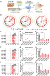
High‐throughput crRNA design for broad‐spectrum targeting of IAV. A) A workflow of Cas13d crRNA analysis for broad‐spectrum targeting of IAV. B) Phylogenetic trees to visualize the evolutionary relationships between H1, H3, H5, H7, and H9 subtype IAV strains isolated from human, avian, and swine sources during the years 2018 to 2022. C–K) Left and middle: mCherry expression as measured by flow cytometry. Right: mRNA abundance of viral sequences as measured by quantitative real‐time PCR. Relative RNA expression was calculated by normalization to the nontarget sample. Data are expressed as the mean ± SD from three biologically independent replicates (n = 3). Statistical analysis was performed using unpaired two‐tailed Student's t‐tests.
Figure 3
Figure 3
Preparation and characterization of sLNP(CRI). A) Schematic representation of Cas13d mRNA modification and synthesis. B) TEM images of sLNP(CRI). C) Confocal images showing the localization of C6‐labeled sLNP(CRI) relative to lysosomes after incubation with A549 cells for 0.5, 4, 6, and 8 h. The lysosomes were stained with Lysotracker Red DND‐99. D,E) Size distribution and zeta potential of sLNP(CRI). F) Changes in the diameter of sLNP(CRI) over 30 days. G) Quantitative analysis of free C6, C6‐LNP(CRI), and C6‐sLNP(CRI) taken up by A549 cells using flow cytometry. H) Mean fluorescence intensity (MFI) of A549 cells after treatment with free C6, C6‐LNP(CRI), and C6‐sLNP(CRI) for 4 h. I) Western blot analysis showing Cas13d protein expression.
Figure 4
Figure 4
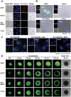
Susceptible cell‐selective delivery of sLNP(CRI). A) Identification of sialic acid receptor expression in A549, HaCaT, CCC‐HEH‐2, and 3T3L1 cells. Biotinylated SSA bound to SAα2,6 Gal and was then detected using Dylight 549‐labeled streptavidin. B) The ability of IAV to infect A549, HaCaT, CCC‐HEH‐2, and 3T3L1 cells. C) Inhibition of the cellular uptake of sLNP(CRI) in a sialic acid receptor competition binding assay. The A549 cells were preincubated with 10 µm lectins (with high affinity for sialic acid α2,6‐galactose or sialic acid α2,3‐galactose), and the cells were subsequently incubated with C6‐labeled sLNP(CRI) for 2 h. Nuclei were stained with Hoechst 33342. D) The specific binding and penetration of sLNP(CRI) in 3D spheroids of A549, HaCaT, CCC‐HEH‐2, and 3T3L1 cells. After incubating for 4 h, confocal microscopy images were obtained by scanning the spheroids from top to bottom at a depth of 20 µm per image.
Figure 5
Figure 5
Antiviral activity of sLNP(CRI) in vitro. A) Effects of the indicated drug on plaque formation by PR8 virus were determined at different concentrations. CRI: free crRNA and Cas13d mRNA. sLNP group: the lipid concentration is equivalent to sLNP(CRI). B) Reduction in virus titers at 10 µg mL−1 of the indicated drug in A549 cells. Oseltamivir: 10 µg mL−1; CRI, LNP(CRI), or sLNP(CRI): 10 µg mL−1 of RNA. C) NHBE cells were infected with PR8 virus and treated with 10 µg mL−1 of the indicated drug. Virus titers in supernatants were determined by a TCID50 assay. D) A549 cells were infected with PR8 virus and treated with 10 µg mL−1 of the indicated drug. Cells were stained with anti‐NP (green) and DAPI (blue) at 36 hpi. E) The effectiveness of sLNP(CRI) against various subtypes of influenza viruses. Data are expressed as the mean ± SD from three biologically independent replicates (n = 3). Statistical analysis was performed using unpaired two‐tailed Student's t‐tests.
Figure 6
Figure 6
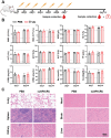
In vivo safety of sLNP(CRI). A) Schematic of the safety evaluation process in vivo. B) Hematology evaluation (WBC, white blood cell; RBC, red blood cell; HCT, hematocrit; HGB, hemoglobin), and blood biochemistry analysis (ALT, alanine transferase; AST, aspartate transferase; TP, total protein; ALP, alkaline phosphatase; CREA, creatinine; UREA, blood urea nitrogen; Na; Ca) of mice on day 7 and day 14 after injection of sLNP(CRI) with 20 µg RNA; the normal range is marked by a dashed line. C) H&E staining of the brain, liver, heart, lung, kidney, and spleen of mice from different groups on day 14. Data are expressed as the mean ± SD from three biologically independent replicates (n = 3). Statistical analysis was performed using unpaired two‐tailed Student's t‐tests.
Figure 7
Figure 7
Biodistribution and antiviral activity of sLNP(CRI) in vivo. A) In vivo and ex vivo IVIS imaging for verifying the distribution and respiratory (lung and nasopharynx) targeting ability of sLNP(CRI). Mice were administered either DiR‐labeled LNP(CRI) or DiR‐labeled sLNP(CRI) via the tail vein at a dose of 20 µg DiR per mouse. B) Timeline of the lethal mouse model of influenza. C) The body weights (n = 10) and survival rates of the mice. Mice were inoculated with PR8 virus on day 0, and their body weights were recorded for 14 days. When weight loss accounted for more than 25% of the initial body weight, the mouse was euthanized. D) Viral titers in the lung homogenates were quantified as the TCID50 value at 3 dpi (n = 6). E) Representative photographs of mice and mice lungs administered different treatments. H&E staining of lungs: thick solid arrow indicates alveolar wall thickening, and a black hollow triangle indicates immune cell infiltration. Immunohistochemical staining with anti‐NP monoclonal antibodies shows the positive detection rate of viral NP protein; thick open arrows indicate positive areas. F) Levels of inflammatory cytokines in the lungs of mice were measured. Samples were collected at 3 dpi. Statistical analysis was performed using unpaired two‐tailed Student's t‐tests.
Figure 8
Figure 8
Antiviral activity of sLNP(CRI) and Oseltamivir in vivo. A) The timeline for administration and sample collection. B) The body weights (n = 10) and survival rates of the mice. Mice were inoculated with PR8 virus on day 0, and their body weights were recorded for 14 days. C) Viral titers (n = 6) in the lung. D) Representative photographs of mice and mice lungs administered different treatments. H&E staining of lungs: thick solid arrows indicate alveolar wall thickening and black hollow triangles indicate immune cell infiltration. Immunohistochemical staining with anti‐NP monoclonal antibodies shows the positive detection rate of viral NP protein; thick open arrows indicate positive areas. Statistical analysis was performed using unpaired two‐tailed Student's t‐tests.
Figure 9
Figure 9
Antiviral activity with delayed administration of sLNP(CRI) and Oseltamivir in vivo. A) The timeline for delayed administration and sample collection. B) The body weights (n = 10) and survival rates of the mice. Mice were inoculated with PR8 virus on day 0, and their body weights were recorded for 14 days. C) Viral titers (n = 6) in the lung. D) Representative photographs of mice and mice lungs following delayed administration. H&E staining of lungs: thick solid arrows indicate alveolar wall thickening and black hollow triangles indicate immune cell infiltration. Immunohistochemical staining with anti‐NP monoclonal antibodies shows the positive detection rate of viral NP protein; thick open arrows indicate positive areas. Statistical analysis was performed using unpaired two‐tailed Student's t‐tests.

Similar articles

References

    1. Lu L., Su S., Yang H., Jiang S., Cell 2021, 184, 1604. - PubMed
    1. Taubenberger J. K., Reid A. H., Lourens R. M., Wang R., Jin G., Fanning T. G., Nature 2005, 437, 889. - PubMed
    1. Yang R., Sun H., Gao F., Luo K., Huang Z., Tong Q., Song H., Han Q., Liu J., Lan Y., Qi J., Li H., Chen S., Xu M., Qiu J., Zeng G., Zhang X., Huang C., Pei R., Zhan Z., Ye B., Guo Y., Zhou Y., Ye W., Yao D., Ren M., Li B., Yang J., Wang Y., Pu J., et al., Lancet Microbe 2022, 3, e824. - PubMed
    1. Taubenberger J. K., Kash J. C., Morens D. M., Sci. Transl. Med. 2019, 11, eaau5485. - PMC - PubMed
    1. Gu C., Maemura T., Guan L., Eisfeld A. J., Biswas A., Kiso M., Uraki R., Ito M., Trifkovic S., Wang T., Babujee L., Presler R. Jr., Dahn R., Suzuki Y., Halfmann P. J., Yamayoshi S., Neumann G., Kawaoka Y., Nature 2024, 636, 711. - PubMed

LinkOut - more resources