. 2025 Jan 10;19(1):e0012792.
doi: 10.1371/journal.pntd.0012792. eCollection 2025 Jan.

Insect-specific RNA viruses detection in Field-Caught Aedes aegypti mosquitoes from Argentina using NGS technology

Affiliations

Insect-specific RNA viruses detection in Field-Caught Aedes aegypti mosquitoes from Argentina using NGS technology

Lucas Ripoll et al. PLoS Negl Trop Dis. .

Abstract

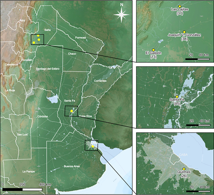
Mosquitoes are the primary vectors of arthropod-borne pathogens. Aedes aegypti is one of the most widespread mosquito species worldwide, responsible for transmitting diseases such as Dengue, Zika, and Chikungunya, among other medically significant viruses. Characterizing the array of viruses circulating in mosquitoes, particularly in Aedes aegypti, is a crucial tool for detecting and developing novel strategies to prevent arbovirus outbreaks. In this study, we address the implementation of a sequencing and analysis pipeline based on the Oxford Nanopore Technologies MinION Mk1b system, for arboviral detection in field-caught mosquitoes from Argentina. Full genome of Humaita Tubiacanga Virus (HTV), Phasi Charoen-like Phasivirus (PCLV), Aedes aegypti totivirus (AaeTV) has been sequenced in three distinct regions of Argentina comprising Buenos Aires province, Santa Fe province and the northern province of Salta. Viral sequences enriched by SISPA and coupled with Nanopore sequencing can be a useful tool for viral surveillance, not only for detecting viruses that have a high impact on human and animal health, but also for detecting insect-specific viruses that could promote the transmission of arboviruses.


Conflict of interest statement

The authors have declared that no competing interests exist.

Figures

Fig 1
Fig 1. Map of sample collection sites: Distribution of sample collection sites in three provinces of Argentina, Buenos Aires, Santa fe and Salta.
Each collection site shows the number of mosquitoes processed in each sample. Basemap data were sourced from Argenmap by the Instituto Geográfico Nacional de Argentina (IGN, https://www.ign.gob.ar) and OpenStreetMap (https://www.openstreetmap.org). Both datasets are provided under the Open Database License (ODbL, https://opendatacommons.org/licenses/odbl/1-0/).
Fig 2
Fig 2. Genome coverage plots of Phasi Charoen-like Phasivirus Segments.
Sequencing reads from different samples were mapped on to the reference sequences for S (small, NC_038263.1), M (medium, NC_038261.1) and L (large, NC_038262.1) segments, respectively, retrieved from GenBank. The X-axis indicates genome position, Y axis indicates sequencing depth. Below each graph, the ORFs of each segment are schematically represented. Collection sites are indicated with different colors.
Fig 3
Fig 3. Genome coverage plots of Humaita Tubiacanga Virus genomic segments and Aedes aegypti Totivirus.
Sequencing reads from different samples were mapped on to the reference sequences for Humaita Tubiacanga Virus–RdRp (MN053809.1), Humaita Tubiacanga Virus–Capsid (MN053810.1), retrieved from GenBank. The X-axis indicates genome position, Y axis indicates sequencing depth. Below each graph, the ORFs of each segment are schematically represented. Collection sites are indicated with different colors.
Fig 4
Fig 4. Phylogeny inference Phasi Charoen-like phasivirus (PCLV) based on its three genomic segments (S, M and L).
Maximum-likelihood tree based on the nucleotide sequences of full-length S (Panel A), M (Panel B) and L (Panel C) genomic segments of PCVL virus. Prototypes and sequences obtained in this work were used. Each virus is identified by the genome ID (Genbank), the name of the virus with its corresponding strain or isolate, and the country of origin in parentheses. The three Argentine PCLV isolations, identified in this study, are in red. Bootstrap values greater than 50 are indicated. Maximum-likelihood phylogenetic tree was inferred using the GTR+F+R2 model and 1000 replicates in every case.
Fig 5
Fig 5. Phylogeny inference of Humaita-Tubiacanga virus based on RNA-dependent RNA polymerase protein.
Maximum-likelihood tree based on full-length RNA-dependent RNA polymerase (RdRp) aminoacidic sequences from Humaita-Tubiacanga virus prototypes and the genome obtained in this work. Each virus is identified by the genome ID (Genbank), the name of the virus with its corresponding strain or isolate, and the country of origin in parentheses. The Argentinian HTV isolate, identified in this study, is in red. Bootstrap values greater than 50 are indicated. Maximum-likelihood phylogenetic tree was inferred using the HIVb (HIV between-patient matrix HIV-Bm) + F + I model and 1000 replicates.
Fig 6
Fig 6. Phylogeny inference of Totivirus based on RNA-dependent RNA polymerase gene.
Maximum-likelihood tree based on full-length RNA-dependent RNA polymerase (RdRp) nucleotide sequences from Totivirus prototypes and the genome obtained in this work. Each virus is identified by the genome ID (Genbank), the name of the virus with its corresponding strain or isolate, and the country of origin in parentheses. The Argentinian Totivirus isolate, identified in this study, are in red. The branches of each main phylogenetic clades (A, B, C) are colored. Bootstrap values greater than 50 are indicated. Maximum-likelihood phylogenetic tree was inferred using the General Time Reversible (GTR) +F + G4 distribution model, Gamma shape alpha of 0.1301 and 1000 replicates.

References

    1. Finlay C. J. (2012). The Mosquito Hypothetically Considered as the Transmitting Agent of Yellow Fever. MEDICC review, 14(1), 56–59. doi: 10.37757/MR2012V14.N1.10 - DOI - PubMed
    1. Weaver S. C., Charlier C., Vasilakis N., & Lecuit M. (2018). Zika, Chikungunya, and Other Emerging Vector-Borne Viral Diseases. Annual review of medicine, 69, 395–408. doi: 10.1146/annurev-med-050715-105122 - DOI - PMC - PubMed
    1. Chala B., & Hamde F. (2021). Emerging and Re-emerging Vector-Borne Infectious Diseases and the Challenges for Control: A Review. Frontiers in public health, 9, 715759. doi: 10.3389/fpubh.2021.715759 - DOI - PMC - PubMed
    1. Kraemer M. U. G., Reiner R. C., Jr, Brady O. J., Messina J. P., Gilbert M., Pigott D. M., et al. (2019). Past and future spread of the arbovirus vectors Aedes aegypti and Aedes albopictus. Nature microbiology, 4(5), 854–863. doi: 10.1038/s41564-019-0376-y - DOI - PMC - PubMed
    1. Robert M. A., Stewart-Ibarra A. M., & Estallo E. L. (2020). Climate change and viral emergence: evidence from Aedes-borne arboviruses. Current opinion in virology, 40, 41–47. doi: 10.1016/j.coviro.2020.05.001 - DOI - PMC - PubMed

LinkOut - more resources