. 2025 Jan 7;122(1):e2411720121.
doi: 10.1073/pnas.2411720121. Epub 2024 Dec 30.

Binding mode-guided development of high-performance antibodies targeting site-specific posttranslational modifications

Affiliations

Binding mode-guided development of high-performance antibodies targeting site-specific posttranslational modifications

Mariapia Riso et al. Proc Natl Acad Sci U S A. .

Abstract

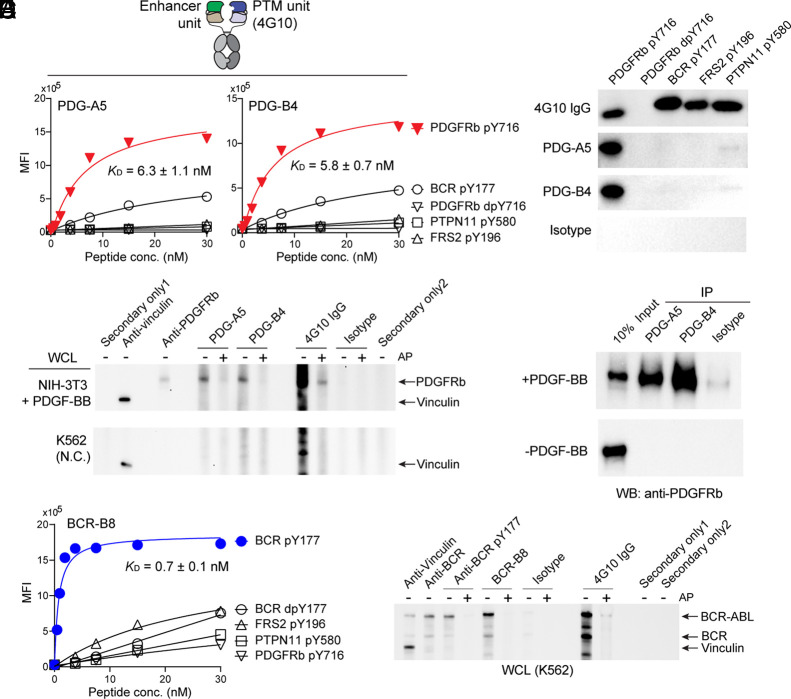
Posttranslational modifications (PTMs) of proteins play critical roles in regulating many cellular events. Antibodies targeting site-specific PTMs are essential tools for detecting and enriching PTMs at sites of interest. However, fundamental difficulties in molecular recognition of both PTM and surrounding peptide sequence have hindered the efficient generation of highly sequence-specific anti-PTM antibodies. Furthermore, the widespread use of potentially inconsistent, nonrenewable, and molecularly undefined antibodies presents experimental challenges thought to contribute to the reproducibility problem in biomedical research. In this study, we describe the binding mode-guided development of a platform that efficiently generates potent and selective recombinant antibodies to PTMs that are molecularly defined and renewable. Our platform is built on our previous discovery of an unconventional binding mode of anti-PTM antibodies, antigen clasping, where two antigen binding sites cooperatively sandwich a single antigen, creating extensive interactions with the antigen and leading to high selectivity and potency. We designed the platform that generates clasping antibodies with two distinct binding units, resulting in efficient generation of antibodies to a set of trimethylated histone H3 with high levels of specificity and affinity. Performance comparison in chromatin immunoprecipitation, a common application in epigenomics, revealed that a clasping antibody to trimethylated histone H3 at lysine 27 exhibited superior specificity to a widely used conventional antibody and captured symmetric and asymmetric nucleosomes in a less biased manner. We further generated clasping antibodies to phosphotyrosine antigens by using the same principle. These results suggest the broad applicability of our platform to generating high-performance clasping antibodies to diverse PTMs.

Keywords: affinity reagents; epigenetics; histone modifications; protein engineering; signaling.


Conflict of interest statement

Competing interests statement:A.J.R. holds equity in EpiCypher. A.K., S.K., and T.H. are listed as inventors of a pending patent on the technology and antibodies developed in this work filed by New York University. A.K., S.K., and T.H. are inventors of US10208110B2 on recombinant histone PTM antibodies managed by the University of Chicago. A.J.R. is a co-inventor of the ICeChIP technology US10732185B2 managed by the University of Chicago and licensed to EpiCypher. A.J.R. is a scientific advisor at EpiCypher. R.N.S. has received consulting fees from EpiCypher. S.K. is a co-founder, receives consulting fees and holds equity in Aethon Therapeutics; is a co-founder and holds equity in Revalia Bio; has received research funding from Aethon Therapeutics, Argenx BVBA, Black Diamond Therapeutics, and Puretech Health, all outside of the current work.

Figures

Fig. 1.
Fig. 1.
Binding mode-guided design of heterodimeric clasping antibodies. (A) Schematic representation of IgG antibody. (B) The structure of homodimeric clasping antibody (PDB ID: 4YHP, Left) and its schematic representation (Middle cartoon). The Right cartoon represents heterodimeric antigen clasping that is composed of two different antigen-binding units, PTM and enhancer units, resembling the distinct roles of Fabs in homodimeric antigen clasping. (C) Binding analysis of PTM and enhancer Fabs to the H3K56me3 peptide with the peptide-IP assay. The peptide concentration used here was 100 nM. The peptide binding was observed only when both PTM and enhancer Fabs were coimmobilized on beads, indicating cooperative binding of both Fabs in an antigen-clasping manner. (D) Binding titration curves of PTM Fab alone and coimmobilized Fabs with the peptide IP assay. The apparent KD values, calculated from the curve fitting of a 1:1 binding model, to the target peptide are shown. Sequence specificity (Top) and methylation-state specificity (Bottom) are assessed. Data shown here are from triplicate measurements. Error bars indicate the SD.
Fig. 2.
Fig. 2.
Efficient identification of clasping antibodies by yeast display and characterization of anti-H3K27me3 antibody in ICeChIP. (A) Schematic representation of the yeast-display format where the enhancer unit is tethered to the PTM unit with a long linker. (B) Characterization of clasping antibodies with the peptide IP assay in the yeast-display format harboring the 22-residue linker. Binding titration curves using the target peptides (Top) and specificity test using off-target peptides (Bottom) are shown. The apparent KD values were calculated from the curve fitting of a 1:1 binding model. The highest peptide concentrations used in the binding titration curves were utilized in the specificity test (Bottom). Data shown here are from triplicate measurements. Error bars indicate the SD. See also SI Appendix, Figs. S2 and S3 for other clones. (CE) Performance comparison of the widely used antibody and the clasping antibody to H3K27me3 in native ICeChIP. (C) Specificity of antibodies assessed by ICeChIP. The Left cartoons represent the nucleosome standards bearing H3K4me3, H3K27me3, or both. Note that the different standards bearing the H3K27me3 have one or two instances of the mark per nucleosome. Antibody specificity was calculated as the percentage of off-target to on-target (H3K27me3) enrichment of nucleosome standards. The 3C2-H2 clasping antibody captured symmetric and asymmetric nucleosome standards in a less biased manner compared with C36B11 IgG. (D) Metagene and heatmap performance comparison of antibodies. Histone modification density (HMD) of H3K27me3 across all promoters in mESCs rank-ordered by their RNA-expression level (RNA-seq from ENCODE) was plotted. (E) Representative locus showing apparent H3K27me3 HMD with antibodies.
Fig. 3.
Fig. 3.
Characterization of clasping antibodies to H3K27me3 in the long-neck scFv-Fc format. (A) Schematic representation of the heterodimeric long-neck scFv-Fc format. Hetero-dimerization is facilitated by the knobs-into-holes mutations in the CH3 domain. (B) Binding titration curves of the clasping antibodies against the target and the off-target peptides with the peptide IP assay. The apparent KD values calculated from the curve fitting of a 1:1 binding model to the target peptide are shown. (C) Kinetic analysis of clasping antibodies with BLI. The H3K27me3 peptide was immobilized, and the binding of soluble long-neck scFv-Fc antibodies was measured. The apparent KD values are from the global fit with a 1:1 binding model to the data. (D and E) Validation of clasping antibodies in Western blot. (D) Whole-cell lysate of K562 cells was blotted with the indicated long-neck scFv-Fc antibodies at three different concentrations (30, 10, and 3.3 nM, respectively). The isotype composes 4-5 (PTM unit) and a nonbinding antibody (in replacement of an enhancer unit) and consequently has weak affinity to Kme3. The “secondary only” lanes refer to no primary antibody with anti-mouse Fc-HRP (secondary only 1) or with anti-rabbit IgG-HRP (secondary only 2). The arrow indicates the location of histone H3. (E) Whole-cell lysates from K562 cells treated with and without GSK126, an EZH2 methyltransferase inhibitor, were blotted with indicated long-neck scFv-Fc antibodies. The vinculin protein was detected as a loading control. (F) Validation of 3C2-H2 long-neck scFv-Fc in native SNAP-ChIP. The enrichment (percentage of H3K27me3 nucleosome recovered after immunoprecipitation relative to input, Top) and specificity (percentage of off-target immunoprecipitation relative to the on-target, Bottom) assessed by qPCR are shown. The clasping antibody specifically captured the nucleosome bearing H3K27me3. Data shown in B and F are from triplicate measurements. Error bars indicate the SD. See SI Appendix, Fig. S6 for the complete datasets for Western blot.
Fig. 4.
Fig. 4.
Characterization of anti-pY clasping antibodies (AD) Characterization of clasping antibodies targeting PDGFRb pY716 in the long-neck scFv-Fc format. (A) Binding titration curves of the clasping antibodies with the peptide IP assay. (B and C) Validation of clasping antibodies in Western blot. SUMO-fused peptides (B) and WCLs from platelet-derived growth factor (PDGF)-BB-treated NIH 3T3 cells (C, Top blot) and K562 cells that do not or minimally express PDGFRb (C, Bottom blot) were blotted with indicated clasping antibodies and control IgGs. (D) Validation of clasping antibodies in IP-Western. The lysates from NIH 3T3 cells treated with or without PDGF-BB were immunoprecipitated with the indicated clasping antibodies, and the immunoprecipitated samples were subsequently blotted with anti-PDGFRb antibody. The clasping antibodies specifically detected and captured PDGFRb in a phosphorylation-dependent manner. (E and F) Characterization of clasping antibodies targeting BCR pY177 in the long-neck scFv-Fc format with the peptide IP assay (E) and Western blot using WCL (F). The isotype, used in BD and F, composes 4G10 (PTM unit) and a nonbinding antibody (in place of an enhancer unit) and consequently has weak affinity to pY. The apparent KD values, calculated from the curve fitting of a 1:1 binding model to the target peptide, are shown in A and E. The abbreviation “dpY” stands for dephosphorylated tyrosine, indicating that the phosphorylation of pY peptides was removed prior to the experiments. The abbreviation “AP” in Western blot (C and F) stands for alkaline phosphatase and each lane displaying “+” was treated with AP prior to being blotted with antibodies. The “secondary only” lanes refer to no primary antibody with anti-mouse Fc-HRP (secondary only 1) or with anti-rabbit IgG-HRP (secondary only 2). See SI Appendix, Figs. S8 and S9 for the complete datasets for Western blot.

Similar articles

Cited by

References

    1. Strahl B. D., Allis C. D., The language of covalent histone modifications. Nature 403, 41–45 (2000). - PubMed
    1. Olsen J. V., et al. , Global, in vivo, and site-specific phosphorylation dynamics in signaling networks. Cell 127, 635–648 (2006). - PubMed
    1. Hochstrasser M., Ubiquitin-dependent protein degradation. Annu. Rev. Genet. 30, 405–439 (1996). - PubMed
    1. Ohtsubo K., Marth J. D., Glycosylation in cellular mechanisms of health and disease. Cell 126, 855–867 (2006). - PubMed
    1. Greer E. L., Shi Y., Histone methylation: A dynamic mark in health, disease and inheritance. Nat. Rev. Genet. 13, 343–357 (2012). - PMC - PubMed

LinkOut - more resources