. 2025 Jan 7;122(1):e2415976121.
doi: 10.1073/pnas.2415976121. Epub 2024 Dec 30.

Genesis and regulation of C-terminal cyclic imides from protein damage

Affiliations

Genesis and regulation of C-terminal cyclic imides from protein damage

Wenqing Xu et al. Proc Natl Acad Sci U S A. .

Abstract

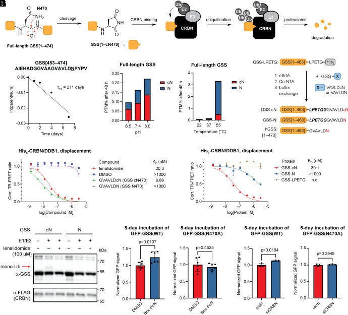
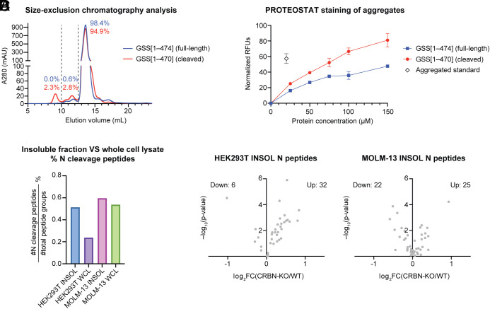
C-Terminal cyclic imides are posttranslational modifications that can arise from spontaneous intramolecular cleavage of asparagine or glutamine residues resulting in a form of irreversible protein damage. These protein damage events are recognized and removed by the E3 ligase substrate adapter cereblon (CRBN), indicating that these aging-related modifications may require cellular quality control mechanisms to prevent deleterious effects. However, the factors that determine protein or peptide susceptibility to C-terminal cyclic imide formation or their effect on protein stability have not been explored in detail. Here, we characterize the primary and secondary structures of peptides and proteins that promote intrinsic formation of C-terminal cyclic imides in comparison to deamidation, a related form of protein damage. Extrinsic effects from solution properties and stressors on the cellular proteome additionally promote C-terminal cyclic imide formation on proteins like glutathione synthetase that are susceptible to aggregation if the protein damage products are not removed by CRBN. This systematic investigation provides insight into the regions of the proteome that are prone to these unexpectedly frequent modifications, the effects of this form of protein damage on protein stability, and the biological role of CRBN.

Keywords: cereblon; deamidation; post-translational modification; protein aggregation; protein damage.


Conflict of interest statement

Competing interests statement:Harvard University has filed a PCT patent application on April 13, 2022 covering the chemical structures and their use. C.M.W. and W.X. are inventors of this patent. The Woo lab receives or has received support from Merck and Ono Pharmaceuticals.

Figures

Fig. 1.
Fig. 1.
Regulatory mechanisms and consensus sequences of asparagine residues that are susceptible to cleavage or deamidation. (A) Schematic of the formation and regulation of C-terminal cyclic imide modifications. (B) Schematic of the formation and regulation of protein damage arising from deamidation. (C) Frequency charts of the sequence alignment spanning the –3 to +3 residues flanking random asparagine sites extracted from the human proteome, C-terminal aspartimide sites, or deamidation sites observed in CPTAC datasets. (D) Frequency charts of the sequence alignment for the three clusters generated from hierarchical clustering of the +1 and +2 residues of the cN sites observed in CPTAC datasets based on similarity of amino acid properties. The percentage of each cluster out of the total population is noted. All alignments were generated with weblogo.berkeley.edu. Amino acids are colored according to their chemical properties: N and Q are in purple, basic amino acids (K, R, H) in blue, acidic amino acids (D, E) in red, polar amino acids (G, S, T, Y, C) in green, and hydrophobic amino acids (A, V, L, I, P, W, F, M) in black.
Fig. 2.
Fig. 2.
Impact of primary sequences of peptides and proteins on C-terminal cyclic imide formation. (A and B) Quantification of cN formation with a peptide library carrying variable +2 (A) or +1 (B) residues after 48 h incubation in 100 mM Na2HPO4 (pH 7.4) at 37 °C. (C) Time-course formation of cN and N from the indicated peptides in 100 mM Na2HPO4 (pH 7.4) at 37 °C. (D) Derivation of the half-life for the indicated peptides in 100 mM Na2HPO4 (pH 7.4) at 37 °C. (E) Design of engineered GFPs to evaluate the impact of protein primary structure on intramolecular cleavage. (F and G) Quantification of cN and N formation from GFP model proteins with varying +1 (F) or +2 (G) residues after 48 h incubation in 100 mM Na2HPO4 (pH 7.4) at 37 °C. (H) Time-course formation of cN and N from the indicated proteins in 100 mM Na2HPO4 (pH 7.4) at 37 °C. (I) Derivation of the half-life for the indicated proteins in 100 mM Na2HPO4 (pH 7.4) at 37 °C.
Fig. 3.
Fig. 3.
Effect of buffer properties on C-terminal cyclic imide formation. (A) Comparison of C-terminal cyclic imide and hydrolysis products for peptides with a differing central N or Q residue after 48 h incubation in 100 mM Na2HPO4 (pH 7.4) at 37 °C. (B) Quantification of cN and N formation from four representative peptides after 48 h incubation in 100 mM NH4OAc, Tris-Cl, or Na2HPO4 (all pH 7.4) at 37 °C. (C) Time-course and half-life derivation for the indicated peptides in 100 mM NH4OAc, Tris-Cl, or Na2HPO4 (all pH 7.4) at 37 °C. (D and E) Quantification of cN and N species from ACTB [96–113] peptide after 48 h incubation in 100 mM Na2HPO4 with variable pH at 37 °C with variable pH (D) or containing 1 mM chloride salts of the noted metal ion at pH 7.4 (E).
Fig. 4.
Fig. 4.
Influence of protein secondary structures on the propensity for cleavage. (A) Workflow of the computational analysis to derive protein secondary structure features. (B) Distribution of relative surface exposure for frequently observed deamidation and cleavage sites in comparison to two background datasets composed of randomly extracted Asn sites. Median values are indicated by a red line and the 25% and 75% percentiles by dotted lines. (C) Comparison of mean values of relative surface exposure across the four evaluated groups. (D) Distribution of distance between Asn amide and backbone carbonyl for frequent deamidation and cleavage sites in comparison to two background datasets composed of randomly extracted Asn sites. Median values are indicated by a red line and the 25% and 75% percentiles by dotted lines. (E) Comparison of mean values of distance across the four evaluated groups. All comparisons were performed using a one-way ANOVA with Šidák’s multiple comparisons test and P-values are noted. (F) Overlay of the original experimental structure in yellow and the predicted structure after cleavage at the indicated asparagine residue in teal for HBB N58 and ALB N154. The calculated values of surface exposure (SE) and distance (dist) and the pLDDT scores for the prediction of asparagine cleavage sites are noted. (G) Dose titration of the indicated peptides in TR-FRET assay against the His6-CRBN/DDB1 complex with the determined KD values noted. The experiment was performed with 3 technical replicates.
Fig. 5.
Fig. 5.
Effect of external stressors on cleavage in vitro and in cells. (A) Quantification of cN and N species from the GFP model protein after 24 h exposure to heat stress (55 °C), 100 µM H2O2, or vortexing. The protein was incubated in 100 mM Na2HPO4 buffer, pH 7.4 at 37 °C unless otherwise noted. (B) Quantification of cN and N species from the GFP model protein after 48 h incubation in 100 mM Na2HPO4 buffer containing varying concentrations of H2O2 at 37 °C, pH 7.4. (C and D) Quantification of cN and N species from the GFP model protein after 48 h incubation in 100 mM Na2HPO4 buffer and variable pH at 37 °C (C) or at pH 7.4 with variable temperature (D). (E) Venn diagrams representing the numbers of significantly upregulated semitryptic peptides ending with N in CRBN-KO HEK293T cells upon treatment with 100 µM H2O2 for 40 min, heat stress at 55 °C for 40 min, or pH elevation at pH 7.8 for 4 h compared to untreated cells incubated at pH 7.3, 37 °C. (F) Volcano plots of semitryptic peptides ending with N in CRBN-KO HEK293T cells upon treatment with 100 µM H2O2 for 40 min, heat stress at 55 °C for 40 min, or pH elevation at pH 7.8 for 4 h compared to untreated cells incubated at pH 7.3, 37 °C. The experiment was performed with 4 biological replicates. P-values for the abundance ratios were calculated by one-way ANOVA with the Tukey HSD post hoc test. Dotted lines represent the cutoff for FC > 1.1 and P < 0.05.
Fig. 6.
Fig. 6.
CRBN regulates GSS upon cleavage. (A) Schematic of the cleavage at N470, recognition by CRBN and removal by the ubiquitin–proteasome system of GSS. (B) Derivation of the cleavage half-life for the GSS parent peptide in 100 mM Na2HPO4 (pH 7.4) at 37 °C. (C) Quantification of cN and N species from full-length GSS protein after 48 h incubation in 100 mM Na2HPO4 buffer at the indicated pH at 37 °C. (D) Quantification of cN and N species from full-length GSS protein after 48 h incubation in 100 mM Na2HPO4 (pH 7.4) at the indicated temperature. (E) Dose titration of the indicated compounds in TR-FRET assay against the His6-CRBN/DDB1 complex with the determined KD values noted. (F) Schematic of sortase system used to generate tagged GSS from GSS-LPETG-His6 and the comparison to the native sequence of human GSS (hGSS). (G) Dose titration of GSS protein tagged with C-terminal cyclic or acyclic asparagine in TR-FRET assay against the His6-CRBN/DDB1 complex with the determined KD values noted. All TR-FRET experiments were performed with 3 technical replicates. (H) In vitro ubiquitination of GSS protein tagged with C-terminal cyclic or acyclic asparagine with K0 ubiquitin. (I) Quantification of GFP level in cells stably expressing GFP fusion of full-length wild-type (WT) GSS or GSS bearing N470A mutation by flow cytometry. Cells were treated with DMSO or 200 µM Boc-FcN across six biological replicates for 5 d with media renewal every 2 d. (J) Quantification of GFP level in cells stably expressing GFP fusion of full-length WT GSS or GSS bearing N470A mutation by flow cytometry. Cells were transfected with 30 nM nontargeting siRNA or siRNA targeting CRBN with three biological replicates and incubated for 5 d posttransfection. Comparisons for the reporter cell lines were performed using an unpaired two-tailed t test and P values are noted.
Fig. 7.
Fig. 7.
Association between cleavage products and protein aggregation. (A) SEC of the affinity-purified GSS proteins. Percentages of the peak areas are noted for different ranges of elution volumes. (B) Quantification of aggregated proteins in the affinity-purified GSS proteins by PROTEOSTAT staining. The experiment was performed with three technical replicates. (C) Quantification of percentage of unique semitryptic peptides ending with N out of all peptides identified in the insoluble fraction or whole cell lysate of HEK293T and MOLM-13 cells. (D) Volcano plots of semitryptic peptides ending with N in the insoluble fractions of cells after genetic knockout of CRBN compared to WT for HEK293T and MOLM-13 cells. The number of upregulated and downregulated peptides are noted. The experiment was performed with four technical replicates. P-values for the abundance ratios were calculated by one-way ANOVA with the Tukey HSD post hoc test.

Update of

Similar articles

Cited by

References

    1. Harmel R., Fiedler D., Features and regulation of non-enzymatic post-translational modifications. Nat. Chem. Biol. 14, 244–252 (2018). - PubMed
    1. Kehm R., Baldensperger T., Raupbach J., Hohn A., Protein oxidation - Formation mechanisms, detection and relevance as biomarkers in human diseases. Redox Biol. 42, 101901 (2021). - PMC - PubMed
    1. Brownlee M., Vlassara H., Cerami A., Nonenzymatic glycosylation and the pathogenesis of diabetic complications. Ann. Intern. Med. 101, 527–537 (1984). - PubMed
    1. Radi R., Protein tyrosine nitration: Biochemical mechanisms and structural basis of functional effects. Acc. Chem. Res. 46, 550–559 (2013). - PMC - PubMed
    1. Perler F. B., Xu M. Q., Paulus H., Protein splicing and autoproteolysis mechanisms. Curr. Opin. Chem. Biol. 1, 292–299 (1997). - PubMed

MeSH terms

LinkOut - more resources