

Spinal impostor: Metastatic cervical paraganglioma presenting with paraparesis, a case report
- PMID: 39793333
- PMCID: PMC11864149
- DOI: 10.1016/j.ijscr.2025.110821
Spinal impostor: Metastatic cervical paraganglioma presenting with paraparesis, a case report
Abstract
Introduction and importance: Paragangliomas are rare neuroendocrine tumors, typically arising from extra-adrenal chromaffin cells. Primary intra-spinal paragangliomas are uncommon, and metastatic spinal paragangliomas without paraneoplastic symptoms are even rarer. This case highlights the diagnostic challenges posed by such rare tumors.
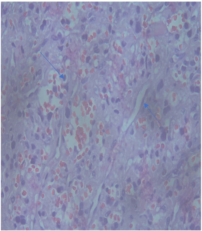
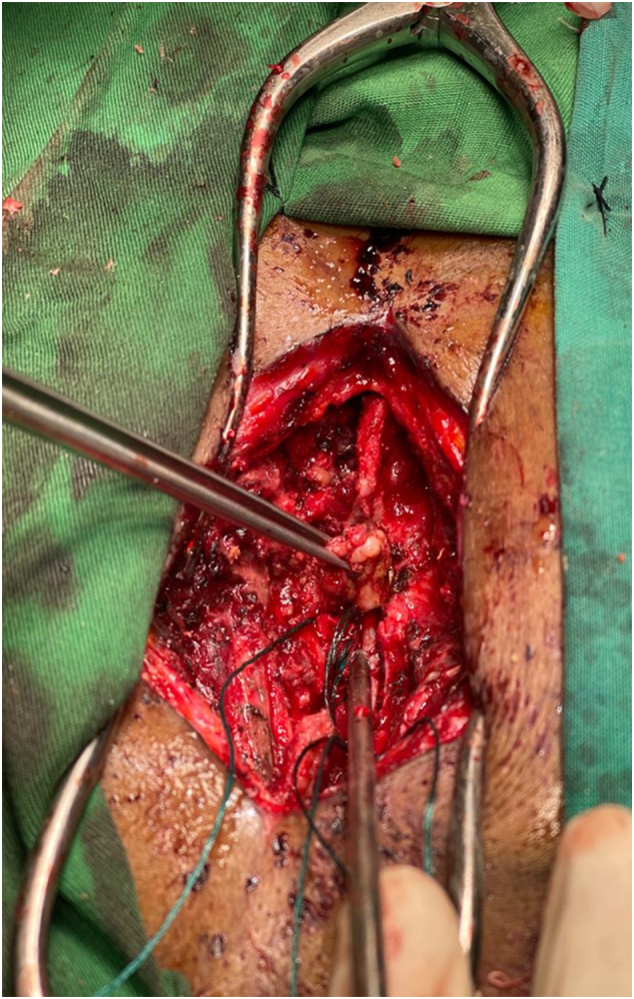
Case presentation: A 28-year-old male soldier from the Comoros Islands presented with a neck mass, initially suspected to be Hodgkin's lymphoma based on imaging. Biopsy of two cervical nodes revealed reactive lymphadenopathy. Later, he developed progressive lower limb weakness and numbness, prompting further investigation. Imaging showed an extradural spinal tumor at T6 with cord compression. Laminectomy and tumor excision relieved compression, revealing a highly vascularized tumor. Histopathology and immunohistochemistry confirmed a paraganglioma, which was consistent with the metastatic nature confirmed by a repeat biopsy of the neck mass.
Clinical discussion: Metastatic spinal paragangliomas are rare and challenging to diagnose, especially without paraneoplastic symptoms. This case underscores the importance of thorough histopathological evaluation when spinal lesions and neck masses present with unusual features and highlights the need for a multidisciplinary approach.
Conclusion: This case emphasizes the diagnostic difficulty of metastatic spinal paragangliomas, particularly when they mimic more common conditions like Hodgkin's lymphoma. It stresses the importance of considering rare differential diagnoses and a collaborative approach to managing such cases.
Keywords: Carotid body tumor; Case report; Diagnostic uncertainty; Neoplastic cord compression; Spinal paraganglioma.
Copyright © 2025 The Authors. Published by Elsevier Ltd.. All rights reserved.
Conflict of interest statement
Declaration of competing interest The author(s) declared no conflicts of interest regarding the research, authorship, and/or publication of this article.
Figures


References
-
- Gelabert-González M. Paragangliomas of the lumbar region. Report of two cases and review of the literature. J. Neurosurg. Spine. 2005;2(3):354–365. - PubMed
-
- Sabiston D.C., Jr., Kim M.K., Koss M.N. In: Sabiston Textbook of Surgery. 21st ed. Townsend C.M. Jr., Evers B.M., Mattox K.L., editors. Elsevier; 2022. Neurosurgery; p. 1895.
-
- Louis DN, Ohgaki H, Wiestler OD, et al. WHO Classification of Tumors of the Central Nervous System. 4th ed. Revised. ISBN: 9789283244929.
-
- Erickson D., Kudva Y.C., Ebersold M.J., Thompson G.B., Grant C.S., van Heerden J.A., Young W.F. Benign paragangliomas: clinical presentation and treatment outcomes in 236 patients. J. Clin. Endocrinol. Metab. 2001;86(11):5210–5216. - PubMed
-
- Zileli M., Kalayci M., Basdemir G. Paraganglioma of the thoracic spine. J. Clin. Neurosci. 2008;15(7):823–827. - PubMed
Publication types
LinkOut - more resources
Full Text Sources
