

Functional outcomes may vary over time after patellar tendon and knee intra-articular heterotopic ossification excision: A case report
- PMID: 39793335
- PMCID: PMC11780159
- DOI: 10.1016/j.ijscr.2024.110773
Functional outcomes may vary over time after patellar tendon and knee intra-articular heterotopic ossification excision: A case report
Abstract
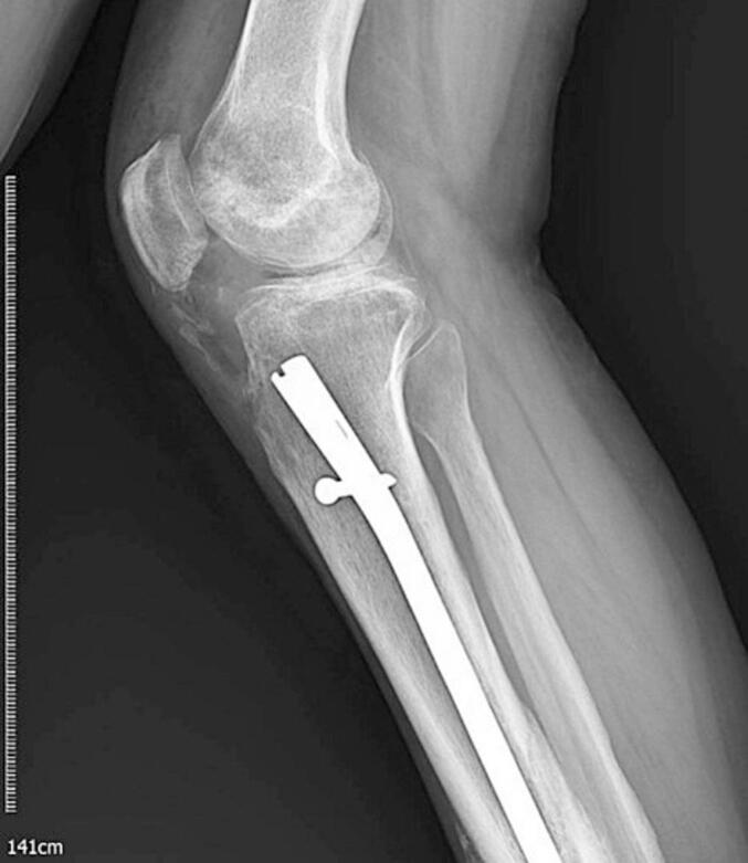
Introduction: Heterotopic ossification (HO) is the formation of mature bone in soft tissue, often occurring after fractures and trauma. Patients with HO experience pain, joint stiffness, and other complications. Treatment aims to improve function; surgical procedures have succeeded in 83.3 % of cases. Existing literature has extensively documented instances of HO occurring in the acetabulum, elbow, and during total hip arthroplasty (THA). HO formation is rare around the knee and intraarticularly after tibia nailing. A unique aspect of our case is the period of restricted ROM in the knee following surgical excision. Additionally, physical therapy played a crucial role in restoring full ROM after this period.
Case: A 38-year-old man presented to our department with right knee pain and restricted ROM resulting from a tibia fracture nailing performed three years earlier in another hospital. Radiographic imaging revealed HO in the retro patellar and intraarticular areas. He underwent surgical excision to remove the HO but continued to experience limited ROM during the follow-up period. Despite undergoing various treatments, including chemoprophylaxis and under anesthesia manipulation, his ROM did not significantly improve. Eventually, after long-term physical therapy, his condition improved, and at the two-year follow-up, he was pain-free with enhanced ROM.
Discussion: Surgical resection is recommended when HO significantly affects joint range of motion, but incomplete resection is linked to recurrence. After surgical removal of HO, prophylaxis with indomethacin or single fraction radiotherapy is used. However, limitations in range of motion may persist after surgical excision, and recurrence can occur despite preventive measures. Physical therapy may play a crucial role in restoring range of motion and achieving optimal treatment outcomes.
Conclusion: Tibia nailing may contribute to the formation of HO and can lead to a restricted ROM in the knee. It is essential to diagnose HO early and consider a multidisciplinary approach that includes surgical excision, chemoprophylaxis, and physical therapy. The role of physical therapy might be more significant than previously thought.
Keywords: Heterotopic ossification; Knee contracture; Physical therapy; Surgical management; Tibia nailing.
Copyright © 2025 The Authors. Published by Elsevier Ltd.. All rights reserved.
Conflict of interest statement
Conflict of interest statement There are no conflicts of interest to declare. All co-authors have agreed with the final manuscript's contents, and no financial interest remains to be announced.
Figures


References
-
- Nadar A.C., Seligson D. Heterotopic ossification in the knee following retrograde nailing of a femur fracture. Eur. J. Orthop. Surg. Traumatol. 2023;33(7):3181–3184. - PubMed
-
- Howell R.D., Park J.H., Egol K.A. Late symptomatic heterotopic ossification of the patellar tendon after medial parapatellar intramedullary nailing of the tibia. Orthopedics. 2011;34(3) - PubMed
-
- Eisenstein N., Stapley S., Grover L. Post-traumatic heterotopic ossification: an old problem in need of new solutions. J. Orthop. Res. 2018;36(4):1061–1068. - PubMed
Publication types
LinkOut - more resources
Full Text Sources
