. 2024 Dec 27;30(1):57.
doi: 10.3390/molecules30010057.

Indocarbocyanine-Indodicarbocyanine (sCy3-sCy5) Absorptive Interactions in Conjugates and DNA Duplexes

Affiliations

Indocarbocyanine-Indodicarbocyanine (sCy3-sCy5) Absorptive Interactions in Conjugates and DNA Duplexes

Evgeny L Gulyak et al. Molecules. .

Abstract

Sulfonated indocyanines 3 and 5 (sCy3, sCy5) are widely used to label biomolecules. Their high molar absorption coefficients and lack of spectral overlap with biopolymers make them ideal as linker components for rapid assessment of bioconjugate stoichiometry. We recently found that the determination of the sCy3:sCy5 molar ratio in a conjugate from its optical absorption spectrum is not straightforward, as the sCy3:sCy5 absorbance ratio at the maxima tends to be larger than expected. In this work, we have investigated this phenomenon in detail by studying the spectral properties of a series of sCy3-sCy5 conjugates in which the dyes are separated by linkers of various lengths, including DNA duplexes. It was found that when sCy3 and sCy5 are located in close proximity, they consistently exhibit an "abnormal" absorbance ratio. However, when the two dyes are separated by long rigid DNA-based spacers, the absorbance ratio becomes consistent with their individual molar absorption coefficients. This phenomenon should be taken into account when assessing the molar ratio of the dyes by UV-Vis spectroscopy.

Keywords: absorption spectra; dyes interaction; energy transfer; oligonucleotide duplex; sulfocyanine dyes.


Conflict of interest statement

The authors declare no conflicts of interest.

Figures

Figure 1
Figure 1
Structures of cyanine dyes (s)Cy3/5; the chromophore is highlighted by color.
Figure 2
Figure 2
The structures of sCy3-sCy5 conjugates and their maximum absorbance ratios as reported by Conley et al. [38].
Figure 3
Figure 3
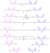
The sCy3–sCy5 conjugates used in this study: covalently linked (14) and based on oligonucleotide duplexes (D1, D2).
Figure 4
Figure 4
Structures of sCy3-sCy5 conjugates 14 and model compounds 4af and 5, 6.
Figure 5
Figure 5
Structures of sCy3-sCy5 oligonucleotide conjugates D1 and D2.
Figure 6
Figure 6
Absorption spectra of 6 and the equimolar mixture 5 + 6 in PBS. Inset: absorption spectra of 5 in PBS and 5 calculated by subtracting the spectrum of 6 from the 5 + 6 mixture spectrum.
Figure 7
Figure 7
Fluorescence decay kinetics of 5 (A) and 6 (B) measured in PBS for individual solutions and an equimolar mixture. (A) sCy3 fluorescence, ex. 510 nm, em. 560 nm; (B) sCy5 fluorescence, ex. 640 nm, em. 680 nm.
Figure 8
Figure 8
Normalized absorption spectra of (A) 4, 4a, 4c, 4e, 5 and (B) 4, 4b, 4d, 4f, 6 in 10 mM PBS.
Figure 9
Figure 9
Fluorescence decay kinetics of an equimolar 4, 4af, 5, and 6 measured in 10 mM PBS. (A) sCy3 fluorescence, ex. 510 nm, em. 560 nm; (B) sCy5 fluorescence, ex. 640 nm, em. 680 nm.
Figure 10
Figure 10
Absorption spectra of conjugate 4 at constant concentration in PBS in the presence of NaCl at various concentrations. Inset: absorbance ratios (sCy3 0–0 band vs. sCy5 0–0 band) at various NaCl concentrations.
Figure 11
Figure 11
Absorption spectra of conjugate 4 at constant concentrations in deionized water in the presence of various DMSO concentrations. The arrows show change in spectral properties of the conjugate with increasing DMSO concentration. Inset: ratio of absorbances at maxima in the same sample as a function of DMSO concentration in solution.
Figure 12
Figure 12
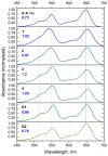
Absorption spectra of the 5 + 6 equimolar mixture, low molecular weight sCy3–sCy5 conjugates 14, and oligonucleotide conjugates D1 and D2 in PBS. Blue numbers indicate the maximum sCy3/sCy5 absorbance ratio.
Figure 13
Figure 13
Normalized fluorescence spectra of conjugates 14 and D1, D2 in PBS (excitation at 520 nm).
Figure 14
Figure 14
Fluorescence decay kinetics of 16, an equimolar 5+6 mixture, D1 and D2 in PBS; (A) sCy3 fluorescence, ex. 510 nm, em. 560 nm; (B) sCy5 fluorescence, ex. 640 nm, em. 680 nm.

Similar articles

References

    1. Levitus M., Ranjit S. Cyanine Dyes in Biophysical Research: The Photophysics of Polymethine Fluorescent Dyes in Biomolecular Environments. Quart. Rev. Biophys. 2011;44:123–151. doi: 10.1017/S0033583510000247. - DOI - PubMed
    1. Li Y., Zhou Y., Yue X., Dai Z. Cyanine Conjugate-Based Biomedical Imaging Probes. Adv. Healthcare Mater. 2020;9:2001327. doi: 10.1002/adhm.202001327. - DOI - PubMed
    1. Liu Y., Zhang D., Qu Y., Tang F., Wang H., Ding A., Li L. Advances in Small-Molecule Fluorescent pH Probes for Monitoring Mitophagy. Chem. Biomed. Imag. 2024;2:81–97. doi: 10.1021/cbmi.3c00070. - DOI - PMC - PubMed
    1. Mu J., Xiao M., Shi Y., Geng X., Li H., Yin Y., Chen X. The Chemistry of Organic Contrast Agents in the NIR-II Window. Angew. Chem. Int. Ed. 2022;61:e202114722. doi: 10.1002/anie.202114722. - DOI - PubMed
    1. Li K., Duan X., Jiang Z., Ding D., Chen Y., Zhang G.-Q., Liu Z. J-Aggregates of Meso-[2.2]Paracyclophanyl-BODIPY Dye for NIR-II Imaging. Nat. Commun. 2021;12:2376. doi: 10.1038/s41467-021-22686-z. - DOI - PMC - PubMed

MeSH terms

LinkOut - more resources