. 2025 Jan 3;30(1):155.
doi: 10.3390/molecules30010155.

Imaging the Ultrastructure of Isolated Peptidoglycan Sacculi from Rod-Shaped Helicobacter pylori J99 Cells by Atomic Force Microscopy

Affiliations

Imaging the Ultrastructure of Isolated Peptidoglycan Sacculi from Rod-Shaped Helicobacter pylori J99 Cells by Atomic Force Microscopy

Daniel Amiteye et al. Molecules. .

Abstract

Peptidoglycan is the basic structural polymer of the bacterial cell wall and maintains the shape and integrity of single cells. Despite years of research conducted on peptidoglycan's chemical composition, the microscopic elucidation of its nanoscopic architecture still needs to be addressed more thoroughly to advance knowledge on bacterial physiology. Apart from the model organism Escherichia coli, ultrastructural imaging data on the murein architecture of Gram-negative bacteria is mostly missing today. This study therefore intended to further our understanding of bacterial physiology by the isolation of peptidoglycan sacculi from the Gram-negative bacterium Helicobacter pylori J99 and the subsequent nanoscopic imaging of the murein network by Atomic Force Microscopy (AFM). With the ability to purify peptidoglycan sacculi from H. pylori J99 for AFM by a modified peptidoglycan isolation protocol, nanoscopic imaging of the murein network by intermittent-contact AFM in air and under liquid yielded ultrastructural insights into the H. pylori J99 cell wall architecture. High-resolution data acquisition on isolated peptidoglycan from H. pylori J99 by AFM under liquid was performed and revealed a molecular network similar to available data from E. coli. Subsequent enzymatic digestion of the isolated H. pylori J99 sacculi and analysis of the resulting fragments by +ESI-LCMS confirmed the presence of N-acetylglucosamine as an additional marker for successful peptidoglycan isolation. By comparison of the nanoscopic sacculus dimensions of H. pylori J99 to E. coli NU14, this study also identified specific differences in the sacculus morphology of both Gram-negative pathogenic bacteria.

Keywords: Atomic Force Microscopy; Helicobacter pylori J99; cell wall; peptidoglycan; ultrastructural imaging.


Conflict of interest statement

The authors declare no conflicts of interest.

Figures

Figure 1
Figure 1
Representative phase-contrast light microscopic image of a H. pylori J99 suspension culture at an OD600 of 0.2. The predominant cell shape under typical high-passage in vitro cultivation conditions was a rod-like morphology.
Figure 2
Figure 2
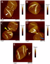
Representative AFM images of isolated, purified, and immobilized H. Pylori J99 (AD) and E. coli NU14 (E) peptidoglycan sacculi recorded by intermittent-contact mode AFM under ambient conditions in air. Intermittent-contact mode AFM imaging in air yielded valuable ultrastructural insights into general H. Pylori J99 peptidoglycan sacculus morphology. The expected nanoscopic murein structure with typical septal morphology (exemplary septum marked with white arrow in (A)) and delicate bands with perpendicular orientation to the growth axis of single bacteria cells (blue arrow in (D)) were clearly recognized. Note the overall rather low number of intracellular remnants as a result of the modified isolation protocol employed for H. Pylori J99 sacculus purification (representative intracellular remnant marked by yellow arrow in (C)). A representative peptidoglycan sacculus from E. coli NU14 of this study is shown in (E).
Figure 3
Figure 3
Representative morphological measurements on an isolated, purified, and immobilized H. Pylori J99 sacculus employing data collected by intermittent-contact mode AFM in air. Measurements are represented by typical section planes for each analyzed parameter (a and b). Dashed blue lines in a and b indicate reference points for the different measurements. Prior to any sectioning measurement, AFM images were flattened employing the 0th-order algorithm implemented in the software Nanoscope Analysis 3.0 (Bruker, Karlsruhe, Germany).
Figure 4
Figure 4
Violin plots representing measurements of morphological characteristics from isolated murein sacculi of H. pylori J99 compared to corresponding measurements on E. coli NU14 sacculi prepared by the same isolation protocol (AC). Altogether, nine isolated sacculi from both species were analyzed for each parameter (three sacculi for each of the three replicates per species). Each parameter was measured at least thrice for every sacculus. Statistical variance was analyzed employing the Student t-test algorithm implemented in the software GraphPad Prism 9 (n.s. = no significant difference, ** = p < 0.01, **** = p < 0.0001; GraphPad Prism 9, GraphPad Software, Boston, MA, USA). No significant differences were detected between the overall septum heights of H. pylori J99 and E. coli NU14 (A). Measurements of sacculus height (B) as well as sacculus roughness (C) data showed significant differences between the two Gram-negative species, indicating differing physiology of their peptidoglycan architecture and/or composition.
Figure 5
Figure 5
Representative AFM height channel images of isolated H. pylori J99 peptidoglycan sacculi imaged by intermittent-contact mode AFM under liquid conditions. The modified isolation protocol of this study reproducibly enabled lower nm resolution on isolated sacculi from H. pylori J99, although true molecular resolution on single peptidoglycan strands was not reached. Nanoscopic insights into the peptidoglycan architecture of H. pylori J99 were established by AFM imaging in TRIS/HCl buffer. Representative image of two complete sacculi is shown in (A), details in images (BD). Note the evenly distributed peptidoglycan network consisting of crosslinked macromolecules ((BD), single strand-like macromolecule indicated with blue arrow in (D)). Additionally, typical dense and less dense regions of peptidoglycan architecture were recognized in the AFM data, attributable to the highly dynamic ultrastructure of peptidoglycan (more dense area highlighted in (B), less dense area highlighted in (D)).
Figure 6
Figure 6
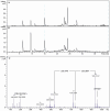
+ESI-LCMS of digested muropeptide of Helicobacter pylori J99. (A): Overview base peak chromatogram, full mass range (m/z 50–1500). (B): Extracted ion chromatogram indicative of the presence of the N-acetylglucosamine fragment at m/z 204.0866 [C8H13NO5 + H]+. (C): +ESI-MS spectrum of the peak at 15 min. The theoretical ion mass of the protonated N-acteylglucosamine-fragment [C8H14NO5]+ deviates from the measured ion mass of 204.0899 u by 3.2 mu, mΣ is 9.4.

References

    1. Hooi J.K.Y., Lai W.Y., Ng W.K., Suen M.M.Y., Underwood F.E., Tanyingoh D., Malfertheiner P., Graham D.Y., Wong V.W.S., Wu J.C.Y., et al. Global Prevalence of Helicobacter pylori Infection: Systematic Review and Meta-Analysis. Gastroenterology. 2017;153:420–429. doi: 10.1053/j.gastro.2017.04.022. - DOI - PubMed
    1. Fung T.C., Vuong H.E., Luna C.D.G., Pronovost G.N., Aleksandrova A.A., Riley N.G., Vavilina A., McGinn J., Rendon T., Forrest L.R., et al. Intestinal serotonin and fluoxetine exposure modulate bacterial colonization in the gut. Nat. Microbiol. 2019;4:2064–2073. doi: 10.1038/s41564-019-0540-4. - DOI - PMC - PubMed
    1. Cave D.R. How is Helicobacter pylori transmitted? Gastroenterology. 1997;113((6 Suppl.)):S9–S14. doi: 10.1016/S0016-5085(97)80004-2. - DOI - PubMed
    1. Dunne C., Dolan B., Clyne M. Factors that mediate colonization of the human stomach by Helicobacter pylori. World J. Gastroenterol. 2014;20:5610–5624. doi: 10.3748/wjg.v20.i19.5610. - DOI - PMC - PubMed
    1. Mobley H.L.T., Mendz G.L., Hazell S.L. Helicobacter Pylori. ASM Press; Washington, DC, USA: 2001. - DOI - PubMed

MeSH terms

LinkOut - more resources