Review
. 2025 Jan 2;17(1):108.
doi: 10.3390/polym17010108.

Leveraging Microneedles for Raised Scar Management

Affiliations
Review

Leveraging Microneedles for Raised Scar Management

Zhengyun Jin et al. Polymers (Basel). .

Abstract

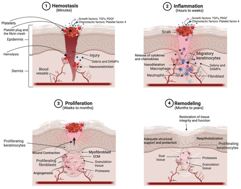
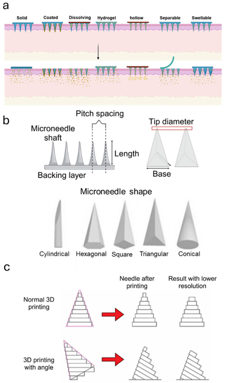
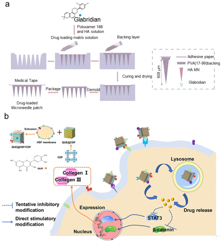
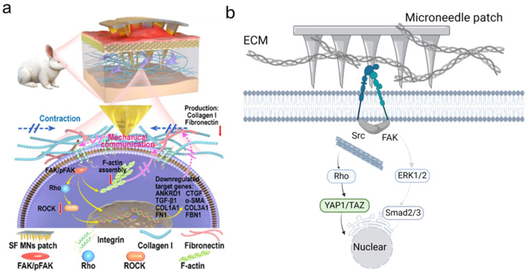
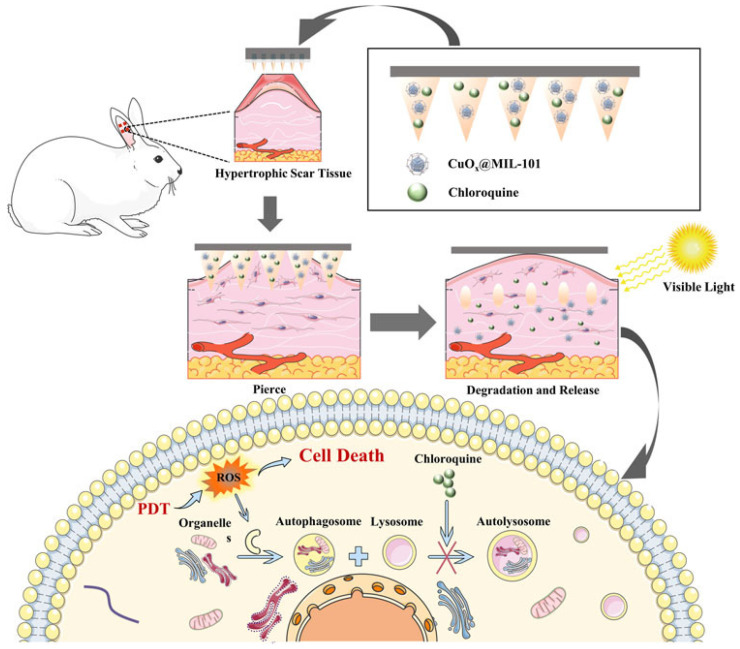
Disruption of the molecular pathways during physiological wound healing can lead to raised scar formation, characterized by rigid, thick scar tissue with associated symptoms of pain and pruritus. A key mechanical factor in raised scar development is excessive tension at the wound site. Recently, microneedles (MNs) have emerged as promising tools for scar management as they engage with scar tissue and provide them with mechanical off-loading from both internal and external sources. This review explores the mechanisms by which physical intervention of drug-free MNs alleviates mechanical tension on fibroblasts within scar tissue, thereby promoting tissue remodeling and reducing scar severity. Additionally, the role of MNs as an efficient cargo delivery system for the controlled and sustained release of a wide range of therapeutic agents into scar tissue is highlighted. By penetrating scar tissue, MNs facilitate controlled and sustained localized drug administration to modulate inflammation and fibroblastic cell growth. Finally, the remaining challenges and the future perspective of the field have been highlighted.

Keywords: drug delivery; hydrogels; hypertrophic scar; keloids; microneedle.


Conflict of interest statement

The authors declare no conflict of interest.

Figures

Figure 1
Figure 1
Schematic representation of different stages of wound healing [31]. Wound healing begins with hemostasis. Subsequently, damaged cells and platelet secretions recruit immune cells to the site, initiating the inflammatory phase. During this phase, immune cells release cytokines and chemokines to attract fibroblasts to the wound site and activate them. Activated fibroblasts differentiate into myofibroblasts, which deposit ECM components to form the structural framework for new tissue. Finally, in the remodeling phase, cellular activity significantly decreases, and myofibroblasts undergo programmed cell death (apoptosis).
Figure 2
Figure 2
Schematic representation of microneedle patch design, material selection, and fabrication processes. (a) Different types of MN [72]. (b) Key design factors, including microneedle shape, tip diameter, base size, pitch spacing, length, and tip angle, significantly influence penetration efficiency and functionality. Various shapes, such as cylindrical, hexagonal, square, triangular, and conical, are depicted. (c) Comparison of standard 3D printing and angled 3D printing methods, highlighting the impact of resolution and needle angle [60].
Figure 4
Figure 4
Corticosteroid-loaded MN patches for drug delivery. (a) Schematic representation of the bilayer MN patch fabrication process using the micromolding approach, illustrating the sequential loading of different drug formulations into the MN tips and shafts [46]. (b) Confocal microscopy images demonstrating the capability of MNs to compartmentalize different drugs between the tip and shaft regions for target delivery. (c) Photographic, optical coherence tomography (OCT), and histological images confirming effective MN penetration into the skin layers, showcasing their structural integrity and delivery efficiency. (d) Sequential photographs from the in vivo study highlighting the therapeutic efficacy of drug-loaded MN patches. Comparison of treated groups (drug-loaded MNs) with MNs alone and sham groups illustrates the superior performance and clinical potential of the drug-loaded patches in reducing symptoms. (eg) The commercialized design of MNs for drug delivery [90]. (e) Commercially available soluble MNs for transdermal drug delivery. (f) Scanned 3D photos of the MNs-treated keloid. (c) Photographs of keloid scars before and after 6 weeks of applying the drug-loaded MNs show improvement in scar elevation index.
Figure 5
Figure 5
Schematic illustration of the fabrication process and therapeutic mechanism of plant-extract-loaded MNs for raised scar management. (a) Plant-extract-based MNs loaded with MNs for keloid treatment [99]. (b) Engineered MNs leverages cell membrane-cloaked QUE@ CDF particles to target collagen deposition in hypertrophic scars by modulating Wnt/β-catenin and JAK2/STAT3 signaling pathways [101].
Figure 3
Figure 3
Schematic illustration of the physical intervention mechanism of microneedle patches. (a) Microneedle patches disrupt mechanical communication between cells and ECM by interfering with the integrin-FAK-mediated signaling pathway [23]. (b) Detailed representation of integrin-mediated mechanotransduction pathways, highlighting how microneedle-induced physical disruption affects mechanosensation. This intervention downregulates fibrotic gene transcription by inhibiting the integrin/FAK/ERK1-2 pathway, thereby reducing fibrotic responses and fibroproliferative activity (created with BioRender).
Figure 6
Figure 6
Schematic illustration of CuO₂@MIL-101 and chloroquine-loaded MNs for a synergistic strategy combining PDT and autophagy inhibition to treat HTSs [113]. The dissolving MNs penetrate the hypertrophic scar tissue, enabling localized delivery of CuO₂@MIL-101 and chloroquine. Upon visible light irradiation to nanoparticles, destructive ROS are generated through PDT, leading to cell death. Concurrently, chloroquine disrupts autophagy by inhibiting lysosome–autophagosome fusion, enhancing the therapeutic effect.

Similar articles

Cited by

References

    1. Lin X., Lai Y. Scarring skin: Mechanisms and therapies. Int. J. Mol. Sci. 2024;25:1458. doi: 10.3390/ijms25031458. - DOI - PMC - PubMed
    1. Monstrey S., Middelkoop E., Vranckx J.J., Bassetto F., Ziegler U.E., Meaume S., Téot L. Updated scar management practical guidelines: Non-invasive and invasive measures. J. Plast. Reconstr. Aesthetic Surg. 2014;67:1017–1025. doi: 10.1016/j.bjps.2014.04.011. - DOI - PubMed
    1. Pradhan M., Pethe P. Focus: Cell Fate: The Molecular Mechanisms Involved in the Hypertrophic Scars Post-Burn Injury. Yale J. Biol. Med. 2023;96:549. doi: 10.59249/RHUF5686. - DOI - PMC - PubMed
    1. Ogawa R. Update on Hypertrophic Scar Management in Burn Patients. Clin. Plast. Surg. 2024;51:349–354. doi: 10.1016/j.cps.2024.02.001. - DOI - PubMed
    1. Dohi T., Kuribayashi S., Tosa M., Aoki M., Akaishi S., Ogawa R. Z-plasty and postoperative radiotherapy for upper-arm keloids: An analysis of 38 patients. Plast. Reconstr. Surg.–Glob. Open. 2019;7:e2496. doi: 10.1097/GOX.0000000000002496. - DOI - PMC - PubMed

LinkOut - more resources