

Comparison of Non-Contrast CT vs. Contrast-Enhanced CT with Both Intravenous and Rectal Contrast Application for Diagnosis of Acute Colonic Diverticulitis: A Multireader, Retrospective Single-Center Study
- PMID: 39795557
- PMCID: PMC11719699
- DOI: 10.3390/diagnostics15010029
Comparison of Non-Contrast CT vs. Contrast-Enhanced CT with Both Intravenous and Rectal Contrast Application for Diagnosis of Acute Colonic Diverticulitis: A Multireader, Retrospective Single-Center Study
Abstract
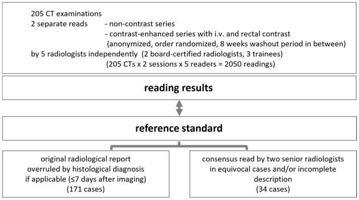
Objectives: To evaluate the non-inferiority of non-contrast CT compared to contrast-enhanced CT with both intravenous and rectal contrast application for the diagnosis of acute colonic diverticulitis. Methods: Five readers retrospectively evaluated the non-contrast and contrast-enhanced series of CTs of 205 consecutive patients with clinical suspicion of acute diverticulitis. Two randomized reading sessions, both containing all 205 cases as either contrast-enhanced or non-contrast (1:1) series, were performed with ≥8 weeks washout between them. The non-inferiority margin was set to 0.1. Results: The pooled prevalence (all readers) of diverticulitis was similar for non-contrast CT (63.9%, range: 60.5-65.0%) and contrast-enhanced CT (64.4%, 61.5-67.8%). Non-contrast CT was non-inferior for the diagnosis of diverticulitis (accuracy 0.90 [95% confidence interval: 0.89, 0.92]) compared to contrast-enhanced CT (0.92 [0.90, 0.94]; the difference in accuracy: -0.01 [-0.04, 0.01]) (normal deviate test: p-valueone-sided = 5.20 × 10⁻⁶). Sensitivities for perforation and abscess were slightly but significantly lower for the non-contrast CT than for the contrast-enhanced CT (differences: -0.15 [-0.20, -0.05], -0.17 [-0.27, -0.07]), while no differences in accuracies and specificities were observed. Conclusions: Non-contrast CT is non-inferior to contrast-enhanced CT (intravenous and rectal contrast) for the diagnosis of acute colonic diverticulitis. Contrast-enhanced CT is associated with significantly higher sensitivities for the presence of an abscess or perforation.
Keywords: colonic diverticulitis; computed tomography; contrast enema; non-contrast computed tomography; reader study.
Conflict of interest statement
The authors declare no conflicts of interest.
Figures


References
-
- Bharucha A.E., Parthasarathy G., Ditah I., Fletcher J.G., Ewelukwa O., Pendlimari R., Yawn B.P., Melton J.L., Schleck C., Zinsmeister A.R. Temporal Trends in the Incidence and Natural History of Diverticulitis: A Population-Based Study. Am. J. Gastroenterol. 2015;110:1589–1596. doi: 10.1038/ajg.2015.302. - DOI - PMC - PubMed
-
- Vennix S., Morton D.G., Hahnloser D., Lange J.F., Bemelman W.A., The Research Committee of the European Society of Coloproctocology Systematic Review of Evidence and Consensus on Diverticulitis: An Analysis of National and International Guidelines. Color. Dis. 2014;16:866–878. doi: 10.1111/codi.12659. - DOI - PubMed
-
- Andersen J.C., Bundgaard L., Elbrønd H., Laurberg S., Walker L.R., Støvring J., Danish Surgical Society Danish National Guidelines for Treatment of Diverticular Disease. Dan. Med. J. 2012;59:C4453. - PubMed
-
- National Institute for Health and Care Excellence (NICE) Diverticular Disease: Diagnosis and Management. National Institute for Health and Care Excellence (NICE); London, UK: 2019. NG 147. - PubMed
-
- Schultz J.K., Azhar N., Binda G.A., Barbara G., Biondo S., Boermeester M.A., Chabok A., Consten E.C.J., Van Dijk S.T., Johanssen A., et al. European Society of Coloproctology: Guidelines for the Management of Diverticular Disease of the Colon. Color. Dis. 2020;22:5–28. doi: 10.1111/codi.15140. - DOI - PubMed
LinkOut - more resources
Full Text Sources
Research Materials
