. 2024 Dec 24;26(1):51.
doi: 10.3390/ijms26010051.

Single Disulfide Bond in Host Defense Thanatin Analog Peptides: Antimicrobial Activity, Atomic-Resolution Structures and Target Interactions

Affiliations

Single Disulfide Bond in Host Defense Thanatin Analog Peptides: Antimicrobial Activity, Atomic-Resolution Structures and Target Interactions

Swaleeha Jaan Abdullah et al. Int J Mol Sci. .

Abstract

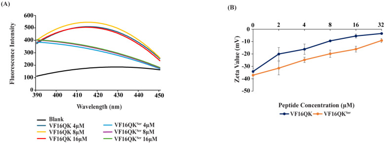
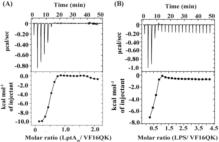
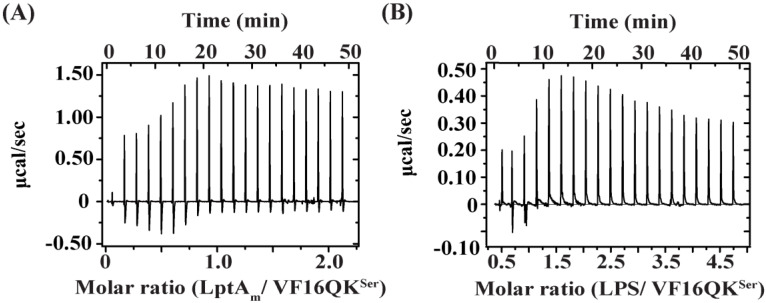
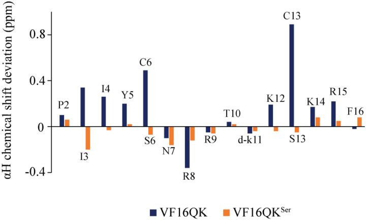
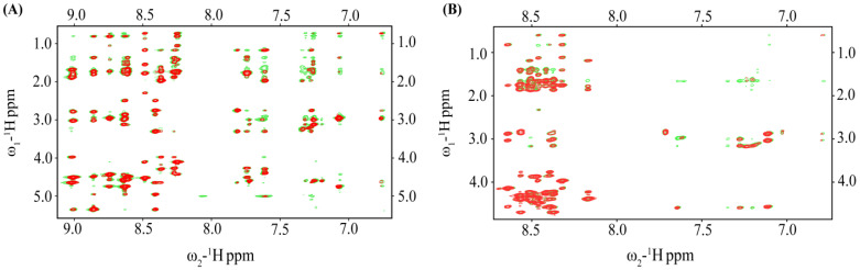
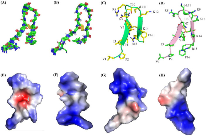
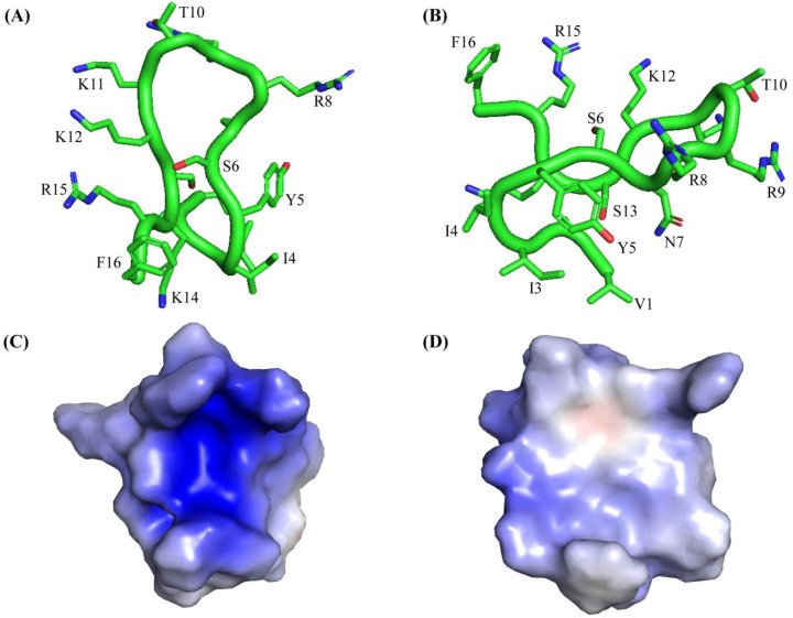
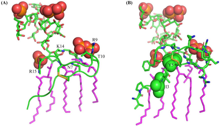
Host defense antimicrobial peptides (AMPs) are promising lead molecules with which to develop antibiotics against drug-resistant bacterial pathogens. Thanatin, an inducible antimicrobial peptide involved in the host defense of Podisus maculiventris insects, is gaining considerable attention in the generation of novel classes of antibiotics. Thanatin or thanatin-based analog peptides are extremely potent in killing bacterial pathogens in the Enterobacteriaceae family, including drug-resistant strains of Escherichia coli and Klebsiella pneumoniae. A single disulfide bond that covalently links two anti-parallel β-strands in thanatin could be pivotal to its selective antibacterial activity and mode of action. However, potential correlations of the disulfide covalent bond with structure, activity and target binding in thanatin peptides are currently unclear to. Here, we examined a 16-residue designed thanatin peptide, namely disulfide-bonded VF16QK, and its Cys to Ser substituted variant, VF16QKSer, to delineate their structure-activity relationships. Bacterial growth inhibitory activity was only detected for the disulfide-bonded VF16QK peptide. Mechanistically, both peptides vastly differ in their bacterial cell permeabilizations, atomic-resolution structures, interactions with the LPS-outer membrane and target periplasmic protein LptAm binding. In particular, analysis of the 3-D structures of the two peptides revealed an altered folded conformation for the VF16QKSer peptide that was correlated with diminished LPS-outer membrane permeabilization and target interactions. Analysis of docked complexes of LPS-thanatin peptides indicated potential structural requirements and conformational adaptation for antimicrobial activity. Collectively, these observations contrast with those for the disulfide-bonded β-hairpin antimicrobial protegrin and tachyplesin peptides, where disulfide bonds are dispensable for activity. We surmise that the atomistic structures and associated molecular interactions presented in this work can be utilized to design novel thanatin-based antibiotics.

Keywords: LPS; LptA; LptAm; NMR; host defense antimicrobial peptide; thanatin.


Conflict of interest statement

The authors declare no conflicts of interest.

Figures

Figure 1
Figure 1
(A) Fluorescence emission spectra of NPN as a function of concentrations of the VF16QK and VF16QKser peptides in E. coli cell solutions. 10 μM of fluorescence probes were prepared in 10 mM of sodium phosphate buffer with a fixed bacterial cell density of 0.5. Fluorescence spectra were recorded with an excitation wavelength of 350 nm and emission of 390–450 nm. (B) Plot showing z potential changes of E. coli cells as a function of concentrations, 2 to 32 μM, of the VF16QK and VF16QK peptides.
Figure 2
Figure 2
Isothermal titration calorimetry (ITC) of binding interactions of VF16QK with (A) LptAm, the periplasmic LPS transport protein of E. coli, and (B) the LPS-outer membrane. LptAm-peptide ITC studies were carried out in 50 mM of sodium phosphate buffer and 150 mM of NaCl at pH 7.0 and 25 °C. LptAm proteins in sample cells were titrated with 2 μL aliquots of peptides and the heat exchange was measured. For LPS–peptide interactions, LPS in sample cells were titrated with 2 μL aliquots of peptides in 10 mM of sodium phosphate buffer, pH 7.0, 37 °C.
Figure 3
Figure 3
Isothermal titration calorimetry (ITC) of binding interactions of (A) LptAm and (B) the LPS-outer membrane with the VF16QKser peptide.
Figure 4
Figure 4
Bar diagram showing secondary chemical shifts of αH resonances of residues of the VF16QK and VF16QKser peptides.
Figure 5
Figure 5
(A) Overlay of partial 1H-1H two-dimensional NOESY (red contour) and tr-NOESY (green contour) spectra of the VF16QK peptide and (B) overlay of partial 1H-1H two-dimensional NOESY (red contour) and tr-NOESY (green contour) spectra of the VF16QKser peptide. NOESY/tr-NOESY spectra show NOEs involving downfield shifted amide and aromatic proton resonances along the ω2 dimension with the upfield shifted aliphatic proton resonances along the ω1 dimension.
Figure 6
Figure 6
Superpositions of twenty CYANA-derived low-energy structures of the VF16QK peptide (A) in free solution (B) and in complex with LPS micelles. Ribbon representation showing backbone and sidechain orientations of the VF16QK peptide (C) in free solution and (D) in complex with LPS micelles. Electrostatic surface potential of the VF16QK peptide (E,F) in free solution and (G,H) in complex with LPS micelles.
Figure 7
Figure 7
(A,B) Ribbon representation of the structure of the VF16QKser peptide in free solution showing backbone folding and sidechain dispositions in two different orientations. (C,D) Electrostatic surface potential of the structure of the VF16QKser peptide in free solution in two different orientations.
Figure 8
Figure 8
Docked structure of LPS-VF16QK showing (A) potential ionic and polar interactions among residues R9, N7, T10, K14 and R15 with the phosphate groups (in red sphere) of the lipid A moiety of LPS and (B) potential non-polar packing interactions of the residues I3 and Y5 with the acyl chain of LPS.

Similar articles

References

    1. Roope L.S.J., Smith R.D., Pouwels K.B., Buchanan J., Abel L., Eibich P., Butler C.C., Tan P.S., Walker A.S., Robotham J.V., et al. The challenge of antimicrobial resistance: What economics can contribute. Science. 2019;364:eaau4679. doi: 10.1126/science.aau4679. - DOI - PubMed
    1. O’Neill J. The Review on Antimicrobial Resistance (AMR) Government of the United Kingdom; London, UK: 2016. Tackling drug-resistant infections globally: Final report and recommendations.
    1. Laxminarayan R., Sridhar D., Blaser M., Wang M., Woolhouse M. Achieving global targets for antimicrobial resistance. Science. 2016;353:874–875. doi: 10.1126/science.aaf9286. - DOI - PubMed
    1. Antimicrobial Resistance Collaborators Global burden of bacterial antimicrobial resistance in 2019: A systematic analysis. Lancet. 2022;399:629–655. doi: 10.1016/S0140-6736(21)02724-0. - DOI - PMC - PubMed
    1. European Antimicrobial Resistance Collaborators The burden of bacterial antimicrobial resistance in the WHO European region in 2019: A cross-country systematic analysis. Lancet Public Health. 2022;7:e897–e913. doi: 10.1016/S2468-2667(22)00225-0. - DOI - PMC - PubMed

MeSH terms

LinkOut - more resources