. 2024 Dec 26;26(1):113.
doi: 10.3390/ijms26010113.

Direct Vascular Effects of Angiotensin II (A Systematic Short Review)

Affiliations

Direct Vascular Effects of Angiotensin II (A Systematic Short Review)

György L Nádasy et al. Int J Mol Sci. .

Abstract

The octapeptide angiotensin II (Ang II) is a circulating hormone as well as a locally formed agonist synthesized by the angiotensin-converting enzyme (ACE) of endothelial cells. It forms a powerful mechanism to control the amount and pressure of body fluids. All main effects are directed to save body salt and water and ensure blood pressure under basic conditions and in emergencies. All blood vessels respond to stimulation by Ang II; the immediate response is smooth muscle contraction, increasing vascular resistance, and elevating blood pressure. Such effects are conveyed by type 1 angiotensin receptors (AT1Rs) located in the plasma membrane of both endothelial and vascular smooth muscle cells. AT1Rs are heterotrimeric G protein-coupled receptors (GPCRs), but their signal pathways are much more complicated than other GPCRs. In addition to Gq/11, the G12/13, JAK/STAT, Jnk, MAPK, and ERK 1/2, and arrestin-dependent and -independent pathways are activated because of the promiscuous attachment of different signal proteins to the intracellular G protein binding site and to the intracellular C terminal loop. Substantial changes in protein expression follow, including the intracellular inflammation signal protein NF-κB, endothelial contact proteins, cytokines, matrix metalloproteinases (MMPs), and type I protocollagen, eliciting the inflammatory transformation of endothelial and vascular smooth muscle cells and fibrosis. Ang II is an important contributor to vascular pathologies in hypertensive, atherosclerotic, and aneurysmal vascular wall remodeling. Such direct vascular effects are reviewed. In addition to reducing blood pressure, AT1R antagonists and ACE inhibitors have a beneficial effect on the vascular wall by inhibiting pathological wall remodeling.

Keywords: angiotensin II; angiotensin receptors; renin-angiotensin system; vascular effects; vascular remodeling.


Conflict of interest statement

The authors declare no conflicts of interest. The funders had no role in the design of the study; in the collection, analyses or interpretation of data; in the writing of the manuscript; or in the decision to publish the results.

Figures

Figure 1
Figure 1
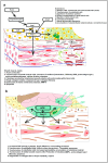
Signal pathways of the angiotensin type 1 receptor (AT1R) in vascular cells. (a). Dimerization of the receptor molecule might cause mutual inhibition. (b). The “classical” heterotrimeric G protein activation pathway (with Gαq/11) induces smooth muscle contraction. Increased intracellular Ca2+ and membrane DAG activate PKC enzyme subtypes with diverse effects. (c). “Promiscuous” association of the intracellular G protein binding site with other heterotrimeric G proteins, altered specificity is supposed to be controlled by different RGS proteins. (d). Receptor internalization, digestion, or recycling to the membrane after β-arrestin binding. threonin (T) and serine (S) phosphorylations by GRK kinases at the intracellular C terminal loop promote β-arrestin binding. The binding of heterotrimeric G proteins to their binding site will be prevented. (e). Diverse intracellular signal pathways are activated by C terminal phosphorylations and β-arresting binding. Abbreviations: Ang II, angiotensin II; AT1R, angiotensin type 1 receptor molecule; N, N terminal; C, C terminal; PLC, phospholipase C; IP3, inositol triphosphate; CaM, calmodulin; MLCK, myosin light chain kinase; Gq/11, G12/13, Gi/o, heterotrimeric G proteins with corresponding α subunits; RGS, regulator of G protein signaling; NO, nitrogen oxide; PG, prostaglandins; RhoA, small GTPase; ROCK, Rho-associated protein kinase; Arr, β-Arrestin; GRK, G protein-coupled receptor kinase; Ras, Raf, MEK, components in the MAP (mitogen-activated protein) kinase cascade; ERK-P, extracellular signal-regulated kinase, phosphorylated (activated) form; JAK/STAT, cytokine signal pathway; NADPH oxidase, main source of reactive oxygen species (ROS) in mammalian tissues; JNK, apoptotic signal protein; ADAM, membrane metalloproteinase, it cleaves off membrane-bound EGFR ligand; EGF, released peptide with EGF (epithelial growth factor) activity; EGFR, epithelial growth factor receptor. Blue color marks signal pathways through the G protein binding site, green color marks signal pathways through the C terminal loop.
Figure 2
Figure 2
Ang II-induced pathologic remodeling of the vascular wall. (a). Remodeling of the endothelium and of the media (b). Role of Ang II in atherosclerotic vessel wall remodeling. Abbreviations: Ang I, angiotensin I; Ang II, Angiotensin II; ACE, angiotensin-converting enzyme; MMP, matrix metalloproteinase; TGFbeta, transforming growth factor beta; ROS, reactive oxygen species.
Figure 3
Figure 3
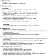
A schematic, concise description of direct angiotensin II effects on the vascular wall. Abbreviations: Ang II, angiotensin II; ACE, angiotensin-converting enzyme; AT1R, angiotensin type 1 receptor; GPCR, G protein-coupled receptor; Gα, α subunit of heterotrimeric G protein; PLC, phospholipase C, IP3, inositol triphosphate; MLCK, myosin light chain kinase; VSMC, vascular smooth muscle cell; NO, nitric oxide; Rho, small GTPase; ROCK, Rho-associated kinase; MLCP, myosin light chain phosphatase; GRB, growth receptor binding protein; MAPK, mitogen-activated protein kinase; ERK1/2, extracellular signal-regulated kinases; Src, nonreceptor tyrosine kinase; JAK, Janus kinase; STAT, signal transducer and activator of transcription; JNK, c-Jun NH2-terminal kinase; NADPH, nicotinamide adenine dinucleotide phosphate, oxidoreductase coenzyme; ROS, reactive oxygen species; TGFß, transforming growth factor type β; EGF, epithelial growth factor; MMP, matrix metalloproteinase.

References

    1. Wu C.H., Mohammadmoradi S., Chen J.Z., Sawada H., Daugherty A., Lu H.S. Renin-Angiotensin system and cardiovascular functions. Arterioscler. Thromb. Vasc. Biol. 2018;38:e108–e116. doi: 10.1161/ATVBAHA.118.311282. - DOI - PMC - PubMed
    1. Nadasy G.L., Balla A., Szekeres M. From living in saltwater to a scarcity of salt and water, and then an overabundance of salt—The biological roller-coaster to which the renin-angiotensin system has had to adapt: An editorial. Biomedicines. 2023;11:3004. doi: 10.3390/biomedicines11113004. - DOI - PMC - PubMed
    1. Castrop H., Höcherl K., Kurtz A., Schweda F., Todorov V., Wagner C. Physiology of kidney renin. Physiol. Rev. 2010;90:607–673. doi: 10.1152/physrev.00011.2009. - DOI - PubMed
    1. Sparks M.A., Crowley S.D., Gurley S.B., Mirotsou M., Coffman T.M. Classical renin-angiotensin system in kidney physiology. Compr. Physiol. 2014;4:1201–1228. - PMC - PubMed
    1. Broeker K.A.E., Schrankl J., Fuchs M.A.A., Kurtz A. Flexible and multifaceted: The plasticity of renin-expressing cells. Pfluegers Arch. Eur. J. Physiol. 2022;474:799–812. doi: 10.1007/s00424-022-02694-8. - DOI - PMC - PubMed

Publication types

MeSH terms

Substances

LinkOut - more resources