Review
. 2024 Dec 27;26(1):157.
doi: 10.3390/ijms26010157.

Recent Advances in the Search for Effective Anti-Alzheimer's Drugs

Affiliations
Review

Recent Advances in the Search for Effective Anti-Alzheimer's Drugs

Martyna Ogos et al. Int J Mol Sci. .

Abstract

Alzheimer's disease, the most common form of dementia, is characterized by the deposition of amyloid plaques and neurofibrillary tangles in the brain, leading to the loss of neurons and a decline in a person's memory and cognitive function. As a multifactorial disease, Alzheimer's involves multiple pathogenic mechanisms, making its treatment particularly challenging. Current drugs approved for the treatment of Alzheimer's disease only alleviate symptoms but cannot stop the progression. Moreover, these drugs typically target a single pathogenic mechanism, leaving other contributing factors unaddressed. Recent advancements in drug design have led to the development of multi-target-directed ligands (MTDLs), which have gained popularity for their ability to simultaneously target multiple pathogenic mechanisms. This paper focuses on analyzing the activity, mechanism of action, and binding properties of the anti-Alzheimer's MTDLs developed between 2020 and 2024.

Keywords: AChE inhibitors; Alzheimer’s disease; BChE inhibitors; donepezil; multi-target-directed ligands; neurodegenerative disorders; tacrine; β-amyloid aggregation inhibitors.


Conflict of interest statement

The authors declare no conflicts of interest.

Figures

Figure 3
Figure 3
Comparison of current therapies for Alzheimer’s disease with MTDL approaches.
Figure 4
Figure 4
Structure of tacrine.
Figure 5
Figure 5
Compound 1 derived from vilazodone and donepezil. Green shapes indicate fragments that contribute to the highest huAChE inhibition (four-methylene linker, unsubstituted phenyl ring, and para-substituted benzoyl).
Figure 6
Figure 6
Structure of compound 2. The green color indicates fragments that contribute to the highest huAChE inhibition (two methoxy groups and chlorine atom).
Figure 7
Figure 7
Structure of compound 3.
Figure 8
Figure 8
Structure of compound 4.
Figure 9
Figure 9
Structure of compound 5.
Figure 10
Figure 10
Structures of compounds 6, 7, and 8.
Figure 11
Figure 11
Structure of compound 9.
Figure 12
Figure 12
Structure of compound 10.
Figure 13
Figure 13
Structure of compound 11.
Figure 14
Figure 14
Structure of compound 12.
Figure 15
Figure 15
Structure of compound 13.
Figure 16
Figure 16
Structure of compound 14.
Figure 17
Figure 17
Structure of compound 15.
Figure 18
Figure 18
Compound 16 derived from isatin derivatives, triazine derivatives, and donepezil. Colors indicate similar structural motifs.
Figure 19
Figure 19
Structure of compound 17. The green color indicates fragments that contribute to the highest huChE inhibition (methyl substituents at carboxylic and phenolic groups and tacrine).
Figure 20
Figure 20
Structure of compound 18.
Figure 21
Figure 21
Structures of compound 19.
Figure 22
Figure 22
Structure of compound 20.
Figure 23
Figure 23
Structure of compound 21.
Figure 24
Figure 24
Structure of compound 22. The green color indicates fragments that contribute to the highest huAChE inhibition (four-methylene linker and 6-chlorotacrine).
Figure 25
Figure 25
Structure of compound 23.
Figure 26
Figure 26
Structure of compound 24.
Figure 27
Figure 27
Structure of compound 25.
Figure 28
Figure 28
Design of compound 27. Green shapes indicate fragments that contribute to the highest huBChE inhibition (alkyl substituents at carbamate, methoxy substituent of ferulic acid, and 1,2,3,4-tetrahydroisoquinoline).
Figure 29
Figure 29
Structure of compound 28. The green color indicates fragments that contribute to the highest eeAChE inhibition (substitution of tryptanthrin at position 2, ethylene linker, and amine group substituted with two methyl groups).
Figure 30
Figure 30
Structure of compound 29.
Figure 31
Figure 31
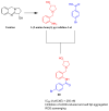
Design of compound 30 based on 1-(2-aminobenzyl)pyrrolidine-3-ol, derived from vasicine.
Figure 32
Figure 32
Effects of compounds 30, 59, and 60 (at doses of 2.5, 5, and 10 mg/kg) on scopolamine-administrated (Scop.) cognitive dysfunction mice, with donepezil (Don.) as reference in 5 mg/kg. Data are shown as mean ± SD. Each compound has its own positive and negative control as well as reference.
Figure 33
Figure 33
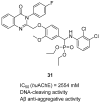
Structure of compound 31.
Figure 34
Figure 34
Structure of compound 32.
Figure 35
Figure 35
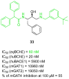
Structure of compound 33. The green color indicates fragments that contribute to the highest eqBChE inhibition (direct connection of benzhydryl moiety with amide group, amide group itself, and bulky substituent at benzylamine).
Figure 36
Figure 36
Design of compound 35 based on pitolisant and compound 34. Colors indicate common structural motifs.
Figure 37
Figure 37
Structure of compound 36. The green color indicates fragments that contribute to the highest huH3R affinity (five-methylene linker, para position of the alkoxyl chain, and fluorine atom).
Figure 38
Figure 38
Structure of compound 37.
Figure 39
Figure 39
Designs of compounds 40 and 41 from molecules 38 and 39. Green shapes indicate fragments that contribute to the highest huH3R affinity (pyrrolidine and piperidine rings).
Figure 40
Figure 40
Structure of compound 42. Green shape indicates fragments that contribute to the highest tau antiaggregating activity (thioxanthen-9-one with methoxy and methyl substituents). The violet color indicates the fragment that contributes to the highest anti-Aβ activity (thioxanthen-9-one).
Figure 41
Figure 41
Structures of compounds 43 and 44.
Figure 42
Figure 42
Structure of compound 45. The green color indicates the fragment that contributes to the highest anti-Aβ aggregation activity (arylalkyl fragment with one methylene group).
Figure 43
Figure 43
Structure of compound 46.
Figure 44
Figure 44
Structure of compound 47.
Figure 45
Figure 45
Design of compound 49 from molecule 48. Green shapes indicate fragments that contribute to the highest GSK-3β inhibitory activity (methoxy group, fluorine atom, and methylene linker). The violet shape shows the fragment that significantly improves DYRK1A inhibitory activity (methoxy substituent).
Figure 46
Figure 46
Design of compound 52 from molecules 50 and 51. Colors represent the main pharmacophoric features.
Figure 47
Figure 47
Structure of compound 53.
Figure 48
Figure 48
Structure of compound 54. The green color indicates fragments that contribute to the highest huMAO-B inhibitory activity (methyl substituent at 3-hydroxypyridin-4-one and methoxy group at position 7 of the coumarin scaffold).
Figure 49
Figure 49
Structure of compound 55. The green color indicates the fragment that contributes to the metal chelating properties (hydroxyphenylbenzimidazole).
Figure 50
Figure 50
Structure of compound 56. The green color indicates the fragment that contributes to the metal chelating activity (amine group).
Figure 51
Figure 51
Structure of compound 57. The carbamate group and methoxy substituent at the indole moiety (highlighted in green) are involved in the significant antioxidant activity of the compound.
Figure 52
Figure 52
Structure of compound 58. The green color indicates fragments that contribute to the highest huBACE1 activity (methoxy groups and styryl-thiazole core).
Figure 53
Figure 53
Structure of compound 59.
Figure 54
Figure 54
Structure of compound 60.
Figure 55
Figure 55
Design of compound 62 from compound 61. Methoxy substituent at β-carboline and complete aromatization (colored in green) contribute to increased NMDAR inhibitory activity.
Figure 56
Figure 56
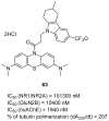
Structure of compound 63.
Figure 1
Figure 1
Pathogenesis of Alzheimer’s disease.
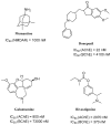
Figure 2
Figure 2
Structures and activities of approved anti-Alzheimer agents.

References

    1. World Health Organisation Dementia. 2023. [(accessed on 30 December 2023)]. Available online: https://www.who.int/news-room/fact-sheets/detail/dementia.
    1. Sheppard O., Coleman M. Alzheimer’s Disease: Drug Discovery. Exon Publications; Brisbane, QLD, Australia: 2020. Alzheimer’s Disease: Etiology, Neuropathology and Pathogenesis; pp. 1–22. - DOI - PubMed
    1. Alarcón-Espósito J., Mallea M., Rodríguez-Lavado J. From Hybrids to New Scaffolds: The Latest Medicinal Chemistry Goals in Multi-target Directed Ligands for Alzheimer’s Disease. Curr. Neuropharmacol. 2021;19:832–867. doi: 10.2174/1570159X18666200914155951. - DOI - PMC - PubMed
    1. Sun X.T., Jin L., Ling P.X. Review of drugs for Alzheimer’s disease. Drug Discov. Ther. 2012;6:285–290. doi: 10.5582/ddt.2012.v6.6.285. - DOI - PubMed
    1. Tiwari S., Atluri V., Kaushik A., Yndart A., Nair M. Alzheimer’s disease: Pathogenesis, diagnostics, and therapeutics. Int. J. Nanomedicine. 2019;14:5541–5554. doi: 10.2147/IJN.S200490. - DOI - PMC - PubMed