. 2024 Dec 30;26(1):230.
doi: 10.3390/ijms26010230.

Systematic Evaluation of Extracellular Coating Matrix on the Differentiation of Human-Induced Pluripotent Stem Cells to Cortical Neurons

Affiliations

Systematic Evaluation of Extracellular Coating Matrix on the Differentiation of Human-Induced Pluripotent Stem Cells to Cortical Neurons

Siyao Li et al. Int J Mol Sci. .

Abstract

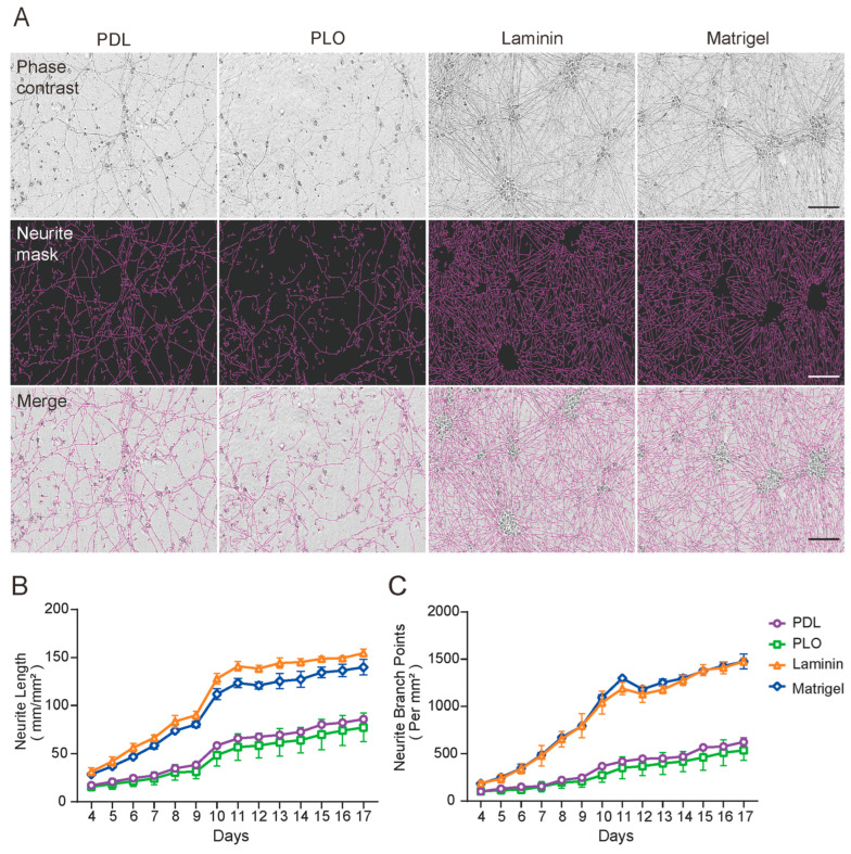
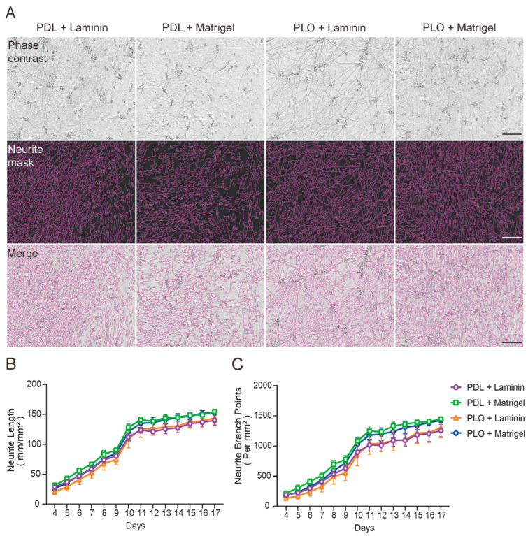
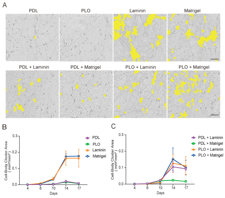
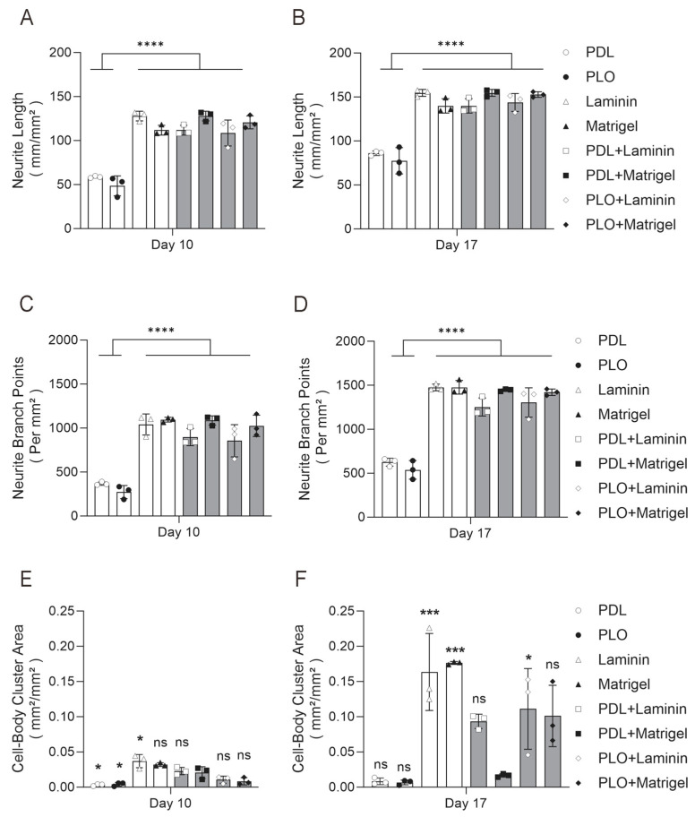
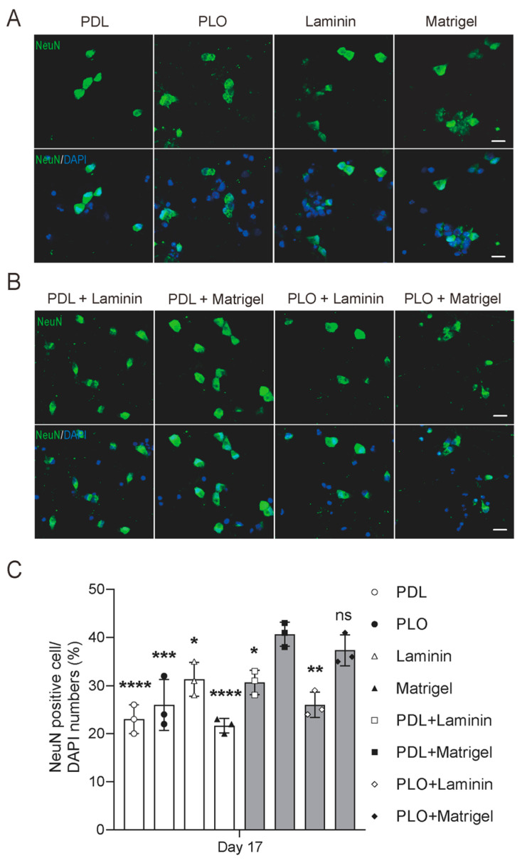
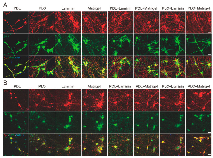
Induced pluripotent stem cell (iPSC)-derived neurons (iNs) have been widely used as models of neurodevelopment and neurodegenerative diseases. Coating cell culture vessels with extracellular matrixes (ECMs) gives structural support and facilitates cell communication and differentiation, ultimately enhances neuronal functions. However, the relevance of different ECMs to the natural environment and their impact on neuronal differentiation have not been fully characterized. In this study, we report the use of four commonly used extracellular matrixes, poly-D-lysine (PDL), poly-L-ornithine (PLO), Laminin and Matrigel, which we applied to compare the single-coating and double-coating conditions on iNs differentiation and maturation. Using the IncuCyte live-cell imaging system, we found that iNs cultured on single Matrigel- and Laminin-coated vessels have significantly higher density of neurite outgrowth and branch points than PLO or PDL but produce abnormal highly straight neurite outgrowth and larger cell body clumps. All the four double-coating conditions significantly reduced the clumping of neurons, in which the combination of PDL+Matrigel also enhanced neuronal purity. Double coating with PDL+Matrigel also tended to improve dendritic and axonal development and the distribution of pre and postsynaptic markers. These results demonstrate that the extracellular matrix contributes to the differentiation of cultured neurons and that double coating with PDL+Matrigel gives the best outcomes. Our study indicates that neuronal differentiation and maturation can be manipulated, to a certain extent, by adjusting the ECM recipe, and provides important technical guidance for the use of the ECM in neurological studies.

Keywords: induced pluripotent stem cells (iPSCs); live-cell imaging; morphology; neurite outgrowth; neuronal differentiation; synaptic markers.


Conflict of interest statement

The authors declare no conflicts of interest.

Figures

Figure 1
Figure 1
iNs cultured on Matrigel- and Laminin-coated vessels have a significantly higher density of neurite and branch points than PLO or PDL. iNs were cultured on a 96-well plate coated with a single matrix and their differentiation was monitored using the IncuCyte live-cell imaging system. (A) Representative images of iNs cultured for 17 days. The top panel shows phase-contrast images, the middle panel shows neurons identified by the Incucyte NeuroTrack algorithm (purple), and the bottom panel shows composite images of the identified neurons and phase-contrast field. Scale bars are 100 μm. (B,C) iNs were monitored continuously from day 4 to 17 and images were collected at 24 h intervals. The neurite length and branch points at each time points were quantified using the NeuroTrack algorithm. The values shown represent the mean (±SD) from triplicate wells for each treatment. Single coatings of Laminin (orange) and Matrigel (blue) resulted in significantly higher neurite length and branch points in iNs compared to PDL (purple) and PLO (green).
Figure 2
Figure 2
iNs cultured under different double-coating conditions yield similar and robust neurite outgrowth. iNs were cultured on a 96-well plate with double-coating matrixes and their differentiation monitored using the IncuCyte live-cell imaging system. (A) Representative images of iNs cultured for 17 days. The top panel shows phase-contrast images, the middle panel shows neurons identified by the Incucyte NeuroTrack algorithm (purple), and the bottom panel shows composite images of the identified neurons and the phase-contrast field. Scale bars are 100 μm. (B,C) iNs were monitored continuously from day 4 to 17 and images were collected at 24 h intervals. Neurite length and branch points at each time points were quantified using the NeuroTrack algorithm. The values shown represent the mean (±SD) from triplicate wells for each treatment. No significant differences in iNs differentiation were observed between double coatings of PDL+Laminin (purple), PDL+Matrigel (green), PLO+Laminin (orange) and PLO+Matrigel (blue).
Figure 3
Figure 3
PDL+Matrigel double coating significantly reduces clumping of induced neurons. Images were collected at 2–4 days intervals from iNs day 4 to 17 and the area of cell body clusters was identified and quantified by the Incucyte NeuroTrack algorithm. (A) Representative images of iNs on day 17 and cell body clusters are masked (yellow). Scale bars are 100 μm. (B) Single-coating comparison shows that Laminin (orange) and Matrigel (blue) led to significantly more cell body clumps than PDL (purple) and PLO (green). (C) Among the four double-coating conditions, PDL+Matrigel (green) resulted in the lowest area of cell body clusters when compared to PDL+Laminin (purple), PLO+Laminin (orange) and PLO+Matrigel (blue). Only the cell body clusters with a size >400 μm2 were counted. The values represent the mean (±SD) from triplicate wells for each treatment.
Figure 4
Figure 4
Comprehensive live-cell comparison of single-coating and double-coating conditions shows that PDL+Matrigel is the optimal substrate for long-term neuronal culture. Images collected at iNs day 10 and 17 (in Figure 1, Figure 2 and Figure 3) were statistically compared for their neurite length, neurite branch points and cell body cluster area. (A,B) Neurite length of iNs cultured at day 10 and 17 under four single-coating and four double-coating conditions. (C,D) Neurite branch points of iNs cultured at day 10 and 17 under four single-coating and four double-coating conditions. (E,F) Cell body cluster area of iNs cultured at day 10 and 17 under four single-coating and four double-coating conditions. Data represent the mean values (±SD) from triplicate wells for each condition. Single-coating conditions included Laminin, Matrigel, PDL and PLO. Double-coating conditions included PDL+Matrigel, PDL+Laminin, PLO+Laminin and PLO+Matrigel. Statistical analysis was conducted using one-way ANOVA followed by Tukey’s multiple comparison test. Significant differences are denoted as **** p < 0.0001; *** p < 0.001; * p < 0.05; ns (not significant) p > 0.05.
Figure 5
Figure 5
PDL+Matrigel double-coating condition enhances the purity of induced neurons. At 17 days post-induction, iNs were fixed and stained for the mature neuronal marker NeuN (green). Cell nuclei were stained with DAPI (blue). Scale bar: 20 μm. (A,B) Representative immunofluorescence images collected at iNs day 17 show co-localization of NeuN and DAPI. (C) Quantification of NeuN-positive cells versus total cells shows the purity of iNs cultured under different coating conditions. Data represent the mean values (±SD) from triplicate wells for each condition. Single-coating conditions included PDL, PLO, Laminin and Matrigel. Double-coating conditions included PDL+Matrigel, PDL+Laminin, PLO+Laminin and PLO+Matrigel. Statistical analysis was conducted using one-way ANOVA followed by Tukey’s multiple comparison test. Significant differences are denoted as **** p < 0.0001, *** p < 0.001, ** p < 0.01, * p < 0.05, ns (not significant) p > 0.05.
Figure 6
Figure 6
Expression of neuronal and synaptic markers tends to be improved in iNs cultured under PDL+Matrigel double-coating condition. At 17 days post-induction, iNs were fixed and stained for an array of neuronal markers. (A) Representative images showing immunostaining for axons (K9JA, red) and dendrites (MAP2, green) under eight coating conditions. (B) Representative images showing immunostaining for presynaptic marker Synapsin-1 (Syn1, red) and postsynaptic marker Postsynaptic Density Protein 95 (PSD-95, green) under eight coating conditions. Scale bar: 20 μm.

Similar articles

Cited by

References

    1. Bayati A., Ayoubi R., Aguila A., Zorca C.E., Deyab G., Han C., Recinto S.J., Nguyen-Renou E., Rocha C., Maussion G., et al. Modeling Parkinson’s disease pathology in human dopaminergic neurons by sequential exposure to α-synuclein fibrils and proinflammatory cytokines. Nat. Neurosci. 2024;27:2401–2416. doi: 10.1038/s41593-024-01775-4. - DOI - PubMed
    1. Sun Z., Kwon J.-S., Ren Y., Chen S., Walker C.K., Lu X., Cates K., Karahan H., Sviben S., Fitzpatrick J.A.J., et al. Modeling late-onset Alzheimer’s disease neuropathology via direct neuronal reprogramming. Science. 2024;385:adl2992. doi: 10.1126/science.adl2992. - DOI - PMC - PubMed
    1. Mehta S.R., Tom C.M., Wang Y., Bresee C., Rushton D., Mathkar P.P., Tang J., Mattis V.B. Human Huntington’s Disease iPSC-Derived Cortical Neurons Display Altered Transcriptomics, Morphology, and Maturation. Cell Rep. 2018;25:1081–1096 e6. doi: 10.1016/j.celrep.2018.09.076. - DOI - PubMed
    1. Tsitkov S., Valentine K., Kozareva V., Donde A., Frank A., Lei S., EVan Eyk J., Finkbeiner S., Rothstein J.D., Thompson L.M., et al. Disease related changes in ATAC-seq of iPSC-derived motor neuron lines from ALS patients and controls. Nat. Commun. 2024;15:3606. doi: 10.1038/s41467-024-47758-8. - DOI - PMC - PubMed
    1. Vierbuchen T., Ostermeier A., Pang Z.P., Kokubu Y., Südhof T.C., Wernig M. Direct conversion of fibroblasts to functional neurons by defined factors. Nature. 2010;463:1035–1041. doi: 10.1038/nature08797. - DOI - PMC - PubMed

LinkOut - more resources